. 2018 Dec 11;115(50):E11780-E11789.
doi: 10.1073/pnas.1813572115. Epub 2018 Nov 14.

DksA-DnaJ redox interactions provide a signal for the activation of bacterial RNA polymerase

Affiliations

DksA-DnaJ redox interactions provide a signal for the activation of bacterial RNA polymerase

Ju-Sim Kim et al. Proc Natl Acad Sci U S A. .

Abstract

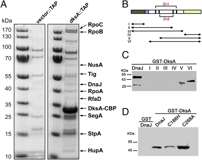
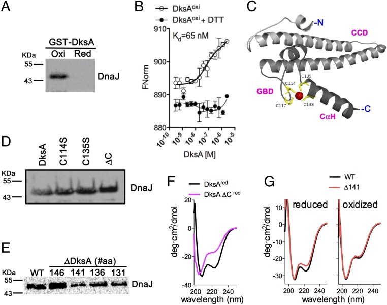
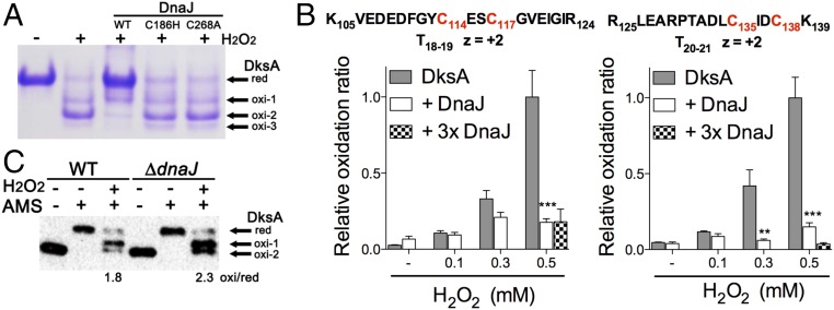
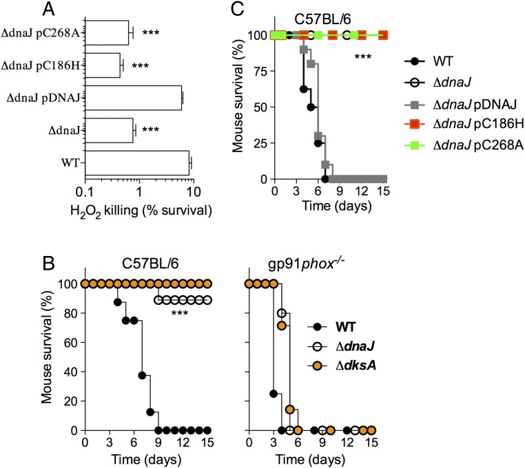
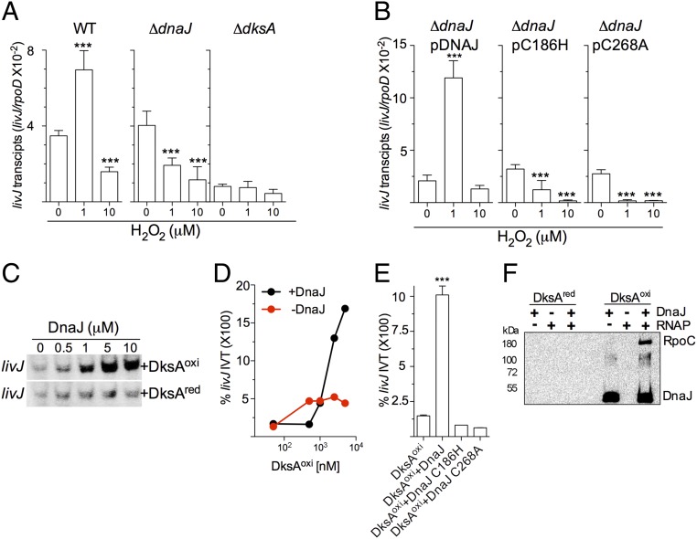
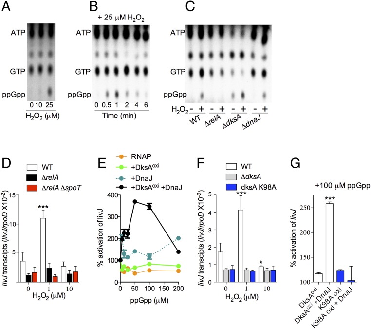
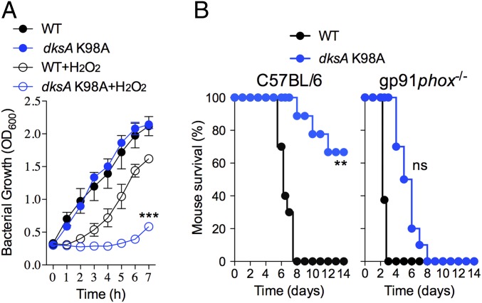
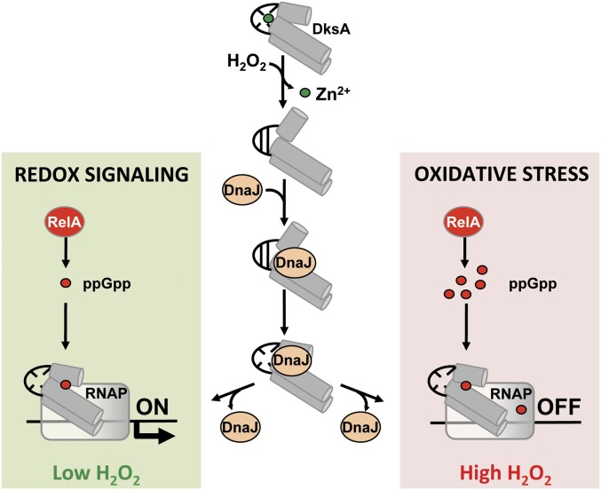
RNA polymerase is the only known protein partner of the transcriptional regulator DksA. Herein, we demonstrate that the chaperone DnaJ establishes direct, redox-based interactions with oxidized DksA. Cysteine residues in the zinc finger of DksA become oxidized in Salmonella exposed to low concentrations of hydrogen peroxide (H2O2). The resulting disulfide bonds unfold the globular domain of DksA, signaling high-affinity interaction of the C-terminal α-helix to DnaJ. Oxidoreductase and chaperone activities of DnaJ reduce the disulfide bonds of its client and promote productive interactions between DksA and RNA polymerase. Simultaneously, guanosine tetraphosphate (ppGpp), which is synthesized by RelA in response to low concentrations of H2O2, binds at site 2 formed at the interface of DksA and RNA polymerase and synergizes with the DksA/DnaJ redox couple, thus activating the transcription of genes involved in amino acid biosynthesis and transport. However, the high concentrations of ppGpp produced by Salmonella experiencing oxidative stress oppose DksA/DnaJ-dependent transcription. Cumulatively, the interplay of DksA, DnaJ, and ppGpp on RNA polymerase protects Salmonella from the antimicrobial activity of the NADPH phagocyte oxidase. Our research has identified redox-based signaling that activates the transcriptional activity of the RNA polymerase regulator DksA.

Keywords: DnaJ; Salmonella; oxidation; redox signaling; stringent response.


Conflict of interest statement

The authors declare no conflict of interest.

Figures

Fig. 1.
Fig. 1.
DnaJ is a binding partner of DksA. (A) Putative DksA binding partners were identified by mass spectrometry of cytoplasmic proteins isolated by TAP from ΔdksA Salmonella harboring pWSK29::TAP (Vector::TAP) or pWSK29::dksA::TAP (dksA::TAP) plasmids. TAP-purified proteins separated in SDS/PAGE gels were visualized by Coomassie Brilliant blue staining. The positions of DksA-binding partners are indicated by arrows. (B) Schematic representation of DnaJ domains and DnaJ truncated proteins (I–VI). N-terminal J (gray) and glycine- and phenylalanine-rich GF domains (blue), followed by two zinc fingers (black and red brackets), Zn1 and Zn2, and the C-terminal domain (green). The biochemical pull-down assays using (C and D) GST-DksA as bait and truncated DnaJ variants I–VI (C) or C186H and C268A DnaJ variants (D) as prey were performed in the absence of DTT. The GST protein (D) was used as a negative control. DnaJ proteins isolated in the pull-down assays were detected by Western blotting. Data are representative of three independent experiments.
Fig. 2.
Fig. 2.
DksA oxidation triggers high affinity binding to DnaJ. (A) Binding of DksA oxidized with H2O2 to DnaJ was assessed in Western blots using pull-down assays, as described in Fig. 1. (B) Binding affinity of the DksA–DnaJ redox couple was estimated by microscale thermophoresis. The data are the mean ± SD from three independent experiments. (C) Diagram of DksA modeled from the E. coli structure PDB ID code 5W1T (7), showing the coiled-coil domain (CCD), globular domain (GBD) with the four cysteines in the zinc finger, and C-terminal α-helix (CαH). Interactions of DnaJ (prey) with DksA variants (bait) bearing mutations in cysteine residues (D) or truncations in the C-terminal domain (E). DnaJ proteins pulled-down in A, D, and E were detected by Western blotting. A DksA variant missing all four cysteine residues (ΔC) was used for comparison. DksA truncated proteins are identified by the position of the deletion at the C-terminal domain. (F and G) The α-helical secondary structure of DksA proteins was analyzed by CD spectroscopy. Some of the proteins in F and G were reduced with DTT before analysis. The data are representative of two to three independent experiments and each spectrum shows the average of three independent scans.
Fig. 3.
Fig. 3.
The oxidoreductase activity of DnaJ resolves disulfide bonds in oxidized DksA. (A) The redox state of 5 μM DksA was evaluated by nonreducing SDS/PAGE and Commassie Brilliant blue staining after reduced cysteine residues were alkylated with the thiol trapper AMS. Where indicated, the specimens were treated with 1 mM H2O2 in the presence or absence of 5 μM DnaJ. (B) Abundance of intramolecular disulfide bonds between DksA Cys114-Cys117 and Cys135-Cys138 residues was determined by mass spectrometry. DksA proteins were alkylated with NEM after treatment with H2O2 in the presence or absence of DnaJ. Select samples were treated with a threefold molar excess of DnaJ (3× DnaJ). (C) Redox state of DksA in anaerobically grown Salmonella was evaluated by AMS-derivatization and Western blotting. Selected samples were treated with 5 μM H2O2 for 5 min before the thiols were derivatized with AMS. The ratio of the sum of oxidized species to reduced DnaJ within H2O2-treated, AMS-derivatized DksA was calculated by densitometry. Data in AC are from three independent experiments; **P < 0.01 and ***P < 0.001, according to two-way ANOVA.
Fig. 4.
Fig. 4.
DnaJ contributes to antioxidant defense and Salmonella pathogenesis. (A) Susceptibility of wild-type Salmonella and the indicated mutants to 200 μM H2O2; ***P < 0.001 as determined by one-way ANOVA (n = 12). (B and C) C57BL/6 and congenic gp91phox−/− mice were challenged with ∼100 CFU of the indicated Salmonella strains. The fraction of mice that survive the challenge was followed over time. The data are from 9 to 10 mice evaluated in two independent experiments; ***P < 0.001 as determined by log-rank analysis.
Fig. 5.
Fig. 5.
DnaJ facilitates interactions of oxidized DksA with RNA polymerase. Normalized abundance of livJ transcripts in WT, ΔdksA, and ΔdnaJ Salmonella (A) or ΔdnaJ bacteria complemented with the indicated dnaJ alleles (B) during exponential growth in an anaerobic chamber. Indicated cultures were left untreated, or treated with 1 or 10 μM H2O2 in EGCA minimal media. ***P < 0.001 as determined by one-way ANOVA compared with untreated controls (n = 6). The abundance of livJ message was normalized to transcripts of the housekeeping gene rpoD. Data are the mean ± SD from six independent experiments collected on 2 d. (C) Autoradiograms of [32-P]-UTP-labeled livJ in vitro transcripts from reactions containing E. coli RNA polymerase together with reduced or oxidized DksA and increasing concentrations of DnaJ. The products of the reactions were separated by SDS/PAGE. The blot is representative of three independent experiments. (D and E) DnaJ- and DksA-dependent activation of livJ in vitro transcription was evaluated by qRT-PCR. Reactions in D contained increasing concentrations of oxidized DksA in the presence or absence of 5 μM DnaJ. Reactions in E contained 5 μM of DnaJ and 1 μM of the indicated DksA variant. Data are the mean ± SD from three independent experiments. (F) Interactions of reduced and oxidized DksA proteins with S. Typhimurium RNA polymerase in the presence or absence of DnaJ. Oxidized and reduced DksA proteins in F were used as bait. DnaJ and the RpoC subunit of RNA polymerase were detected by immunobloting. The data in F are representative of three independent experiments.
Fig. 6.
Fig. 6.
Guanosine tetraphosphate synergizes with the DksA/DnaJ redox couple to regulate transcription. (AC) TLC autoradiograms of nucleotides labeled with inorganic 32-P. Nucleotides were extracted from aerobic wild-type (A and B) or mutant Salmonella (C) at 1 min (A and C) or at the indicated times (B) after H2O2 exposure. All autoradiograms are representative of three independent experiments. (D and F) Abundance of livJ transcripts in log phase anaerobic Salmonella that were treated with the indicated concentrations of H2O2. The data, which are normalized to internal transcripts of the housekeeping gene rpoD, are the mean ± SD from four independent experiments. (E and G) livJ in vitro transcription reactions containing ppGpp and 5 μM of oxidized DksA and/or DnaJ. The ability of recombinant WT and K98A DksA proteins to activate livJ in vitro transcription is compared in G. Data are the mean ± SD from four independent experiments. *P < 0.05 and ***P < 0.001 compared with untreated (D and F) or DksAoxi (G) controls as determined by two-way (D and F) or one-way (G) ANOVA.
Fig. 7.
Fig. 7.
Guanosine tetraphosphate binding to site 2 in the interface formed between DksA and RNA polymerase contributes to the antioxidant defenses of Salmonella. (A) Growth of WT and dksA K98A Salmonella in EGCA minimal medium. Indicated cultures were treated with 500 μM H2O2. ***P < 0.001 compared with WT + H2O2 as determined by two-way ANOVA. (B) C57BL/6 and congenic gp91phox−/− mice were inoculated intraperitoneally with ∼100 CFU of either WT or dksA K98A Salmonella. Mouse survival data are from 9 to 10 mice per bacterial strain and were collected from two independent experiments. **P < 0.01 or nonsignificantly different (ns) as determined by log-rank analysis.
Fig. 8.
Fig. 8.
Model for the redox-dependent, DksA–DnaJ regulation of transcription. Oxidation of cysteine residues destroys the zinc finger of DksA. The consequent conformational changes in the globular domain and C-terminal α-helix of oxidized DksA trigger high-affinity binding to DnaJ. The oxidoreductase and chaperone activities of DnaJ resolve disulfide bonds and help load DksA into RNA polymerase. Concomitantly, the accumulation of deacylated tRNAs following H2O2-induced amino acid auxotrophies stimulates production of guanosine tetraphosphate (ppGpp) from RelA bound to ribosomes. The low concentrations of ppGpp generated at low concentrations of H2O2 binds at site 2 formed in the interface of DksA and RNA polymerase, activating transcription of amino acid biosynthetic genes as part of the adaptation of Salmonella to redox signaling. However, our biochemical data suggest that the high concentrations of ppGpp produced in response to high levels of H2O2 inhibit transcription in Salmonella undergoing oxidative stress, perhaps by binding to sites 1 and 2 of RNA polymerase.

Comment in

Similar articles

Cited by

References

    1. Potrykus K, Cashel M. (p)ppGpp: Still magical? Annu Rev Microbiol. 2008;62:35–51. - PubMed
    1. Paul BJ, Ross W, Gaal T, Gourse RL. rRNA transcription in Escherichia coli. Annu Rev Genet. 2004;38:749–770. - PubMed
    1. Mechold U, Potrykus K, Murphy H, Murakami KS, Cashel M. Differential regulation by ppGpp versus pppGpp in Escherichia coli. Nucleic Acids Res. 2013;41:6175–6189. - PMC - PubMed
    1. Ross W, Vrentas CE, Sanchez-Vazquez P, Gaal T, Gourse RL. The magic spot: A ppGpp binding site on E. coli RNA polymerase responsible for regulation of transcription initiation. Mol Cell. 2013;50:420–429. - PMC - PubMed
    1. Zuo Y, Wang Y, Steitz TA. The mechanism of E. coli RNA polymerase regulation by ppGpp is suggested by the structure of their complex. Mol Cell. 2013;50:430–436. - PMC - PubMed

Publication types

MeSH terms

LinkOut - more resources