. 2019 Jan 17;93(3):e01709-18.
doi: 10.1128/JVI.01709-18. Print 2019 Feb 1.

Conformational Differences between Functional Human Immunodeficiency Virus Envelope Glycoprotein Trimers and Stabilized Soluble Trimers

Affiliations

Conformational Differences between Functional Human Immunodeficiency Virus Envelope Glycoprotein Trimers and Stabilized Soluble Trimers

Luis R Castillo-Menendez et al. J Virol. .

Abstract

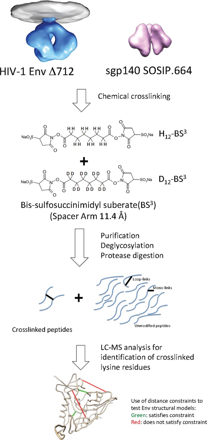
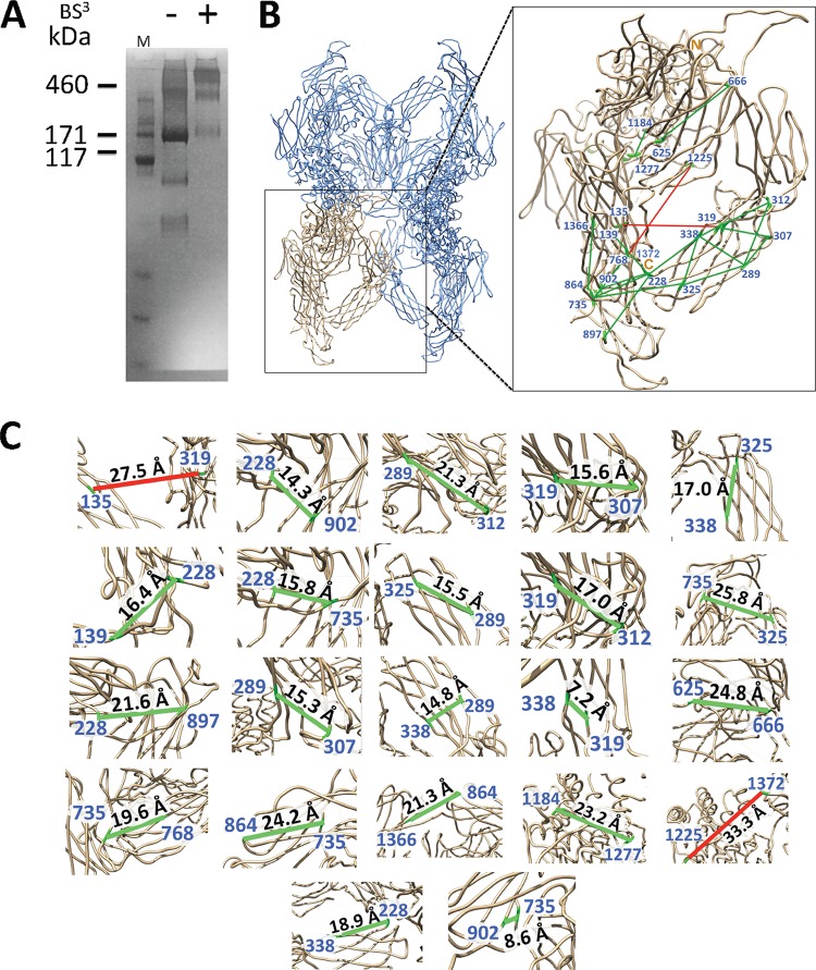
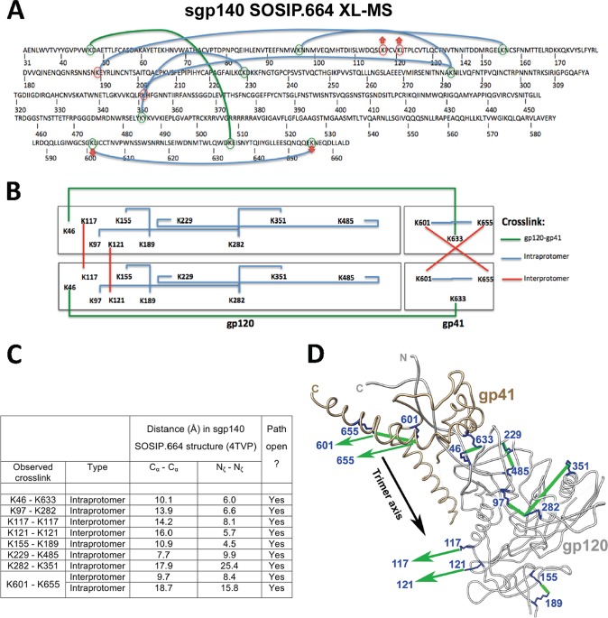
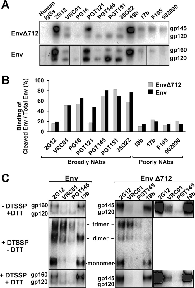
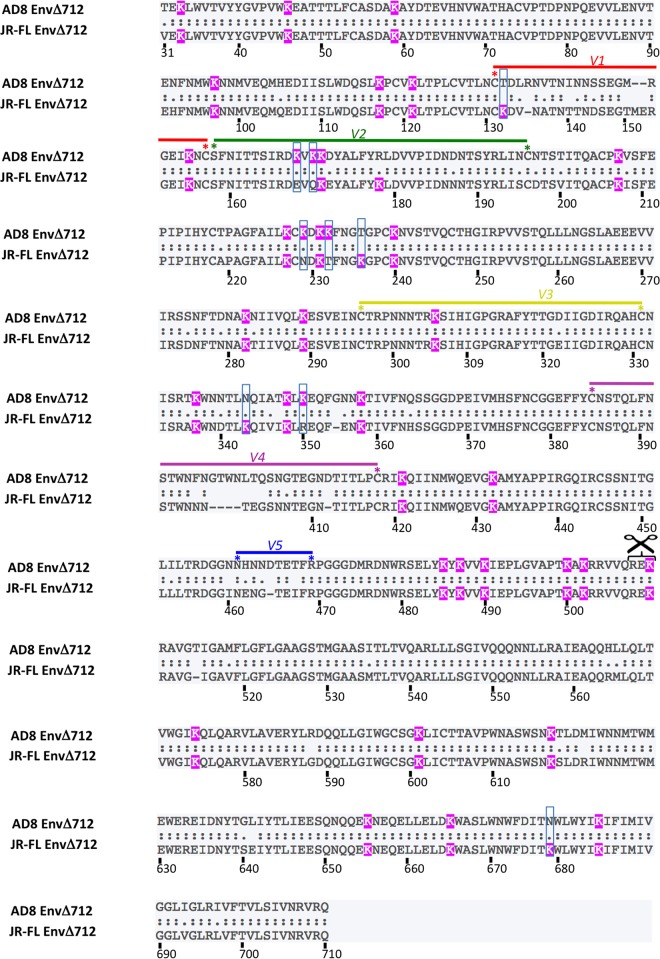
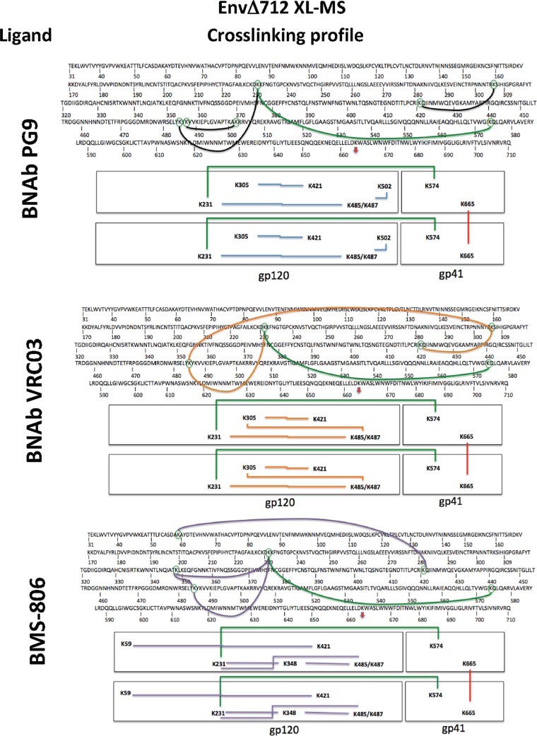
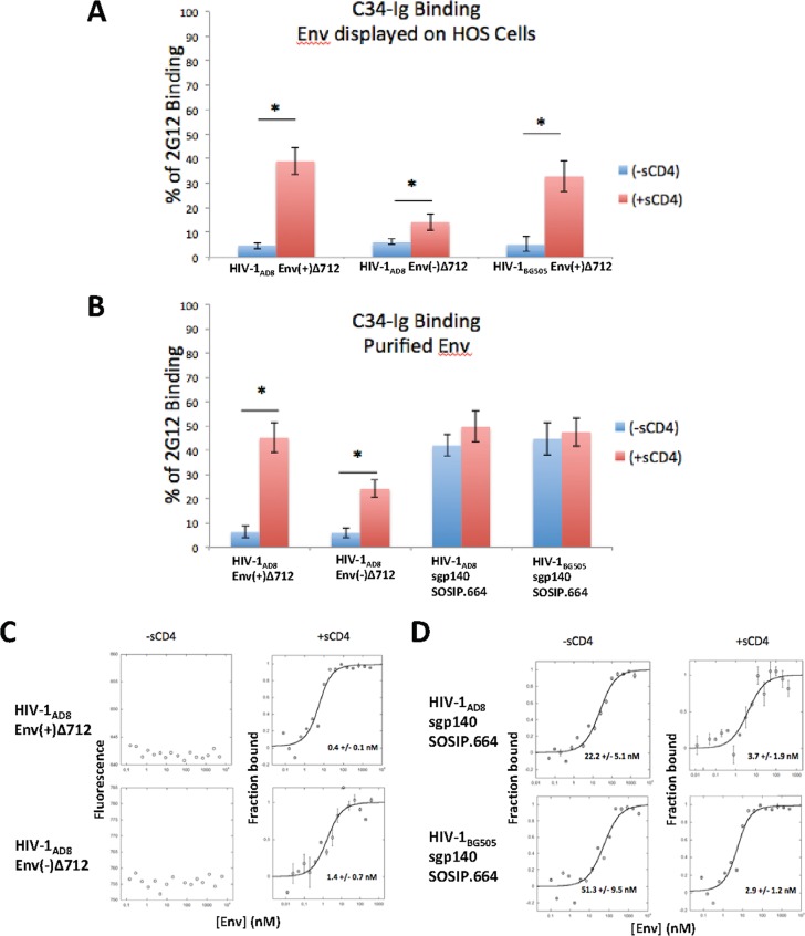
Binding to the receptor CD4 triggers entry-related conformational changes in the human immunodeficiency virus type 1 (HIV-1) envelope glycoprotein (Env) trimer, (gp120/gp41)3 Soluble versions of HIV-1 Env trimers (sgp140 SOSIP.664) stabilized by a gp120-gp41 disulfide bond and a change (I559P) in gp41 have been structurally characterized. Here, we use cross-linking/mass spectrometry to evaluate the conformations of functional membrane Env and sgp140 SOSIP.664. Differences were detected in the gp120 trimer association domain and C terminus and in the gp41 heptad repeat 1 (HR1) region. Whereas the membrane Env trimer exposes the gp41 HR1 coiled coil only after CD4 binding, the sgp140 SOSIP.664 HR1 coiled coil was accessible to the gp41 HR2 peptide even in the absence of CD4. Our results delineate differences in both gp120 and gp41 subunits between functional membrane Env and the sgp140 SOSIP.664 trimer and provide distance constraints that can assist validation of candidate structural models of the native HIV-1 Env trimer.IMPORTANCE HIV-1 envelope glycoprotein spikes mediate the entry of the virus into host cells and are a major target for vaccine-induced antibodies. Soluble forms of the envelope glycoproteins that are stable and easily produced have been characterized extensively and are being considered as vaccines. Here, we present evidence that these stabilized soluble envelope glycoproteins differ in multiple respects from the natural HIV-1 envelope glycoproteins. By pinpointing these differences, our results can guide the improvement of envelope glycoprotein preparations to achieve greater similarity to the viral envelope glycoprotein spike, potentially increasing their effectiveness as a vaccine.

Keywords: conformation; cross-linker; envelope glycoprotein; human immunodeficiency virus; mass spectrometry; stabilization; structure; trimer.


Figures

FIG 1
FIG 1
XL-MS analysis of HIV-1 membrane Env and soluble, stabilized Env trimers. The workflow for XL-MS analysis of the HIV-1 Envs in this study is shown. Two HIV-1 Envs were studied: the mature EnvΔ712 trimer on the surface of HOS cells and soluble gp140 (sgp140) stabilized by the SOSIP.664 changes. The Envs were cross-linked under physiological conditions with a 1:1 mixture of H12-BS3 and D12-BS3. After Env purification, deglycosylation, and proteolysis, the cross-linked peptide pairs were identified by mass spectrometry based on their isotopic signatures. The 11.4-Å spacer arms of the BS3 cross-linker impose distance constraints that were used to test structural models of Env trimers.
FIG 2
FIG 2
XL-MS analysis of a model glycoprotein, alpha-2-macroglobulin. (A) Alpha-2-macroglobulin was purified from fetal bovine serum. The untreated and BS3-cross-linked proteins were analyzed on an SDS-polyacrylamide gel, which was stained with Coomassie brilliant blue. The cross-linked protein consists of a tetramer of ∼725 kDa. (B and C) The BS3 cross-links introduced into bovine alpha-2-macroglobulin were identified by mass spectrometry and mapped onto the structure of the human alpha-2-macroglobulin (PDB entry 4ACQ). The cross-linked lysine residues are numbered according to the bovine alpha-2-macroglobulin sequence (GenBank accession no. NP_001103265.1). (B) The human alpha-2-macroglobulin crystal structure (61) is shown as a Cα worm, with one protomer of the tetramer shown in the inset. The Cα atoms of the cross-linked lysine residues are linked by green lines for Cα-Cα distances less than 26 Å and by red lines for Cα-Cα distances greater than 26 Å. (C) The alpha-2-macroglobulin structure in the vicinity of selected cross-links is shown as a Cα worm. The Cα-Cα distance between the indicated cross-linked lysine residues is shown. The cross-links are colored as described for panel B.
FIG 3
FIG 3
XL-MS analysis of stabilized soluble HIV-1BG505 Env trimers. The BS3 cross-links identified by XL-MS on the HIV-1BG505 sgp140 SOSIP.664 glycoprotein are mapped on the amino acid sequence of the glycoprotein (A) and shown schematically (B). The Env amino acids are numbered according to convention, based on alignment with the HIV-1HXB2 prototype (102). Interprotomer cross-links are designated with red arrows in panel A and with red lines in panel B. Intraprotomer cross-links are shown in blue and gp120-gp41 cross-links in green. (C and D) The BS3 cross-links identified by XL-MS on the HIV-1BG505 sgp140 SOSIP.664 glycoprotein were mapped onto the structure of the HIV-1BG505 sgp140 SOSIP.664 trimer (PDB entry 4TVP) (30). In panel C, the Cα-Cα and Nζ-Nζ distances between the identified cross-linked lysine residues in the sgp140 SOSIP.664 structure are listed, with intraprotomer and interprotomer cross-links designated. The existence of an accessible path for the cross-linker between the lysine residues is indicated. In panel D, the cross-linked lysine residues (in blue) identified by XL-MS on the HIV-1BG505 sgp140 SOSIP.664 Env are mapped onto the structure of one protomer of the HIV-1BG505 sgp140 SOSIP.664 trimer (PDB entry 4TVP). Intraprotomer cross-links are designated by straight lines and interprotomer cross-links by arrows. Cross-links between lysine residues within a 26-Å Cα-Cα distance are compatible with the distance constraints imposed by the BS3 cross-linker and are colored green. Note that no cross-links were observed between lysine residues with Cα-Cα distances greater than 26 Å.
FIG 4
FIG 4
Comparison of antibody binding to HIV-1AD8 full-length and cytoplasmic tail-deleted Envs on the cell surface. (A) HOS cells transiently expressing partially cleaved HIV-1AD8 EnvΔ712 (top) or full-length Env (bottom) on the cell surface were incubated with the indicated antibodies for 1 h at room temperature. 2G12, VRC01, PG16, PGT121, PGT145, PGT151, and 35O22 are broadly neutralizing antibodies, and 19b, 17b, F105, and 902090 are poorly neutralizing antibodies. After washing, cells were lysed and the clarified cell lysates were incubated with protein A-agarose beads. The proteins captured on the beads were Western blotted with a goat anti-gp120 antibody. Some of the lanes in the bottom panel were rearranged to match the order in the top panel. The experiment was repeated with comparable results. (B) Image Lab Software (Bio-Rad) was used to quantify the intensity of the gp145/gp160 (uncleaved Env) and gp120 (cleaved Env) bands in nonsaturated Western blots from the experiment shown in panel A. The percent recognition of cleaved gp120 to the total Env (gp120 + gp145 or gp160) is shown for each antibody for both HIV-1AD8 Env and EnvΔ712. (C) HOS cells expressing HIV-1AD8 full-length Env (left) or EnvΔ712 (right) were treated with DTSSP, a cross-linker with spacer arms similar in length to those of BS3 but that is cleavable with DTT. After the cross-linking reaction was quenched, the untreated and DTSSP-treated cells were incubated with the indicated antibodies. After washing, the cells were lysed and the cell lysates were incubated with protein A-agarose beads. The proteins captured on the beads were run on reducing (+DTT) and nonreducing (−DTT) gels and Western blotted with a goat anti-gp120 antibody. Short and long exposures of the Western blot are shown for the EnvΔ712 glycoprotein without DTSSP cross-linking (upper) and after DTSSP cross-linking and reduction with DTT (lower). The experiment was repeated with comparable results.
FIG 5
FIG 5
Cell-cell fusion activity of HIV-1AD8 EnvΔ712. (A) The syncytium-forming ability of HIV-1AD8 EnvΔ712 was assessed using an α-complementation assay (104). As a negative control, we used EnvΔ712 SOS, which has an artificial disulfide bond between gp120 and gp41 that renders Env nonfunctional except in a reducing environment (85, 86). HOS cells expressing these Envs and α-gal were cocultivated with Cf2Th-CD4/CCR5 cells expressing ω-gal in the absence or presence of increasing concentrations of a reducing agent, DTT. Three hours later, galactosidase activity in the cells was measured. In the absence of DTT, 5.28 × 105 relative light units (RLU) of galactosidase activity was measured for EnvΔ712, whereas 5.93 × 103 RLU was measured for EnvΔ712 SOS. (B) To assess the effect of BS3 on Env-mediated cell-cell fusion, the Env-expressing HOS cells were washed and treated with various concentrations of BS3 for 30 min at room temperature. After quenching the cross-linking reaction with 15 mM Tris-HCl for 15 min, the HOS cells were cocultivated with Cf2Th-CD4/CCR5 cells in 1 mM DTT. Three hours later, galactosidase activity was measured. The means and standard deviations from six samples are shown for a typical experiment.
FIG 6
FIG 6
Conformation of the untreated and cross-linked HIV-1AD8 EnvΔ712 glycoprotein trimer. (A) Cross-linking of the EnvΔ712 trimer by BS3. HOS cells expressing the HIV-1AD8 EnvΔ712 glycoprotein were untreated or cross-linked with a saturating concentration of BS3. Cross-linked cells were lysed in buffer containing 1% Cymal-5 detergent, and Env solubilized from cell membranes was purified by Galanthus nivalis lectin affinity chromatography. The gel was Western blotted with a rabbit anti-gp120-HRP antibody. The results indicate that EnvΔ712 is efficiently processed in HOS cells, BS3 cross-linking is effective, and EnvΔ712 is largely a trimer. (B) Cell-based ELISA of efficiently cleaved HIV-1AD8 EnvΔ712 displayed on the surface of HOS cells. The EnvΔ712-expressing cells were untreated or incubated with 20 µM BMS-806. Half of the cell cultures were then cross-linked with 5 mM BS3. After washing, the cells were analyzed by cell-based ELISA with the indicated primary antibodies. The results are shown relative to the values seen for the 2G12 anti-gp120 antibody (set at 100% for each condition). The means and standard deviations from two experiments are shown. Recognition of untreated and BS3-cross-linked EnvΔ712 by the antibodies did not significantly differ (independent samples t test). (C) HOS cells expressing the HIV-1AD8 EnvΔ712 glycoprotein were treated with 5 mM BS3 for 30 min at 25°C. EnvΔ712 was purified from cell membranes and analyzed by SDS-PAGE. The gel was stained with Coomassie brilliant blue.
FIG 7
FIG 7
XL-MS analysis of the unliganded HIV-1AD8 membrane EnvΔ712 trimer. The BS3 cross-links of the cell surface HIV-1AD8 EnvΔ712 glycoprotein determined by XL-MS are mapped on the EnvΔ712 sequence (A) and shown schematically (B). The cross-links are colored as described for Fig. 3. (C and D) The cross-linked lysine residues identified by XL-MS analysis on the unliganded HIV-1AD8 membrane EnvΔ712 glycoprotein were mapped onto the structure of the HIV-1BG505 sgp140 SOSIP.664 trimer (PDB entry 4TVP). (C) The Cα-Cα and Nζ-Nζ distances between the identified cross-linked lysine residues in the sgp140 SOSIP.664 structure are listed. The existence of an accessible path for the cross-linker between the lysine residues is indicated. (D) The cross-linked lysine residues (in blue) identified by XL-MS on EnvΔ712 are mapped onto the structure of one protomer of the HIV-1BG505 sgp140 SOSIP.664 trimer (PDB entry 4TVP). Interprotomer cross-links are designated by arrows. Cross-links between lysine residues within a 26-Å Cα-Cα distance conform to the distance constraints imposed by the BS3 cross-linker and are colored green. Note that although the Cα-Cα distances between Lys 46 and Lys 231 and between Lys 305 and Lys 421 are less than 26 Å, there is no unimpeded path for the cross-linker between these lysine residues in the sgp140 SOSIP.664 structure; therefore, these lysine pairs are asterisked in panel C and the associated cross-links are colored orange in panel D. Cross-links between lysine residues that are more than 26 Å apart are colored red.
FIG 8
FIG 8
Alignment of HIV-1AD8 EnvΔ712 and HIV-1BG505 sgp140 SOSIP.664 sequences. The sequences of HIV-1AD8 EnvΔ712 and HIV-1BG505 sgp140 SOSIP.664 are aligned, with amino acid residues numbered according to current convention (102). Identical residues are indicated by two dots, conserved residues by one dot. All lysine residues are highlighted in pink. Lysine residues present only in one of the glycoproteins are boxed in blue rectangles. The V1 to V5 variable regions are indicated. The scissor indicates the gp120-gp41 cleavage site. Approximately 65% of the lysine residues in the HIV-1AD8 EnvΔ712 ectodomain are conserved in HIV-1BG505 sgp140 SOSIP.664.
FIG 9
FIG 9
Alignment of HIV-1AD8 EnvΔ712 and HIV-1JR-FL EnvΔCT sequences. The sequences of the HIV-1AD8 EnvΔ712 and HIV-1JR-FL EnvΔCT glycoproteins are aligned, with amino acid residues numbered according to current convention (102). Identical residues are indicated by two dots, conserved residues by one dot. All lysine residues are highlighted in pink. Lysine residues present in only one of the glycoproteins are boxed in blue rectangles. The V1 to V5 variable regions are indicated. The scissor indicates the gp120-gp41 cleavage site. Approximately 86% of the lysine residues in the HIV-1AD8 EnvΔ712 ectodomain are conserved in the HIV-1JR-FL EnvΔCT glycoprotein.
FIG 10
FIG 10
XL-MS analysis of the ligand-bound HIV-1AD8 membrane EnvΔ712 glycoprotein. The BS3 cross-links identified by XL-MS analysis on the HIV-1AD8 membrane EnvΔ712 glycoprotein incubated with the indicated ligands are mapped on the EnvΔ712 sequence (upper) and are shown schematically (lower). The interprotomer cross-link is designated with red arrows and lines and the gp120-gp41 cross-link with green lines.
FIG 11
FIG 11
Conformation of the gp41 ectodomain in membrane and soluble Envs. (A) The binding of C34-Ig, which contains a gp41 HR2 peptide (23), to the HIV-1AD8 or HIV-1BG505 EnvΔ712 glycoproteins or to the cleavage-defective HIV-1AD8 Env(−)Δ712 glycoprotein expressed on the surface of transfected HOS cells is shown, normalized to the binding of the 2G12 anti-gp120 antibody. C34-Ig binding was measured in the absence (blue) or presence (red) of saturating amounts of soluble CD4 (sCD4). (B) C34-Ig binding in the absence and presence of sCD4 is shown to purified HIV-1AD8 or HIV-1BG505 sgp140 SOSIP.664 trimers or BS3-cross-linked HIV-1AD8 EnvΔ712 glycoprotein captured on ELISA plates. The results represent the means and standard deviations from two experiments. The asterisk in panels A and B indicates a P value less than or equal to 0.05 by unpaired t test. (C and D) Microscale thermophoresis on the indicated Envs purified from membranes (C) or prepared as soluble trimers (D) was used to measure C34-Ig binding with and without sCD4 incubation (the molar ratio of sCD4 to Env trimer is 3:1). The dissociation constants associated with C34-Ig-Env binding are reported where measurable.

Similar articles

Cited by

References

    1. Wyatt R, Sodroski J. 1998. The HIV-1 envelope glycoproteins: fusogens, antigens, and immunogens. Science 280:1884–1888. doi:10.1126/science.280.5371.1884. - DOI - PubMed
    1. Karlsson Hedestam GB, Fouchier RA, Phogat S, Burton DR, Sodroski J, Wyatt RT. 2008. The challenges of eliciting neutralizing antibodies to HIV-1 and to influenza virus. Nat Rev Microbiol 6:143–155. doi:10.1038/nrmicro1819. - DOI - PubMed
    1. Hoxie JA. 2010. Toward an antibody-based HIV-1 vaccine. Annu Rev Med 61:135–152. doi:10.1146/annurev.med.60.042507.164323. - DOI - PubMed
    1. Fauci AS. 2016. An HIV vaccine: mapping uncharted territory. JAMA 316:143–144. doi:10.1001/jama.2016.7538. - DOI - PubMed
    1. Klatzmann D, Champagne E, Chamaret S, Gruest J, Guetard D, Hercend T, Gluckman JC, Montagnier L. 1984. T-lymphocyte T4 molecule behaves as the receptor for human retrovirus LAV. Nature 312:767–768. doi:10.1038/312767a0. - DOI - PubMed

Publication types

MeSH terms

Substances