. 2018 Nov 14;9(1):4781.
doi: 10.1038/s41467-018-07225-7.

ΦCrAss001 represents the most abundant bacteriophage family in the human gut and infects Bacteroides intestinalis

Affiliations

ΦCrAss001 represents the most abundant bacteriophage family in the human gut and infects Bacteroides intestinalis

Andrey N Shkoporov et al. Nat Commun. .

Abstract

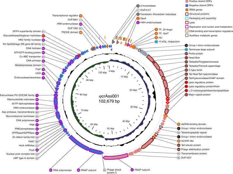
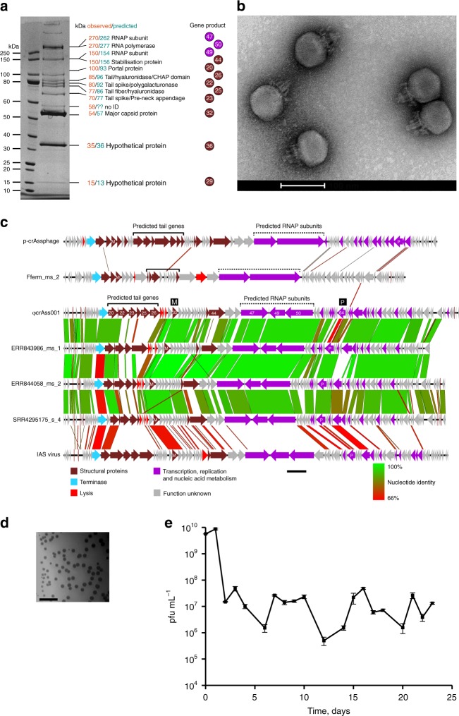
CrAssphages are an extensive and ubiquitous family of tailed bacteriophages, predicted to infect bacteria of the order Bacteroidales. Despite being found in ~50% of individuals and representing up to 90% of human gut viromes, members of this viral family have never been isolated in culture and remain understudied. Here, we report the isolation of a CrAssphage (ΦCrAss001) from human faecal material. This bacteriophage infects the human gut symbiont Bacteroides intestinalis, confirming previous in silico predictions of the likely host. DNA sequencing demonstrates that the bacteriophage genome is circular, 102 kb in size, and has unusual structural traits. In addition, electron microscopy confirms that ΦcrAss001 has a podovirus-like morphology. Despite the absence of obvious lysogeny genes, ΦcrAss001 replicates in a way that does not disrupt proliferation of the host bacterium, and is able to maintain itself in continuous host culture during several weeks.


Conflict of interest statement

The authors declare no competing interests.

Figures

Fig. 1
Fig. 1
Circular map of ΦcrAss001 genome. Inner circle (green and blue), GC skew; middle circle (black), G + C content; outer circle (pink and purple fill), protein-coding genes (ORFs); outermost circle (orange fill), tRNA genes. Stroke colour on ORFs and fill colour on gene numbers corresponds with the general predicted function (see colour legend for details); black stroke, introns; genes with no functional annotations (coding for hypothetical proteins) are left unlabelled
Fig. 2
Fig. 2
Predicted tertiary structure of ΦcrAss001 major capsid protein (MCP). a Homology-based modelling of aa 176405 fragment of ΦcrAss001 MCP. A-domain, axial domain; P-domain, peripheral domain; N-terminal domain and E-loop could not be resolved due to lack of similarity with the used template; colour gradient reflects aa position in the sequence (blue to green to red from N terminus to C terminus). b Staphylococcus phage 812 MCP (PDB 5LII [https://www.rcsb.org/structure/5LII]) used as template in HHPred-MODELLER suit (HHPred probability score 96.87%, e-value 0.00081). c Superposition of φcrAss001 MCP model (red) with p-crAssphage (green) and IAS virus (blue) MCP models (probability scores against PDB 5LII [https://www.rcsb.org/structure/5LII] 99.37, 96.89%, e-values 4e-14, 0.00063, respectively)
Fig. 3
Fig. 3
ΦcrAss001 virion structure, related bacteriophages and persistence in a continuous host co-culture. a SDS-PAGE analysis of protein content in ΦcrAss001 virions and identification of selected polypeptides using MALDI-TOF (see supplementary methods for details). b TEM image of uranyl acetate negatively contrasted ΦcrAss001 virions, scale bar is 100 nm (×62,000 magnification, accelerating voltage of 120 kV). c Genomic synteny and BLASTn nucleotide sequence homology between ΦcrAss001, a group of highly related uncultured bacteriophages (ERR843986_ms_1, ERR844058_ms_2, SRR4295175_s_4, see Guerin et al. for details), and other members of the crAss-like virus family (p-crAssphage, IAS virus, bacteriophage Fferm_ms_2 propagated and visualised from a faecal fermentor system); highly variable tail genes region is marked a solid bracket; region containing giant open reading frames of predicted RNA polymerase subunits is highlighted with dotted bracket; M, major capsid protein; P, DNA polymerase, scale bar is 5 kb. d Plaque morphology of ΦcrAss001 after 48 h incubation in a 0.3% FAA agar overlay with B. intestinalis 919/174 as host, scale bar is 1 cm. e Persistence of ΦcrAss001 in a continuous culture of bacteriophage infected B. intestinalis 919/174 with daily (or bi-daily) transfers for 23 days (pfu per mL, mean ± SD of three independent cultures incubated in parallel)

References

    1. Ogilvie LA, Jones BV. The human gut virome: a multifaceted majority. Front. Microbiol. 2015;6:918. doi: 10.3389/fmicb.2015.00918. - DOI - PMC - PubMed
    1. Carding SR, Davis N, Hoyles L. Review article: the human intestinal virome in health and disease. Aliment. Pharmacol. Ther. 2017;46:800–815. doi: 10.1111/apt.14280. - DOI - PMC - PubMed
    1. Manrique P, et al. Healthy human gut phageome. Proc. Natl Acad. Sci. USA. 2016;113:10400–10405. doi: 10.1073/pnas.1601060113. - DOI - PMC - PubMed
    1. Norman JM, et al. Disease-specific alterations in the enteric virome in inflammatory bowel disease. Cell. 2015;160:447–460. doi: 10.1016/j.cell.2015.01.002. - DOI - PMC - PubMed
    1. Reyes A, et al. Gut DNA viromes of Malawian twins discordant for severe acute malnutrition. Proc. Natl Acad. Sci. USA. 2015;112:11941–11946. doi: 10.1073/pnas.1514285112. - DOI - PMC - PubMed

Publication types