. 2018 Nov 14;9(1):4780.
doi: 10.1038/s41467-018-06813-x.

Presenilin-mediated cleavage of APP regulates synaptotagmin-7 and presynaptic plasticity

Affiliations

Presenilin-mediated cleavage of APP regulates synaptotagmin-7 and presynaptic plasticity

Gaël Barthet et al. Nat Commun. .

Abstract

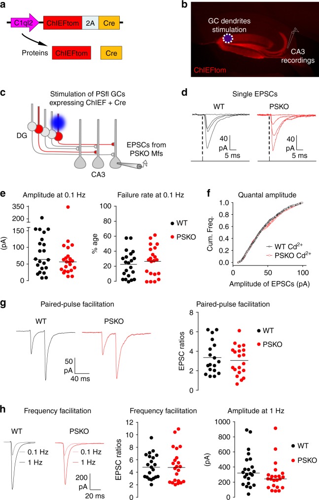
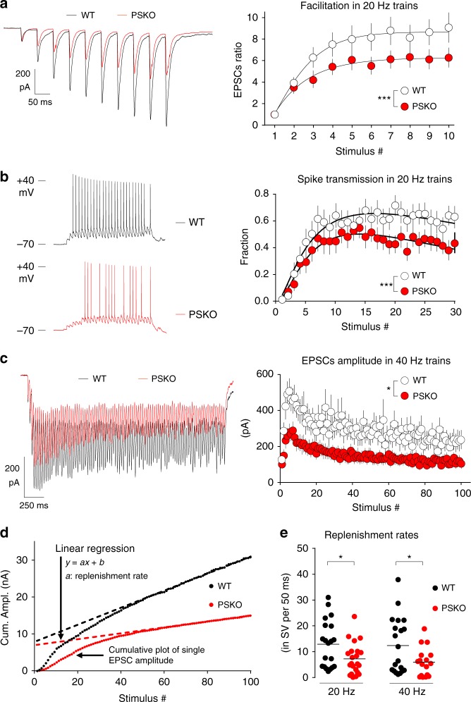
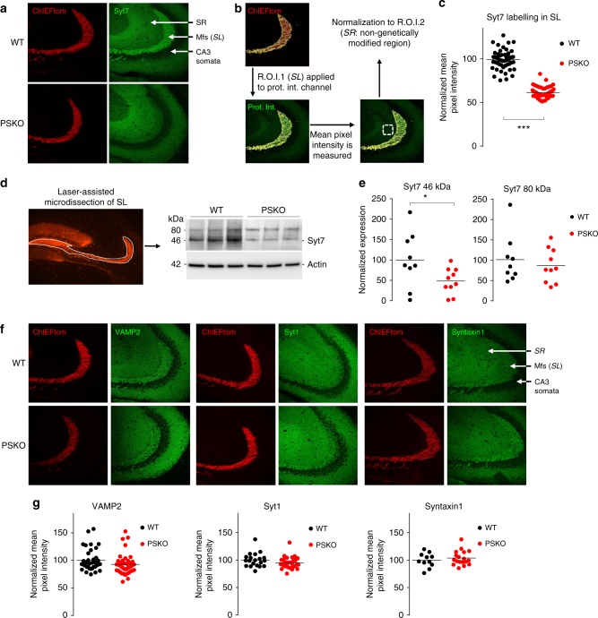
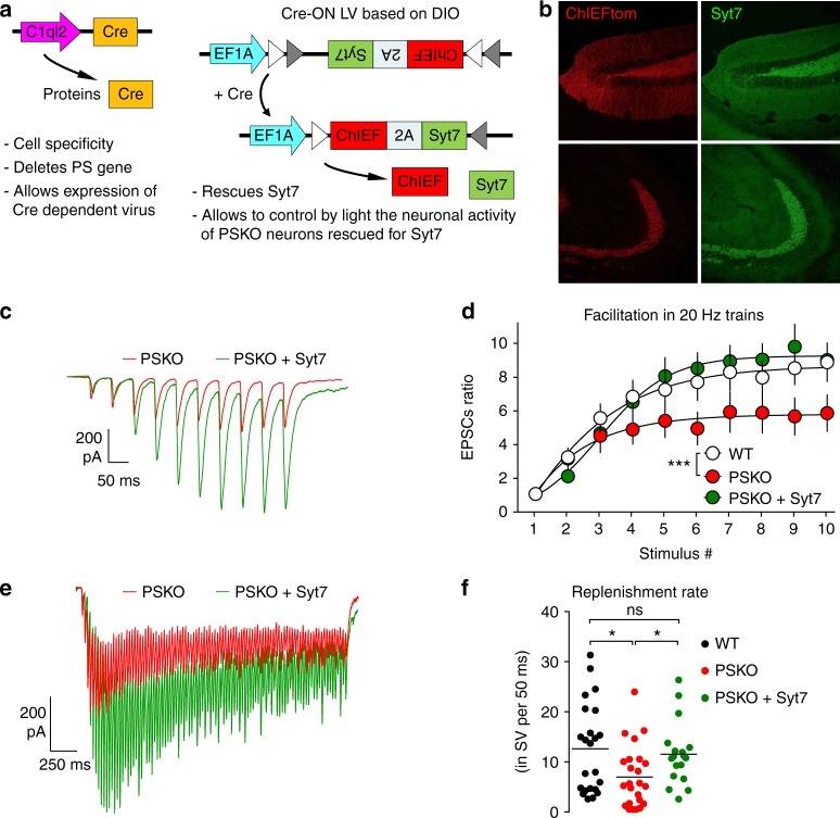
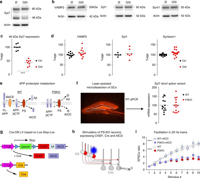
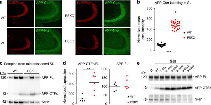
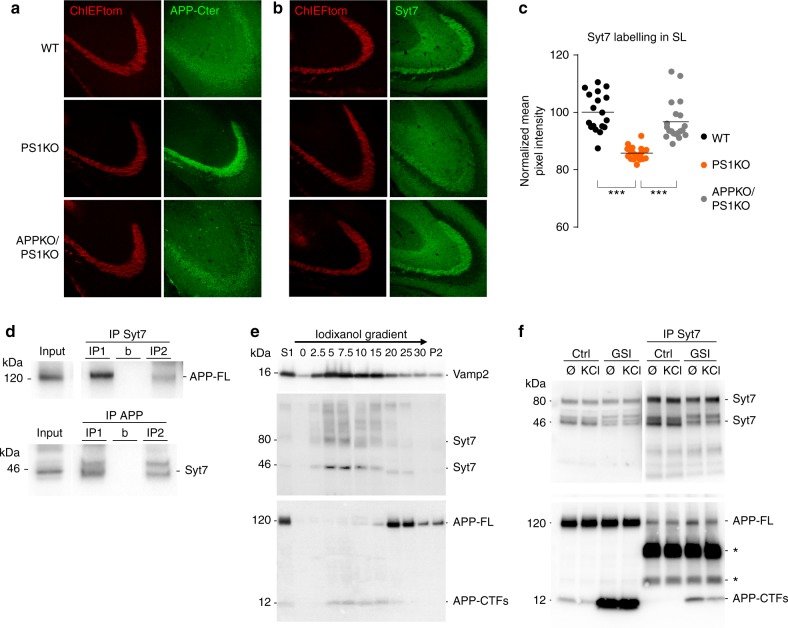
Mutations of the intramembrane protease presenilin (PS) or of its main substrate, the amyloid precursor protein (APP), cause early-onset form of Alzheimer disease. PS and APP interact with proteins of the neurotransmitter release machinery without identified functional consequences. Here we report that genetic deletion of PS markedly decreases the presynaptic levels of the Ca2+ sensor synaptotagmin-7 (Syt7) leading to impaired synaptic facilitation and replenishment of synaptic vesicles. The regulation of Syt7 expression by PS occurs post-transcriptionally and depends on γ-secretase proteolytic activity. It requires the substrate APP as revealed by the combined genetic invalidation of APP and PS1, and in particular the APP-Cterminal fragments which interact with Syt7 and accumulate in synaptic terminals under pharmacological or genetic inhibition of γ-secretase. Thus, we uncover a role of PS in presynaptic mechanisms, through APP cleavage and regulation of Syt7, that highlights aberrant synaptic vesicle processing as a possible new pathway in AD.


Conflict of interest statement

The authors declare no competing interests.

Figures

Fig. 1
Fig. 1
Study of PS presynaptic function at Mf/CA3 synapses using an optogenetic strategy. a Scheme of lentiviral construct which co-expresses ChIEF fused to the fluorescent protein tomato and the Cre recombinase. The promoter C1ql2 restricts expression to DG granule cells (GCs). b Picture of a parasagittal section of a mouse brain injected with a virus coding for ChIEFtom. c Strategy to selectively study Mf/CA3 synapses that are PSKO presynaptically and intact postsynaptically. The blue light triggers the firing of granule cells that express ChIEF and by obligation (bicistronic construct) the Cre recombinase. In PS “floxed” conditional knock-out mice, the activated GCs do not express PS; EPSCs evoked in CA3 neurons by light pulses in the DG arise from presynaptic PSKO Mf terminals. d Representative traces of CA3 EPSCs evoked by single light-pulses focused as in (b). e Basal transmission at Mf/CA3 synapses is not altered by PS genotype. Scatter plots with medians of the amplitude, and means of the failure rate of EPSCs recorded in CA3 neurons at 0.1 Hz in WT or PSKO condition (n = 22/genotype). f Cumulative frequencies of EPSCs amplitude in presence of Cd2+. g Representative recordings of paired-pulses EPSCs evoked by two light-pulses separated by 40 ms, and analyses of paired-pulse ratio. The facilitation is represented by the amplitude of the second EPSC normalized to the amplitude of the first EPSC. h Representative traces of EPSCs evoked by light-pulses at 0.1 or 1 Hz, and analyses of facilitation. The frequency facilitation is represented by the amplitude of EPSCs obtained at 1 Hz normalized to the amplitude of EPSCs obtained at 0.1 Hz. The raw amplitude of EPSCs at 1 Hz does not differ between WT and PSKO
Fig. 2
Fig. 2
PS promotes presynaptic facilitation, spike transmission, and SV replenishment. a Representative traces of EPSCs evoked by 10 repetitive light-pulses at 20 Hz, and analysis of the facilitation within the train. The facilitation is represented by the amplitude of EPSCs normalized to the amplitude of the first EPSC. The facilitation is strongly impaired by absence of presynaptic PS. Two-way ANOVA, n = 19 in WT, 16 in PSKO, p = 0.0004. b Representative recordings of CA3 spikes in response to 30 stimulations at 20 Hz, and plot of the fractions of spike transmission (for 16 recorded neurons per genotype; five sweeps per neuron) in WT or PSKO genotype. The highest rate of transmission (over 50%) is reached around 15 stimuli and the plot in PSKO is lower than that in WT condition (two-way ANOVA, n = 16 in WT, 18 in PSKO p < 0.0001). c Representative EPSCs evoked by 100 stimuli at 40 Hz, and the analysis of their amplitude. After the initial rising phase (facilitation), the amplitude drops and reaches a steady state that is reduced by absence of PS (two-way ANOVA, n = 20 in WT, 17 in PSKO, p = 0.03). d Graph of the cumulative amplitude of Mf-EPSCs in WT or PSKO condition from trains of stimulation plotted against the stimulus number. A linear regression fits the second part of the cumulative plot where a steady state is reached. The slope is proportional of the replenishment rate. e Scatter plots with means of replenishment rates calculated neuron per neuron are represented by genotype and frequency. Absence of PS decreases the rates of replenishment (unpaired t-test p < 0.05; n = 20 in WT, 22 in PSKO). Errors bars represent s.e.m
Fig. 3
Fig. 3
PS supports the presynaptic expression of Syt7. a Confocal images of the stratum lucidum (SL) where the Mfs can be distinguished in the red channel (ChIEFtom) and stainings of Syt7 depicted in green. Syt7 staining in PSKO Mfs was negligible in comparison to WT. The phenotype presented here using antibody 105173 from Synaptic System is reproduced in Supplementary Fig. S3d with the antibody N275/14 from Millipore. b To quantify the intensity of the stainings a mask corresponding to the SL was created in the red channel. This first region of interest (ROI) was applied to the green channel were the mean pixel intensity of the protein of interest was measured only in the SL. To normalize the results, the mean pixel intensity was also measured in a second ROI: the stratum radiatum (SR). The staining intensities in the SL were normalized to the intensities in the SR. c Scatter plots of the mean pixel intensity in the SL normalized to the mean pixel intensity in the SR. A reduction of 40% of intensity is observed while approximately half of the Mfs are projected from PSKO GCs. n = 53 slices from nine mice in WT, n = 50 slices from eight mice in PSKO, p = 0.0004. d Confocal image of the hippocampus depicting the area dissected with a laser to prepare protein lysates, and western blot from these lysates showing a decreased expression of Syt7. e Quantitative analysis of western blot in c. A 50% decrease in the expression of the 46 kDa band of Syt7 was detected. f Confocal images of the stratum lucidum (SL) where the Mfs can be distinguished in the red channel (ChIEFtom) and stainings of VAMP2, Syt1 or Syntaxin1A are seen in green. g Scatter plots of the mean pixel intensity in the SL normalized to the mean pixel intensity in the SR measured in images as in e. Staining patterns and intensities do not differ between genotypes. For VAMP2, n = 38 slices from eight mice in WT, n = 41 slices from nine mice in PSKO. For Syt1, n = 20 slices from five mice in WT, n = 33 slices from seven mice in PSKO. For Syntaxin1, n = 11 slices from four mice in WT, n = 18 slices from seven mice in PSKO
Fig. 4
Fig. 4
Impaired facilitation and SV replenishment in PSKO Mfs is caused by decreased Syt7 expression. a Schemes of the two lentiviruses (LV) used to rescue Syt7 expression in PSKO GCs controlled by light. C1ql2 promoter restricts the expression of Cre to GCs where the floxed PS gene is deleted. The Cre also allows the Cre-ON LV to express Syt7 and ChIEF. b Confocal images showing the rescued expression of Syt7 in the hilus (up) and in the stratum lucidum (down) using the strategy described in a. c Representative traces of EPSCs evoked by 10 repetitive light pulses at 20 Hz. d Analysis of presynaptic facilitation in 20 Hz trains. Facilitation is represented by the amplitude of the EPSCs normalized to the amplitude of the first EPSC. Facilitation, impaired in absence of presynaptic PS, recovers to WT level by re-expression of Syt7. (Two-way ANOVA, n = 24 in WT, 23 in PSKO, 24 in PSKO + Syt7, p = 0.001). Errors bars represent s.e.m.) e Representative EPSCs evoked by 100 stimuli at 40 Hz. The amplitude of EPSCs is rescued by Syt7 re-expression. f Scatter plots with means of replenishment rates calculated per neuron are represented by genotype. The replenishment rate impaired by the absence of PS is rescued by re-expression of Syt7
Fig. 5
Fig. 5
γ-secretase activity regulates Syt7 protein levels. a, b Western blots of protein samples from neuronal cultures treated or not with 10 µM GSI from 10 to 14DIV. The 46 kDa Syt7 band decreases dramatically when γ-secretase is inhibited. γ-secretase inhibition does not impair expression of VAMP2, Syt1, and Syntaxin1A. c, d Scatter plots of the western-blot quantification of samples from neuronal culture treated or not with GSI. Syt7 expression is decreased by half in treated condition as seen in a, while the expression of VAMP2, Syt1, and Syntaxin1A is stable under GSI condition as seen in b. e Schemes of APP proteolytic metabolism in WT and PSKO conditions. Full-length (FL) APP is first cleaved by BACE producing βCTF, a transmembrane stub, further degraded by PS (γ-secretase) into Aβ and APP IntraCellular Domain (AICD). In absence of PS, βCTF cannot be catabolized and thus accumulates. f Confocal image of the hippocampus depicting the GCs layer dissected with a laser to prepare RNA samples, and RT-qPCR analysis of the samples indicates that mRNA level of Syt7 is not impaired in absence of PS. g Schemes of the two lentiviruses (LV) used to rescue AICD expression in PSKO GCs controlled by light. C1ql2 promoter restricts the expression of Cre to GCs where the PS floxed gene is deleted. The Cre also removes the Lox-Stop-Lox sequence allowing the Cre-ON LV to express AICD and ChIEF. h Strategy to selectively study Mf/CA3 synapses that are presynaptically PSKO, yet express AICD. In PS “floxed” conditional knock-out mice, double-infected GCs do not express PS (Cre deletes PSfl gene), yet express AICD (Cre dependently), and ChIEF (bicistronic construct) allowing the control of their activity by light. GCs infected by a single virus do not respond to light. i Facilitation of EPSCs amplitude measured along trains of 10 stimuli at 20 Hz in WT or PSKO-expressing AICD. The impaired facilitation of PSKO is not rescued by expression of AICD. Two-way ANOVA, n = 8 in WT, 11 in PSKO, p = 0.017. Errors bars represent s.e.m
Fig. 6
Fig. 6
γ-secretase proteolytic activity regulates the axonal expression of APP-βCTF. a Confocal images of the Mfs tracts distinguished in the red channel (ChIEFtom) and of stainings of APP Cterminal with Y188 antibody (upper panel) and Nterminal with JRD32 antibody (lower panel). In WT, Mfs are labelled with the Cterminal but barely with the Nterminal antibody. In PSKO, Mfs are strongly labelled with the Cterminal but not with the Nterminal antibody. b Scatter plots of the mean pixel intensity of APP-Cter in the SL normalized to the mean pixel intensity in the SR. A five-time increase of the intensity is observed in PSKO condition compared to WT. c Samples prepared as in (Fig. 3c) show a striking increase in APP-CTFs levels in dissected Mfs detected with Y188 antibody against APP-Cter. d Quantitative analysis of western blot in c. A 400% increase in the expression of APP-CTFs was detected. e Western-blot of samples prepared from neuronal cultures treated with GSI from 6 h to 9 days in vitro. APP-CTFs accumulate while Syt7 drops
Fig. 7
Fig. 7
PS regulation of Syt7 expression depends on APP-CTFs, new interacting partners of Syt7. a Confocal images of ChIEFtom (red channel) and APP-Cterminal (detected with C1/6.1 antibody, green channel) in the Mf tracts in WT, PS1KO, and PS1KO/APPKO conditions. Mfs are faintly labelled with the APP antibody in WT and barely in APPKO/PS1KO. In contrast, PS1KO Mfs display strong labelling of APP. b Confocal images as in a displaying Syt7 labelling. Mfs are strongly labelled in WT and in APPKO/PS1KO but faintly in PS1KO. c Scatter plots of the mean pixel intensity in the SL normalized to the mean pixel intensity in the stratum radiatum report a reduction of 18%. In this condition, 50% of the GCs are infected by a Cre-expressing LV thus ∼50% of the Mfs are projected from non-infected (PS expressing) GCs. d WB from synaptosomal samples prepared from whole brains of adult mice. Top: IP of Syt7 pulled-down APP-FL. Bottom: IP of APP pulled-down Syt7. The characterization of the synaptosomal fraction is depicted in Supplementary Fig.7. b: beads + immuno-precipitating antibody without sample. e Western-blot of cellular fractions from cultured neurons separated on an iodixanol gradient. VAMP2, Syt7, and APP-CTFs are enriched in light fractions while APP-FL is present in heavier fractions. f Samples prepared from cultured neurons treated or not with GSI from 10 to 14 DIV and with or without KCl (30 mM) for 15 min before lysis. In the inputs, detection of APP Cterminal with the Y188 antibody indicates that GSI induces CTFs accumulation. IP of Syt7 pulled-down APP-FL and CTFs. Asterisks indicate light and heavy chains of the immuno-precipitating antibody. Syt7/CTFs interaction is decreased by KCl treatment suggesting that it is regulated by neuronal activity

References

    1. Sherrington R, et al. Cloning of a gene bearing missense mutations in early-onset familial Alzheimer’s disease. Nature. 1995;375:754–760. doi: 10.1038/375754a0. - DOI - PubMed
    1. Mullan M, et al. A pathogenic mutation for probable Alzheimer’s disease in the APP gene at the N-terminus of beta-amyloid. Nat. Genet. 1992;1:345–347. doi: 10.1038/ng0892-345. - DOI - PubMed
    1. De Strooper B, et al. Deficiency of presenilin-1 inhibits the normal cleavage of amyloid precursor protein. Nature. 1998;391:387–390. doi: 10.1038/34910. - DOI - PubMed
    1. Barthet G, Georgakopoulos A, Robakis NK. Cellular mechanisms of γ-secretase substrate selection, processing and toxicity. Prog. Neurobiol. 2012;98:166–175. doi: 10.1016/j.pneurobio.2012.05.006. - DOI - PMC - PubMed
    1. DeKosky ST, Scheff SW. Synapse loss in frontal cortex biopsies in Alzheimer’s disease: correlation with cognitive severity. Ann. Neurol. 1990;27:457–464. doi: 10.1002/ana.410270502. - DOI - PubMed

Publication types

MeSH terms