. 2018 Nov 14;8(1):16792.
doi: 10.1038/s41598-018-35060-9.

Revised role for Hfq bacterial regulator on DNA topology

Affiliations

Revised role for Hfq bacterial regulator on DNA topology

Antoine Malabirade et al. Sci Rep. .

Abstract

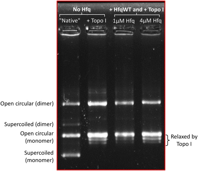
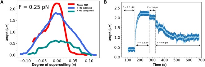
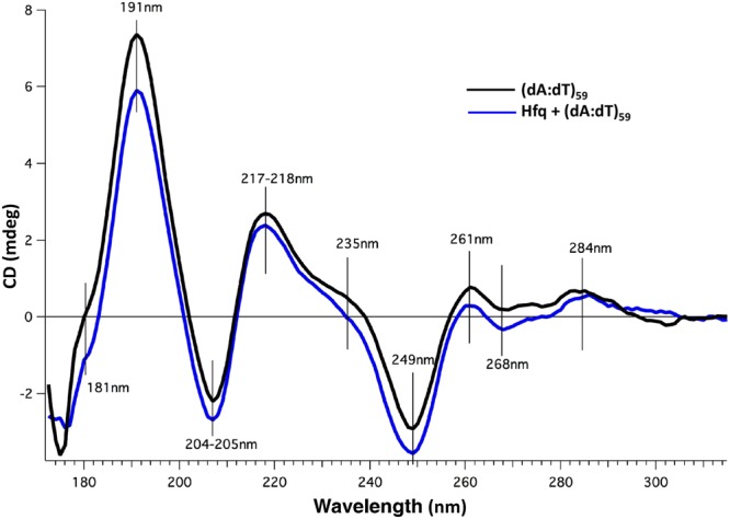
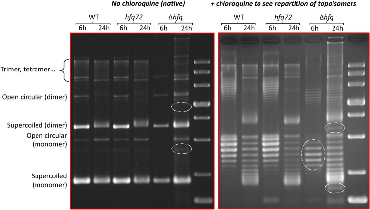
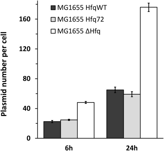
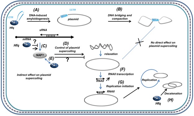
Hfq is a pleiotropic regulator that mediates several aspects of bacterial RNA metabolism. The protein notably regulates translation efficiency and RNA decay in Gram-negative bacteria, usually via its interaction with small regulatory RNA. Besides these RNA-related functions, Hfq has also been described as one of the nucleoid associated proteins shaping the bacterial chromosome. Therefore, Hfq appears as a versatile nucleic acid-binding protein, which functions are probably even more numerous than those initially suggested. For instance, E. coli Hfq, and more precisely its C-terminal region (CTR), has been shown to induce DNA compaction into a condensed form. In this paper, we establish that DNA induces Hfq-CTR amyloidogenesis, resulting in a change of DNA local conformation. Furthermore, we clarify the effect of Hfq on DNA topology. Our results evidence that, even if the protein has a strong propensity to compact DNA thanks to its amyloid region, it does not affect overall DNA topology. We confirm however that hfq gene disruption influences plasmid supercoiling in vivo, indicating that the effect on DNA topology in former reports was indirect. Most likely, this effect is related to small regulatory sRNA-Hfq-based regulation of another protein that influences DNA supercoiling, possibly a nucleoid associated protein such as H-NS or Dps. Finally, we hypothesise that this indirect effect on DNA topology explains, at least partially, the previously reported effect of Hfq on plasmid replication efficiency.


Conflict of interest statement

The authors declare no competing interests.

Figures

Figure 1
Figure 1
SRCD kinetics of Hfq-CTR self-assembly in the presence of DNA. SRCD experiment was performed as a function of time in order to observe progressive amyloid-like structuring. (A) (dA:dT)59: black line; CTR + (dA:dT)59 1 day: green line; CTR + (dA:dT)59 10 days: cyan line; monomeric CTR 10 days: dashed red line monomeric. (B) Controls: CTR 1 day: red line; amyloid CTR: blue line. As shown, the typical signal of amyloids at 210–220 nm appears after 7 days, while it takes more than 2 weeks with peptide alone. Note that DNA induces a structural change of the protein and conversely (see Fig. 6). For this reason we do not substract the spectrum of the peptide alone from the spectrum of DNA:peptide complex.
Figure 2
Figure 2
FTIR spectra of Hfq-CTR in the presence or absence of DNA. D2O buffered samples have been used to avoid the spectral overlaps between Amide I and water bands. Solid black line: peptide alone. Dashed blue line: difference spectrum obtained by subtracting the (dA:dT)59 contribution from the complex spectrum. We clearly observe in the Amide I’ band a contribution at 1615 cm−1, indicative of the formation of the amyloid structure in the presence of DNA. Note in the complex the presence of both Amide II and II′ bands, due to N\H and N\D in-plane bending of the peptidic groups, respectively. Strikingly, the Amide II is present only in the complex, indicating that hydrogens may be entrapped inside the complex and cannot exchange with deuterium.
Figure 3
Figure 3
SAXS curves of Hfq-CTR in the presence or absence of DNA. β-Sheet peaks corresponding to a cross-β structure are clearly observed for Hfq-CTR in the presence of DNA (incubated 10 days, blue curve) and for Hfq-CTR alone incubated for 6 weeks at 20 mg/mL (higher time and concentration are used to ensure the formation of amyloid fibrils in the absence of cofactor, black curve). But no β-Sheet peak is observed for Hfq-CTR alone after 10 days (red curve). Peaks corresponding to cross-β structure can be seen at 0.765 and 1.355 Å−1 for complexed Hfq-CTR, and 0.785 and 1.368 Å−1 for non-complexed Hfq-CTR, corresponding to inter-sheet and inter-strand distances (d) of 8.21 and 4.63 and 8.00 Å and 4.59 Å, respectively (d(Å) = 2π/q(Å−1)).
Figure 4
Figure 4
Topology gel of pHSG298 after incubation with Hfq. The presence of purified Hfq does not influence the repartition of topoisomers, confirming that Hfq has no direct effect on overall plasmid topology.
Figure 5
Figure 5
Real-time Hfq-DNA binding followed by magnetic tweezers. The absence of direct effect of Hfq has also been confirmed by tweezers experiments. Note that in this case we used a linear DNA. (A) Extension-versus-rotation curves at 0.25 pN of naked DNA (red), DNA associated with Hfq (present at a concentration of 60 nM) in extended (blue) and compact states (cyan). Compact conformation is accessible through application of supercoiling larger than +/−0.1, or after letting the molecule at low force (below 0.05 pN) for a few minutes. Asymmetry in the response of naked DNA is due to denaturation of the double helix under negative supercoiling. Hfq interactions prevent this denaturation. (B) Change of the length of DNA interacting with 60 nM of Hfq submitted to different stretching forces (light blue, running average over a 5 s window). At t = 100 s the molecule is compacted under a force of 1.0 +/− 0.1 pN and σ = 0. While the force is increased to 2.2 +/− 0.1 pN DNA undergoes a continuous lengthening until it reaches a plateau at 2.2 µm. Force was then further decreased, when set to 0.6 +/− 0.07 pN molecule relaxed towards a partially compact state with a length of 1.0 µm. As expected, truncated Hfq in the same experiment does not have any effect.
Figure 6
Figure 6
SRCD spectra of DNA interacting with full length Hfq. (dA:dT)59 with Hfq (blue line) or without Hfq (black line). The respective contribution of buffer and protein were subtracted. As shown in Fig. 1, spectral changes indicate that DNA induces a structural change of the protein and conversely. A low Hfq/bp ratio was chosen in order to minimize the signal of the protein sample and to focus on DNA structural change. As previously described, the spectrum of the (dA:dT)59 duplex contains positive bands at ~217 nm and ~191 nm (with a shoulder at ~181 nm), and a negative band at ~205 nm. Spectral band differences for DNA with and without CTR at 181 and 191 nm (charge transfer and pi-pi*, respectively) correspond to local changes in DNA helix conformation. Further spectral differences include a shoulder at 235 nm and amplitude differences at 249, 261, 268 and 284 nm; the later three positions corresponding to well identified peaks in DNA CD spectroscopy reflecting the base pairing and stacking,. Smaller magnitudes reflect a weaker degree of base stacking. Note that the amplitude differences are not proportional, which indicates that structural changes are occurring upon interaction between DNA and the CTR of Hfq.
Figure 7
Figure 7
Topology gel of pHSG298 plasmids extracted from different strains. In the absence of chloroquine (native gel) relaxed plasmid goes slower, while in the presence of chloroquine (to see repartition of topoisomers) relaxed plasmid goes faster. Note that left and right parts are different gels stained separately. From this analysis we can conclude that plasmids are usually more relaxed at 24 h (stationary phase), that there is no difference between hfqWT and hfq72, indicating that strong compaction due to the CTR does not affect plasmid topology. Nevertheless, we also confirm that plasmids are more relaxed in Δhfq strain than in others (see dashed ovals). The small bimodal population seen in Δhfq strain at 24 h is comparable to the effect observed previously. The presence of multimers is due to the use of MG1655 strain, which is recA+.
Figure 8
Figure 8
Evaluation of pHSG298 plasmid copy number in hfq mutated strains. As expected, a great number of plasmids is observed in late exponential and stationary phases and in the total absence of Hfq. This reconfirms the results of Cech et al. and shows additionally that deletion of Hfq CTR does not have an impact on plasmid copy number.
Figure 9
Figure 9
Hfq role in the control of plasmid compaction, supercoiling and replication. Hfq was previously shown to strongly influence DNA compaction (B),. But Hfq also regulates at the post-transcriptionnal level the expression of various proteins, including NAPs such as H-NS or Dps, (C). These NAPs can influence DNA topology (D). Here we demonstrate that DNA induces amyloidogenesis of Hfq-CTR (ΣCTR refers to amyloid self-assembly) (A), resulting in DNA bridging and compaction (B). In parallel, sRNA-dependent post-transcriptional regulation may regulate positively or negatively the expression of one NAP, resulting in changes in plasmid supercoiling (positive or negative). Note that Hfq and some NAP may interact physically or functionally (E). The global effect of Hfq deletion on topology results in plasmid relaxation, possibly due to primer (RNAII) transcription (F) involved in replication initiation (G) or in plasmids decatenation after replication (H). Hence, Hfq deletion influences plasmid replication positively. Plasmids are depicted as grey circles; non coding RNAs as open arrows; mRNAs as thick black lines; 5′ and 3′ of the mRNA are depicted by a “ball and arrow head”, respectively; Hfq as a toroidal hexamer; other proteins (NAPS) as grey ellipses; positive and negative regulation are indicated by arrows and horizontal bars, respectively; dotted line symbolizes Peptidoglycan (PG) between outer (OM) and inner (IM) membranes.

Similar articles

Cited by

References

    1. Franze de Fernandez MT, Hayward WS, August JT. Bacterial proteins required for replication of phage Qb ribonucleic acid. J. Biol. Chem. 1972;247:824–821. - PubMed
    1. Tsui HC, Leung HC, Winkler ME. Characterization of broadly pleiotropic phenotypes caused by an hfq insertion mutation in Escherichia coli K-12. Mol. Microbiol. 1994;13:35–49. doi: 10.1111/j.1365-2958.1994.tb00400.x. - DOI - PubMed
    1. Tsui HC, Feng G, Winkler ME. Negative regulation of mutS and mutH repair gene expression by the Hfq and RpoS global regulators of Escherichia coli K-12. J. Bacteriol. 1997;179:7476–7487. doi: 10.1128/jb.179.23.7476-7487.1997. - DOI - PMC - PubMed
    1. Gottesman S, Storz G. RNA reflections: converging on Hfq. RNA. 2015;21:511–512. doi: 10.1261/rna.050047.115. - DOI - PMC - PubMed
    1. Gottesman S, et al. Small RNA regulators and the bacterial response to stress. Cold Spring Harb Symp Quant Biol. 2006;71:1–11. doi: 10.1101/sqb.2006.71.016. - DOI - PMC - PubMed

Publication types

MeSH terms