. 2018 Nov;563(7731):416-420.
doi: 10.1038/s41586-018-0653-6. Epub 2018 Oct 31.

Metal-free ribonucleotide reduction powered by a DOPA radical in Mycoplasma pathogens

Affiliations

Metal-free ribonucleotide reduction powered by a DOPA radical in Mycoplasma pathogens

Vivek Srinivas et al. Nature. 2018 Nov.

Abstract

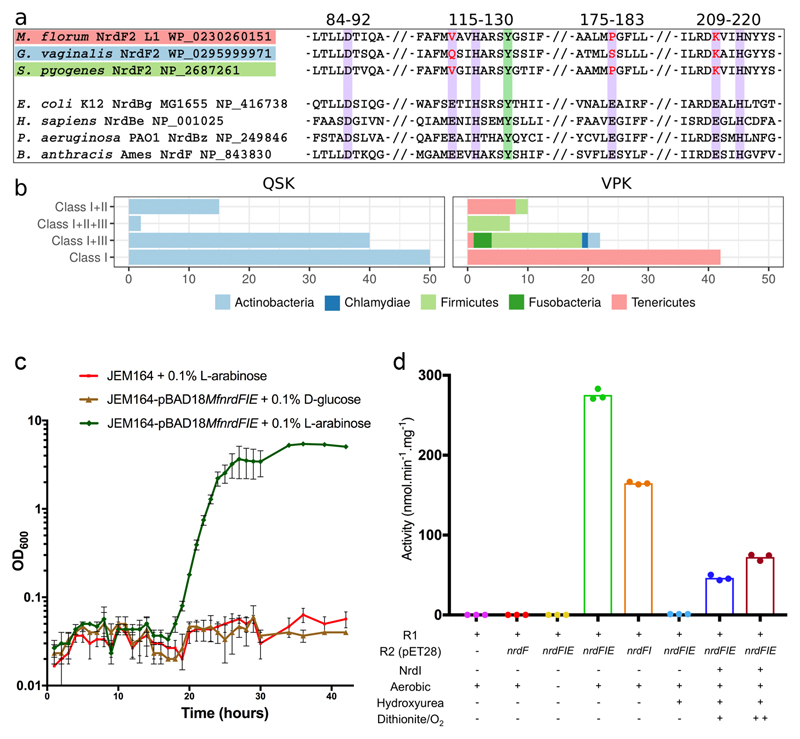
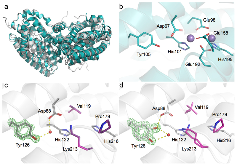
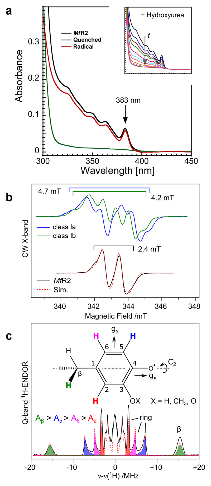
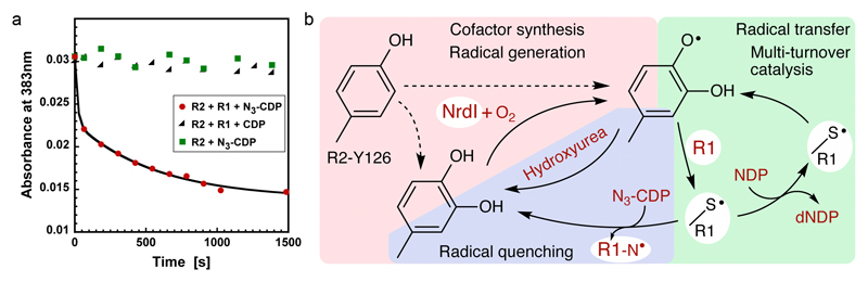
Ribonucleotide reductase (RNR) catalyses the only known de novo pathway for the production of all four deoxyribonucleotides that are required for DNA synthesis1,2. It is essential for all organisms that use DNA as their genetic material and is a current drug target3,4. Since the discovery that iron is required for function in the aerobic, class I RNR found in all eukaryotes and many bacteria, a dinuclear metal site has been viewed as necessary to generate and stabilize the catalytic radical that is essential for RNR activity5-7. Here we describe a group of RNR proteins in Mollicutes-including Mycoplasma pathogens-that possess a metal-independent stable radical residing on a modified tyrosyl residue. Structural, biochemical and spectroscopic characterization reveal a stable 3,4-dihydroxyphenylalanine (DOPA) radical species that directly supports ribonucleotide reduction in vitro and in vivo. This observation overturns the presumed requirement for a dinuclear metal site in aerobic ribonucleotide reductase. The metal-independent radical requires new mechanisms for radical generation and stabilization, processes that are targeted by RNR inhibitors. It is possible that this RNR variant provides an advantage under metal starvation induced by the immune system. Organisms that encode this type of RNR-some of which are developing resistance to antibiotics-are involved in diseases of the respiratory, urinary and genital tracts. Further characterization of this RNR family and its mechanism of cofactor generation will provide insight into new enzymatic chemistry and be of value in devising strategies to combat the pathogens that utilize it. We propose that this RNR subclass is denoted class Ie.


Conflict of interest statement

The authors declare no competing interests.

Figures

Extended data Figure 1
Extended data Figure 1. Unrooted maximum likelihood phylogeny of representative NrdF (RNR subclass Ib radical-generating subunit) sequences.
All RefSeq NrdF sequences were clustered at 75% identity to reduce redundancy and a maximum likelihood phylogeny was estimated. Sequences with non-canonical amino acids in the positions involved in coordinating the metal center of the enzyme formed a well-supported clan in the NrdF2 group of sequences. We identified two variants, one in which three of the glutamates were replaced by Gln, Ser, and Lys (NrdF2.QSK) and the other in which they were replaced by Val, Pro and Lys (NrdF2.VPK). Both variants thus have a substitution of a Lys for the normally metal-bridging Glu (residue 213 in M. florum NrdF2.VPK). Together, the two variants form a well-supported (96% bootstrap support) clan in the phylogeny inside the NrdF2 diversity. The NrdF2.VPK clan appears to be derived from the NrdF2.QSK clan. Behind the sequences in the tree are a set of sequences more than 75% identical to each represented sequence. The VPK and QSK sequences in the phylogeny represent 138 and 182 sequences in RefSeq respectively.
Extended data Figure 2
Extended data Figure 2. Small angle X-ray scattering characterization of the MfR2-NrdI complex.
Solution scattering data for MfR2 (left) and MfR2 incubated with MfNrdI (right). a) Experimental solution scattering profiles (black spheres) for MfR2 alone and incubated with MfNrdI superposed with the theoretical scattering profile of the MfR2 crystal structure (red line) and the theoretical scattering profile from the homology model based on the E. coli R2-NrdI complex structure (blue line). Theoretical scattering curves and goodness of fit values were calculated by CRYSOL. b) Guinier fit and p(r) function of MfR2 alone and incubated with MfNrdI. The fit to the data is shown as an orange line. The shift in invariant parameters Rg and Dmax indicate that an increase in dimensions occurred as MfR2 was incubated with MfNrdI. Radius of gyration statistics were derived from 60 data points within the Guinier region for MfR2 and 55 for MfNrdI-MfR2. c) ab initio models as calculated by DAMMIN of MfR2 alone and with NrdI (grey surface) overlaid with the crystal structure of MfR2 (left) and the homology model based on the E. coli R2-NrdI complex structure model (right).
Extended data Figure 3
Extended data Figure 3. Metal analysis and radical generation in the presence of chelator.
a) Representative TXRF spectra measured for MfR2 (blue, at 664 μM) and Fe-reconstituted class Ia EcR2 (orange, at 635 μM), on the 5 to 10 keV energy range. The spectra have been scaled using the peak size of the Ga internal standard and offset slightly in the Y-direction for clarity. K-level X-ray emission lines are indicated with arrows. For elements, where both Kα and Kβ lines are present, they are specified. Otherwise, arrows indicate Kα lines. Experiments were repeated three times. b) Concentrations of Mn, Fe, Co, Ni, Cu and Zn were measured in the active MfR2, MfNrdI and MfR1 protein solutions and in their respective buffers, as well as in a solution of E. coli class Ia R2 protein reconstituted with Fe in vitro. Mean concentrations and SD of measurements on 3 independently prepared samples for each sample are reported. The concentrations were converted to metal to protein molar ratio. The measurements show that none of the MfRNR proteins contain a significant amount of metal as opposed to EcR2a which as expected contains on the order of 2 metal ions per monomer also after a desalting step. Buffer 1 is the buffer system used for MfR2, i.e. 25 mM HEPES-Na pH 7, 50 mM NaCl. Buffer 2 is the buffer system used for MfR1 and MfNrdI, i.e. 25 mM Tris-HCl pH 8, 50 mM NaCl. The protein purification involves a Ni-affinity step which is likely the reason for nickel being the dominating metal species in the sample. c) HPLC based in vitro assays show that RNR activity can be restored after MfR2 is quenched by hydroxyurea. MfR2 is regenerated by the addition of MfNrdI followed by redox cycling with dithionite and oxygen containing buffer (green) (see main Fig. 2d). Reactivation and activity are observed also in the presence of a metal chelator (EDTA 0.3 mM, blue). Addition of extra metals (0.2 mM of each Mn, Fe, Co, Ni, Cu, Zn) does not improve the activity recovery (pink).
Extended data Figure 4
Extended data Figure 4. Mass spectrometric characterization of intact proteins.
Intact protein mass spectra obtained from purified MfR2 proteins. Inactive protein (above) and active protein (below). Insets represent the decharged and deisotoped mass as calculated by the program Protein Deconvolution (version 4.0) using the ReSpect algorithm therein. The result from the deconvolution of n = 30 consecutive scans in one LC/MS run per protein form is shown. Each protein form was analyzed in duplicate LC/MS runs. Protein intact masses are given as mean +/- standard deviation (SD). SD was 1.4 Da for the “inactive” protein and 2 Da for the “active” form. The results show that the active protein is 17 ± 2 Da heavier than the inactive MfR2.
Extended data Figure 5
Extended data Figure 5. MS2 fragmentation spectra of peptides with oxidized tyrosine.
a) Annotated MS2 fragmentation spectra and respective theoretical fragment ion tables of the doubly charged precursor ion 661.8458 m/z corresponding to peptide VAVHARSY(+15.995)GSIF, and c) the doubly charged precursor ion 458.7279 m/z corresponding to peptide ARSY(+15.995)GSIF, both with the oxidized (+16) Y126 residue. The peptides shown in a) and c) were obtained by proteolytic digestion of the active form of the MfR2 protein with chymotrypsin and pepsin, respectively. The mass error is typically less than 0.01 m/z, in accordance with the high resolution used (15 000). Errors in ppm are indicated for the corresponding fragment ions when detected. Among the fragment ions observed, the most relevant are the b7 and b8 ions for peptide a). The experimental m/z values, the annotation, theoretical m/z values and ppm errors are shown in b), including the peaks for the corresponding isotope envelope. In d) the Y(+O) immonium ion for c) is shown, which demonstrate that Tyr126 is modified by a mass of +15.995 and the absence of the corresponding immonium ion for the unmodified Y. Four independent experiments per peptidase treatment were performed, confirming the modified peptide sequences shown. Figures are taken from one representative experiment.
Extended data Figure 6
Extended data Figure 6. Radical stability and Isotope labeling.
a) Superimposed UV/vis spectra at timepoints between 0 and 400 minutes. Inset, absorbance at 383 nm at 0, 4, 129, 140, 210 and 400 minutes. Experiments were repeated three times. b) X-band spectra of the radical observed in harvested cells grown in minimal media supplemented with deuterated amino acids. EDTA (0.5 mM) was added before induction. From top: non-labeled tyrosine, β,β-D2 tyrosine, 3,5-D2 tyrosine, indole-D5 tryptophan and D5 glycine. The doublet signal collapses to a singlet when β,β-deuterated tyrosine is incorporated in the protein. Furthermore, the additional coupling to the remaining 3/5 proton in the 3,5-D2 tyrosine grown cells disappears, which is also in line with the radical being tyrosine-derived. Finally, the exclusion of tryptophan and glycine as source for the observed radical is evident from the two lower traces in the figure, which are identical to the top spectrum. Five independent cultures were grown each including the indicated deuterated amino acid. Spectra were recorded at 100K in a nitrogen flow system. The spectra have been normalized to the same double integrals, i.e. same number of spins in the cavity.
Extended data Figure 7
Extended data Figure 7. EPR/ENDOR characterization, see Supplementary information.
a) Q-band ENDOR and HYSCORE spectra of the spectral region where an 14N hyperfine coupling should be observed. Experimental parameters are listed in the materials and methods section. b) Full multifrequency (X-/Q-band) EPR dataset and corresponding field dependent Q-band ENDOR spectra. Experimental parameters are listed in the materials and methods section. The red dashed lines represent a simultaneous simulation of all datasets using the spin Hamiltonian formalism. Simulation parameters are listed in Supplementary information Table 1. c) Inferred orientation (θ1, θ2) of the Cβ protons relative to the phenoxyl radical ring plane as determined by the dihedral angle (ϕ) between the ring plane (C1) and Cα. d) Candidates discussed for the MfR2 radical species, see Supplementary information. All ENDOR measurements were repeated at a second microwave frequency (W-band) giving similar results. Pulse EPR and ENDOR measurements represent long data accumulations/averages. EPR: 300 averages (6 scans/50 shots). ENDOR: 600 averages (600 scans/1 shot).
Extended data Figure 8
Extended data Figure 8. EPR saturation.
EPR saturation behavior of the MfR2 radical at 103 and 298 K. Saturation curves at different temperatures determine the microwave power at half saturation P1/2. The temperature dependence of P1/2 gives information about possible relaxing transition metals in the vicinity of the radical. A fast relaxing metal site will give a higher P1/2 than for an isolated radical. The microwave saturation behavior of the MfR2 is similar to that for an hv-irradiated tyrosine solution. Here we evaluate P1/2 ≈ 0.6 mW at 103 K and P1/2 ≈ 30 mW at 298 K for MfR2. This can be compared to hν Tyr• with P1/2 ≈ 0.4mW at 93 K and E. coli Tyr• with P1/2 ≈ 150 mW at 106 K and not possible to saturate at 298K.
Extended data Figure 9
Extended data Figure 9. Primers and operon construct.
a) Primers used in this study. b) Mesoplasma florum class Ie RNR operon construction.
Fig 1
Fig 1. A new RNR subclass able to rescue an Escherichia coli strain lacking aerobic RNR.
a) Sequence alignment of the new R2 protein groups to a number of standard, di-metal containing, R2 proteins. Purple background indicates the 6 normally essential metal-binding residues, only 3 of which are conserved. Two variants are observed in which 3 carboxylate metal ligands are either substituted for valine, proline and lysine (VPK variant) or for glutamine, serine and lysine (QSK variant). The normally radical harboring tyrosine residue is shown with a green background. b) Taxonomic distribution of NrdF2. QSK/VPK encoding organisms and their collected RNR class repertoire. As common for class I RNRs, several genomes encoding the QSK or VPK variant also harbor other RNRs. The QSK clusters are found only in Actinobacteria, whereas the VPK clusters are also found in Firmicutes, Tenericutes, Chlamydiae and Fusobacteria. c) Expression of the MfnrdFIE operon induced by addition of 0.1% v/v L-arabinose (green) rescued the JEM164 double knockout (ΔnrdAB, ΔnrdEF) strain while when gene expression was suppressed with 0.1% v/v D-glucose (brown) the strain failed to recover, as did the strain lacking the vector (red). Growth curves (average of 3 experiments with SD) are shown. d) MfNrdI activates MfR2 in an O2 dependent reaction. HPLC based in vitro RNR activity assays show no activity for R2 protein expressed separately in E. coli (red), while aerobic co-expression with MfnrdI and MfnrdE (green) or MfnrdI (orange) produced an active R2 protein. Anaerobic co-expression (yellow) or incubation of the active R2 with hydroxyurea (light blue) abolishes the activity. Partial activity could be regenerated by the addition of MfNrdI and redox cycling with dithionite and oxygen, blue and maroon for one and two reduction-oxidation cycles respectively. Data points are shown for triplicate experiments.
Fig 2
Fig 2. The active R2 protein is metal-free but covalently modified.
a) Overall structure of the Mesoplasma florum VPK R2 protein (gray) compared to the standard class Ib R2 from Escherichia coli (PDB: 3n37) (cyan). b) Structure of the dinuclear metal site and conserved metal coordinating residues in standard class I RNR R2. c) Structures of inactive MfR2 after expression without MfNrdI (gray) or with MfNrdI under anaerobic conditions (dark gray), both determined to 1.2 Å resolution. These structures are identical within experimental error. The 3 residues substituting the normally conserved carboxylate metal-ligands in canonical class I R2s are shown in pink and purple respectively. In the MfR2 crystal structure, the canonical metal positions are occupied: 1) by a water molecule in a tetrahedral coordination, involving the conserved His216, with distances of 2.8 ± 0.1 Å, as expected for a hydrogen-bonded water, but very unlikely for a metal; 2) by the ε-amino group of Lys213, replacing the conserved metal-bridging glutamate present in all class I R2s. This lysine forms a hydrogen bond with Asp88, the only remaining carboxylate residue. Asp88 also interacts via a H-bonded water with Tyr126, corresponding to the tyrosine harboring the metal-coupled radical in standard class Ia and Ib R2 proteins. d) Structure of the active MfR2 after aerobic co-expression with MfNrdI and MfR1. Here, the tyrosine is covalently modified in the meta-position. Mass spectrometry determined that the tyrosine is hydroxylated in the active protein. Simulated annealing omit Fo-Fc electron density maps for the unmodified or modified Tyr126 is shown in green and contoured at 8σ. Carbons are in cyan, gray/pink for R2 from E. coli and M. florum, respectively. Oxygens and nitrogens are colored red and blue, respectively. Mn(II) ions are represented as purple spheres. H-bond interactions to the Tyr126 are indicated.
Fig 3
Fig 3. Characterization of a novel stable DOPA radical species in MfR2.
a) The UV/vis spectrum of the active blue protein shows a peak at 383 nm and additional structure at lower wavelengths (black). Incubation with 52 mM hydroxyurea for 20 minutes removed all features from the spectrum except the protein-related absorbance peak at 280 nm (green and inset). The red trace represents the active minus quenched spectra. The rate constant for the decay of the absorbance at 348, 364 and 383 nm was identical, consistent with all absorbance features arising from a single radical species. Experiments were repeated three times. b) X-band EPR spectra of tyrosyl radicals observed in R2 proteins (Escherichia coli class Ia R2 and Bacillus cereus class Ib R2 reconstituted with Fe) compared to the M. florum R2 radical species reported here. X-band measurements were performed on isolated MfR2 protein (Fig. 3b) and repeated on crude cell extracts (Extended data Fig. 6b). c) Q-band ENDOR spectrum recorded at the low field edge of the EPR spectrum. The red dashed lines represent a simultaneous simulation of all datasets. Spectral simulations of the multifrequency EPR and ENDOR spectra using the spin Hamiltonian formalism reveal the radical has four resolved non-equivalent proton couplings with isotropic values of 28.7, 9.8 6.5 and 4.4 MHz. The absence of a nitrogen coupling excludes the side-chains of tryptophan and histidine as the site of the stable radical. In addition, the absence of equivalent proton couplings also excludes the side-chains of tyrosine and phenylalanine. Thus, no native aromatic protein residue can explain the observed radical species. The magnitude of coupling constants are smaller than those of tyrosyl (phenoxyl) radicals, and are instead in good agreement with phenoxyl radicals with an additional oxy substituent (O-X). All ENDOR measurements were repeated at a second microwave frequency (W-band) giving similar results. Pulse EPR and ENDOR measurements represent long data accumulations/averages. EPR: 300 averages ENDOR: 600 averages.
Fig 4
Fig 4. Catalytic competency of the radical and proposed mechanistic scheme in class Ie RNR.
a) UV/vis absorbance at 383 nm vs time. The radical signal is quenched in the presence of protein R1 and the mechanism-based inhibitor N3-CDP (red). Protein R2 with N3-CDP alone (green) or turnover conditions with protein R1 and CDP (black) does not quench the MfR2 radical. The experiment shows that the observed radical can be reversibly transferred to the active site and support catalysis in protein R1. Experiments were repeated three times. b) Proposed mechanistic steps in class Ie RNR. The radical-harboring cofactor is first post-translationally generated by hydroxylation of Tyr126 in an NrdI and O2 dependent process. Dashed lines indicate alternative paths for the post-translational modification of Tyr126. It is presently unknown if this reaction also directly forms the radical species. Once the DOPA radical is formed in protein R2 it supports multi-turnover ribonucleotide reduction together with protein R1, presumably analogous to other class I RNR systems. If the radical is lost, activity can be restored in the covalently modified R2 protein by NrdI, again in an O2 dependent process.

Similar articles

Cited by

References

    1. Hofer A, Crona M, Logan DT, Sjöberg B-M. DNA building blocks: keeping control of manufacture. Crit Rev Biochem Mol Biol. 2012;47:50–63. - PMC - PubMed
    1. Nordlund P, Reichard P. Ribonucleotide reductases. Annu Rev Biochem. 2006;75:681–706. - PubMed
    1. Mannargudi MB, Deb S. Clinical pharmacology and clinical trials of ribonucleotide reductase inhibitors: is it a viable cancer therapy? J Cancer Res Clin Oncol. 2017;143:1499–1529. - PMC - PubMed
    1. Aye Y, Li M, Long MJC, Weiss RS. Ribonucleotide reductase and cancer: biological mechanisms and targeted therapies. Oncogene. 2015;34:2011–2021. - PubMed
    1. Brown NC, Eliasson R, Reichard P, Thelander L. Nonheme iron as a cofactor in ribonucleotide reductase from E. coli. Biochem Biophys Res Commun. 1968;30:522–527. - PubMed

Publication types

MeSH terms

LinkOut - more resources