. 2018 Oct 31:9:2510.
doi: 10.3389/fimmu.2018.02510. eCollection 2018.

Early Induction of Human Regulatory Dermal Antigen Presenting Cells by Skin-Penetrating Schistosoma Mansoni Cercariae

Affiliations

Early Induction of Human Regulatory Dermal Antigen Presenting Cells by Skin-Penetrating Schistosoma Mansoni Cercariae

Béatrice M F Winkel et al. Front Immunol. .

Abstract

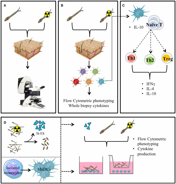
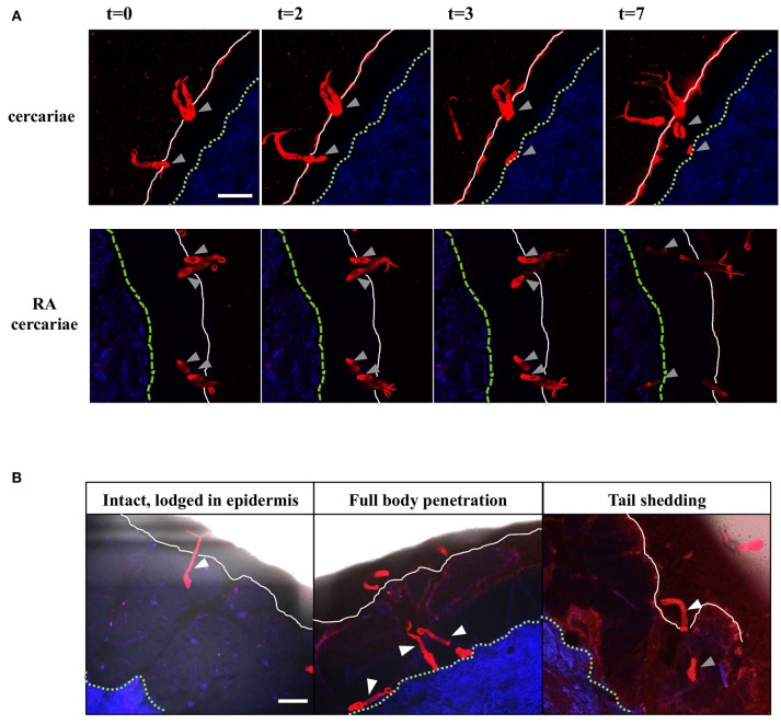
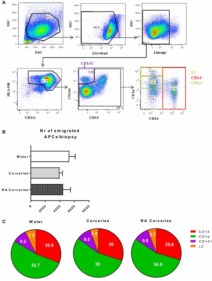
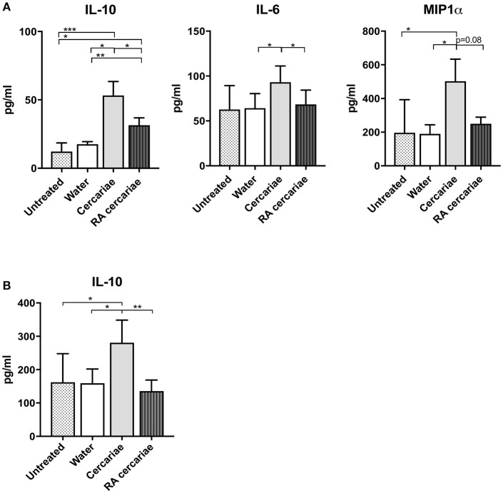
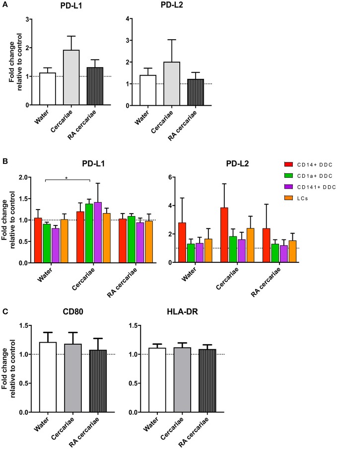
Following initial invasion of Schistosoma mansoni cercariae, schistosomula reside in the skin for several days during which they can interact with the dermal immune system. While murine experiments have indicated that exposure to radiation-attenuated (RA) cercariae can generate protective immunity which is initiated in the skin stage, contrasting non-attenuated cercariae, such data is missing for the human model. Since murine skin does not form a reliable marker for immune responses in human skin, we used human skin explants to study the interaction with non-attenuated and RA cercariae with dermal innate antigen presenting cells (APCs) and the subsequent immunological responses. We exposed human skin explants to cercariae and visualized their invasion in real time (initial 30 min) using novel imaging technologies. Subsequently, we studied dermal immune responses and found an enhanced production of regulatory cytokine interleukin (IL)-10, pro-inflammatory cytokine IL-6 and macrophage inflammatory protein (MIP)-1α within 3 days of exposure. Analysis of dermal dendritic cells (DDCs) for their phenotype revealed an increased expression of immune modulators programmed death ligand (PD-L) 1 and 2, and increased IL-10 production. Ex vivo primed DDCs suppress Th1 polarization of naïve T-cells and increase T-cell IL-10 production, indicating their regulatory potential. These immune responses were absent or decreased after exposure to RA parasites. Using transwells, we show that direct contact between APCs and cercariae is required to induce their regulatory phenotype. To the best of our knowledge this is the first study that attempts to provide insight in the human dermal S. mansoni cercariae invasion and subsequent immune responses comparing non-attenuated with RA parasites. We reveal that cercariae induce a predominantly regulatory immune response whereas RA cercariae fail to achieve this. This initial understanding of the dermal immune suppressive capacity of S. mansoni cercariae in humans provides a first step toward the development of an effective schistosome vaccine.

Keywords: PD-L1; Schistosoma mansoni; bilharzia; cercaria; human; macrophage; schistosome; skin.


Figures

Figure 1
Figure 1
General presentation of the skin invasion set up and immunological markers studied. Skin invasion imaging setup: S. mansoni (RA) cercariae are irradiated or kept at RT. Human skin explant pieces are mounted on confocal microscopy dishes and cercariae are added to the epidermal side. Explants are imaged with a confocal microscope. (A) Skin explants are exposed to (RA) cercariae in water and cultured to harvest emigrating DDCs (B). DDCs are co-cultured with naïve T cells to assess functional effects (C). Monocyte derived dendritic cells (D) (MoDCs) are differentiated from human monocytes. Three hour ES products are generated from (RA) schistosomula. Stimulation of MoDCs with (RA) cercariae or their ES products in presence or absence of a transwell.
Figure 2
Figure 2
Visualization of cercarial invasion into a human skin explant. Cercariae attach and penetrate the epidermis of the human skin explant. Cercariae depicted in red, the dermis in blue, the epidermal surface as a white solid line and the basal membrane as a green dashed line T = 0–7 min. Gray arrowheads: cercarial heads/schistosomula. Top panels: non-attenuated cercariae, lower panels: RA cercariae (A). Cercariae penetrate the skin in different ways. White arrowheads: cercariae/tails (B). Scale bar: 200 μm.
Figure 3
Figure 3
S. mansoni cercariae do not induce DDC emigration from the skin. DDC gating strategy. Antigen presenting cells are selected by forward scatter (FSC) and side scatter (SSC) characteristics. Doublets are excluded (not shown). Live cells are gated and selected for the lack of lineage markers (CD56, CD3, CD19, CD20). APCs are selected on the expression of HLA-DR as well as intermediate to high levels of CD11c. HLA-DR+, CD11c+ cells can be divided into the different DDC populations: CD141+, LC, CD1a+ and CD14+ (A). Total emigrated HLA-DR+, CD11c+ antigen presenting cells from dermal biopsies at 3 days post exposure to S. mansoni cercariae or water control. Mean ± SEM, n = 7 (B). Subset distribution of emigrated cells (C).
Figure 4
Figure 4
Increased production of IL-6 and IL-10 and MIP1α in skin exposed to cercariae. Whole biopsy cytokine analysis at 3 days post exposure to non-attenuated Sm cercariae show an increase in IL-6, IL-10 and MIP1α. The effect is less pronounced in radiation attenuated cercariae. (A). IL-10 production by DDCs after co-culture of emigrated DDCs with CD40L expressing cell line, 7 donors. (B). Data shown in pg/ml, mean ± SEM. *p < 0.05, **p < 0.01, ***p < 0.001 using paired Student's T-test on log transformed data.
Figure 5
Figure 5
DDCs do not increase activation markers upon cercaria exposure but do show a trend of increased levels of immunoregulatory markers PD-L1 and PD-L2. Immunoregulatory markers PD-L1 and 2 are up regulated in DDCs when exposed to Cercariae but not RA Cercariae (n = 3) (A). PD-L1 expression primarily occurs in CD1a+ DDCs. (n = 3) (B). CD80 and HLA-DR expression in total emigrated HLA-DR+, CD11c+ antigen presenting cells from dermal biopsy at 84 h post exposure to S. mansoni cercariae or water control (n = 7) (C). Mean ± SEM. *p < 0.05.
Figure 6
Figure 6
Stimulation with cercariae reduces pro-inflammatory potential of DDCs. Emigrated DDCs were co-cultured with CD4+ T cells to assess immunomodulatory potential. Data shown as percentages IFNγ or IL4 producing CD4+ T cells cultured with ex vivo stimulated DDCs relative to CD4+ T cells cultured with DDCs from untreated skin after PMA/ionomycin stimulation. Mean ± SEM. *p < 0.05, using paired Student's T-test (A). IL-10 production by CD4+ T cells after co-culture with DDCs. ELISA on culture supernatants after 24 h stimulation with anti CD3/28 (B). Data shown in pg/ml. Mean ± SEM.
Figure 7
Figure 7
Cercaria stimulation activates MoDCs but also induces up regulation of immunoregulatory markers PD-L1 and 2 and regulatory cytokine IL-10 production. Stimulation of MoDCs with (RA) cercariae up regulates activation markers CD80, CD86 and CD40. (A). PD-L1 and PD-L2 up regulation after cercaria stimulation. (B) IL-10 production by MoDCs after 48 h stimulation, data shown as pg/ml (C) Mean ± SEM. *p < 0.05, **p < 0.005, ***P < 0.0005, ****p < 0.0001 using paired student's T-test (on log transformed data for IL-10).
Figure 8
Figure 8
Direct contact with cercariae but not cercarial ES products increases immunoregulatory markers PD-L1 and 2 and IL-10 secretion. PD-L1 and PD-L2 up regulation after cercaria stimulation in presence or absence of direct contact (TW = transwell) (A). PD-L1 expression after stimulation with cercariae or cercarial ES alone. The ES dose was matched to the number of cercariae used for stimulation (B). IL-10 production of MoDCs after stimulation with cercariae or their products (C). Mean ± SEM. *p < 0.05, **p < 0.005, ***P < 0.0005, ****p < 0.0001 using paired student's T-test (on log transformed data for IL-10).

References

    1. Wheater PR, Wilson RA. Schistosoma mansoni: a histological study of migration in the laboratory mouse. Parasitology (1979) 79:49–62. 10.1017/S0031182000051970 - DOI - PubMed
    1. Mangold BL, Dean DA. Autoradiographic analysis of Schistosoma mansoni migration from skin to lungs in naive mice. Evidence that most attrition occurs after the skin phase. Am J Trop Med Hyg. (1983) 32:785–9. 10.4269/ajtmh.1983.32.785 - DOI - PubMed
    1. Wilson RA, Coulson PS, Sturrock RF, Reid GD. Schistosome migration in primates: a study in the olive baboon (Papio anubis). Trans R Soc Trop Med Hyg. (1990) 84:80–3. 10.1016/0035-9203(90)90391-Q - DOI - PubMed
    1. Di Meglio P, Perera GK, Nestle FO. The multitasking organ: recent insights into skin immune function. Immunity (2011) 35:857–69. 10.1016/j.immuni.2011.12.003 - DOI - PubMed
    1. Mountford AP, Trottein F. Schistosomes in the skin: a balance between immune priming and regulation. Trends Parasitol. (2004) 20:221–6. 10.1016/j.pt.2004.03.003 - DOI - PubMed

Publication types

MeSH terms