. 2018 Oct 5;8(18):5025-5038.
doi: 10.7150/thno.26837. eCollection 2018.

Novel skin patch combining human fibroblast-derived matrix and ciprofloxacin for infected wound healing

Affiliations

Novel skin patch combining human fibroblast-derived matrix and ciprofloxacin for infected wound healing

Muhammad Suhaeri et al. Theranostics. .

Abstract

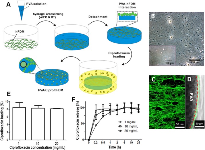
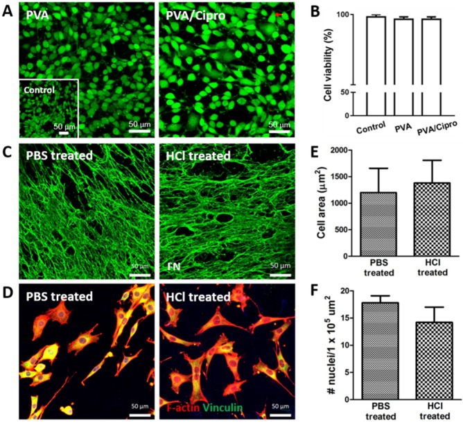
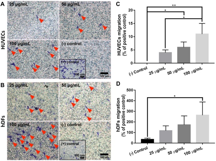
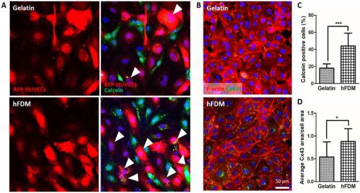
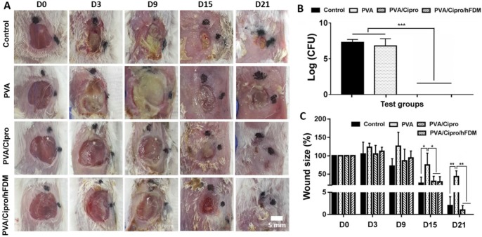
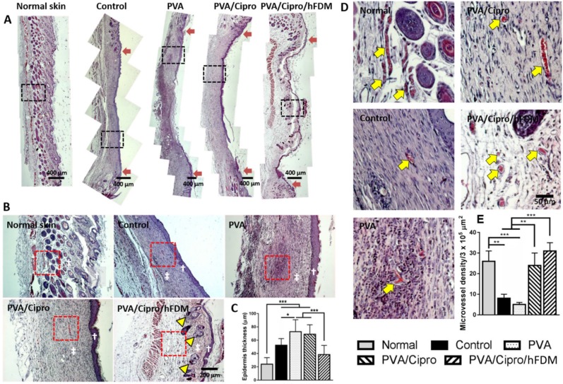
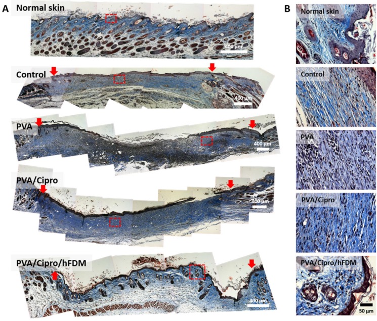
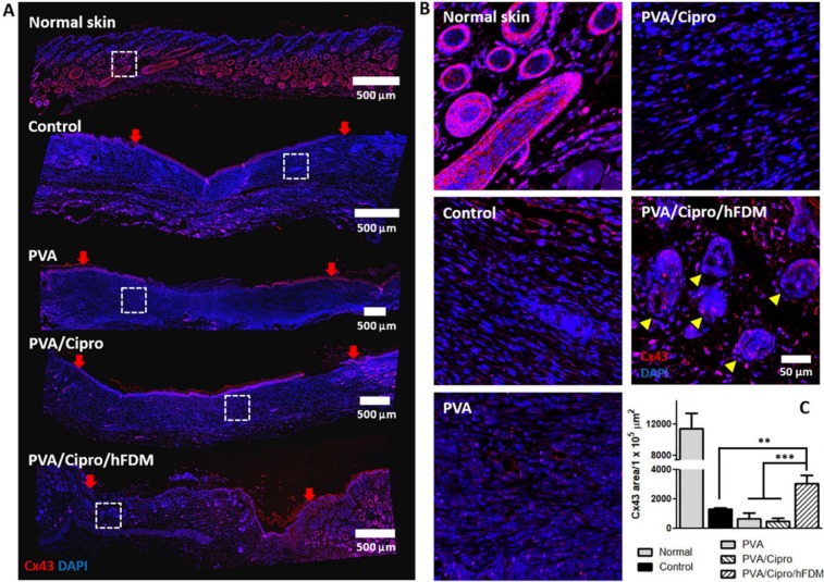
Skin injuries are frequently encountered in daily life, but deep wounds often poorly self-heal and do not recover completely. In this study, we propose a novel skin patch that combines antibiotic, cell-derived extracellular matrix (ECM) and biocompatible polyvinyl alcohol (PVA) hydrogel. Methods: Decellularized human lung fibroblast-derived matrix (hFDM) was prepared on tissue culture plate (TCP) and PVA solution was then poured onto it. After a freeze-thaw process, PVA was peeled off from TCP along with hFDM tightly anchored to PVA. Subsequently, ciprofloxacin (Cipro)-incorporated PVA/hFDM (PVA/Cipro/hFDM) was fabricated via diffusion-based drug loading. Results: In vitro analyses of PVA/Cipro/hFDM show little cytotoxicity of ciprofloxacin, stability of hFDM, rich fibronectin in hFDM, and good cell attachment, respectively. In addition, hFDM proved to be beneficial in promoting cell migration of dermal fibroblasts and human umbilical vein endothelial cells (HUVECs) using transwell inserts. The antibacterial drug Cipro was very effective in suppressing colony growth of gram-negative and -positive bacteria as identified via an inhibition zone assay. For animal study, infected wound models in BALB/c mice were prepared and four test groups (control, PVA, PVA/Cipro, PVA/Cipro/hFDM) were administered separately and their effect on wound healing was examined for up to 21 days. The results support that Cipro successfully reduced bacterial infection and thus encouraged faster wound closure. Further analysis using histology and immunofluorescence revealed that the most advanced skin regeneration was achieved with PVA/Cipro/hFDM, as assessed via re-epithelialization, collagen texture and distribution in the epidermis, and skin adnexa (i.e., glands and hair follicles) regeneration in the dermis. Conclusion: This work demonstrates that our skin patch successfully consolidates the regenerative potential of ECM and the antibacterial activity of Cipro for advanced wound healing.

Keywords: ciprofloxacin; human fibroblast-derived matrix; polyvinyl alcohol hydrogel; skin patch; wound healing.


Conflict of interest statement

Competing Interests: The authors have declared that no competing interest exists.

Figures

Figure 1
Figure 1
Preparation of PVA/hFDM and ciprofloxacin-loaded PVA/hFDM. (A) Schematic illustration of the overall process in fabricating PVA/hFDM and ciprofloxacin-loaded PVA/hFDM. (B) Microscopic image of decellularized hFDM on TCP and the surface of TCP right after the transfer of hFDM into PVA hydrogel. Scale bar is 100 μm. (C) Immunofluorescence of FN in the PVA/hFDM. (D) Cross-sectional view of the interface (dashed red line) between PVA and hFDM. Scale bar is 50 μm. (E) Loading of ciprofloxacin with different initial concentrations into PVA hydrogel and (G) the release profile over 24 h in PBS at 37 °C. FN: fibronectin; hFDM: human lung fibroblast-derived matrix; PVA: polyvinyl alcohol; TCP: tissue culture plastic.
Figure 2
Figure 2
Characterization of PVA-based membranes. (A) Cell viability test of mouse fibroblasts after 24 h culture on gelatin coated-PVA (treated with 0.1 N HCl) and gelatin-coated PVA/Cipro (treated with 1 mg/mL ciprofloxacin) as evaluated via live/dead assay. Inset shows the cells cultured on TCP. (B) Quantitative analysis of cell viability based on image analysis of the live/dead assay. (C) Immunofluorescence of hFDM against FN in PVA/hFDM treated with either PBS or 0.1 N HCl. (D) Culture of fibroblasts on PBS-treated and 0.1 N HCl-treated PVA/hFDM, respectively. F-actin is stained red and focal adhesion molecule vinculin is shown in green. (E) Quantitative analysis of cell spreading area and (F) cell density based on image analysis of (D). All scale bars are 50 μm. Cipro: ciprofloxacin; FN: fibronectin; HCl: hydrochloric acid; hFDM: human lung fibroblast-derived matrix; PBS: phosphate buffered saline; PVA: polyvinyl alcohol; TCP: tissue culture plastic.
Figure 3
Figure 3
Chemotactic effect of hFDM. (A-B) Migration of HUVECs and HDFs, respectively, treated with different concentrations of hFDM suspension, as evaluated via transwell migration assay. The migrated cells are spotted using crystal violet staining. Red triangles designate migrated cells. Positive and negative controls are 10% FBS and serum-free conditions, respectively. Scale bars are 100 μm. (C-D) Quantitative comparison of the number of HUVECs and HDFs migrated through the transwell membrane. The cell number is normalized to that of positive control. Statistically significant difference: *p < 0.05 or **p < 0.01. HDFs: human dermal fibroblasts; hFDM: human lung fibroblast-derived matrix; HUVECs: human umbilical vein endothelial cells.
Figure 4
Figure 4
Gap junction-mediated cell-cell communication. (A) Dye transfer assay between calcein AM-labeled HUVECs (green) and RFP-HUVECs (red) after 24 h co-culture on gelatin and hFDM, respectively; the images in the left side show only the RFP-HUVECs, and the merged images of RFP-HUVECs and calcein AM-labeled HUVECs are on the right side. Positive signals (white triangles) are identified via the RFP-HUVECs (red) with green dye (calcein AM) uptake. (B) Expression of Cx43 protein (green) with HUVECs cultured on either gelatin or hFDM for 3 days, along with F-actin staining (red). (C) Percentage of calcein dye-positive RFP-HUVECs as calculated via image analysis after the dye transfer assay; the percentage was averaged by counting the number of dye-positive RFP-HUVECs against total RFP-HUVECs in a given image. (D) Quantitative evaluation of average Cx43 area per cell area. All scale bars are 50 μm. Statistically significant difference: *p < 0.05 or ***p < 0.001. Cx43: connexin 43; HUVECs: human umbilical vein endothelial cells; RFP-HUVECs: red fluorescence protein expressing HUVECs.
Figure 5
Figure 5
Infected skin wound model and transplantation of PVA-based membranes. (A) Infected wounds on the dorsal region of BABL/c mice and gross appearance of wound closure for up to 21 days (D0-D21). Scale bar is 5 mm. (B) Bacterial count (CFU) of the samples collected from the wound sites 6 h post-treatment. (C) Measurement of wound size as shown as percentage normalized to that of the initial wound size at day 0. Statistically significant difference: *p < 0.05, **p < 0.01, or ***p < 0.001. CFU: colony forming unit; PVA: polyvinyl alcohol.
Figure 6
Figure 6
Skin regeneration in epidermis and dermis on day 21. (A) H&E staining of the excised skin tissues collected at 21 days post-treatments. Red arrows indicate the position of the initial wound areas. Dashed black boxes correspond to the enlarged areas shown in (B). Scale bar is 400 μm. (B) Higher magnification images of regenerated skin tissues, where white dagger (†) and double dagger (‡) denote the epidermis and dermis layers, respectively. Yellow triangles indicate the regenerated hair follicles and glands. Red boxes indicate the area magnified in (D). Scale bar is 200 μm. (C) Thickness of new epidermis. (D) Enlarged images show the presence of microvessels in the regenerated dermis as indicated by yellow arrows and the quantitative analysis of microvessel density. Scale bar is 50 μm. Statistically significant difference: *p < 0.05, **p < 0.01 or ***p < 0.001. H&E: hematoxylin and eosin.
Figure 7
Figure 7
Analysis of collagen texture and distribution pattern on day 21. (A) Masson's trichrome staining of the excised skin tissues on day 21 post-treatments. Collagen and nuclei are indicated by blue color and black dots, respectively. Red arrows indicate the position of the initial wound areas. Dashed red boxes indicate the enlarged areas. Scale bar is 400 μm. (B) Higher magnification images show deposition of collagen and their distribution pattern in the epidermis and dermis. Scale bar is 50 μm.
Figure 8
Figure 8
Analysis of gap junction protein expression. (A) Immunofluorescence of Cx43 protein with the skin tissues retrieved at 21 days post-treatments. Cx43 and nuclei (DAPI) are indicated in red and blue color, respectively. Red arrows show the position of the initial wound regions. Dashed white boxes indicate the magnified areas. Scale bar is 500 μm. (B) Higher magnification images present the expression and organization of Cx43 protein in the regenerated dermis. Yellow triangles indicate the regenerated follicles and glands. Scale bar is 50 μm. (C) Quantitative analysis of Cx43-positive area per unit area (1×105 μm2). Statistically significant difference among control, PVA, PVA/Cipro and PVA/Cipro/hFDM: **p < 0.01 or ***p < 0.001. Cipro: ciprofloxacin; Cx43: connexin 43; DAPI: 4', 6-diamidino-2-phenylindole; hFDM: human lung fibroblast-derived matrix; PVA: polyvinyl alcohol.

References

    1. Deng CM, He LZ, Zhao M. et al. Biological properties of the chitosan-gelatin sponge wound dressing. Carbohydr Polym. 2007;69:583–89.
    1. Guo S, Dipietro LA. Factors affecting wound healing. J Dent Res. 2010;89:219–29. - PMC - PubMed
    1. Fan Z, Liu B, Wang J. et al. A novel wound dressing based on Ag/graphene polymer hydrogel: effectively kill bacteria and accelerate wound healing. Adv Funct Mater. 2014;24:3933–43.
    1. Zilberman M, Egozi D, Shemesh M. et al. Hybrid wound dressings with controlled release of antibiotics: structure-release profile effects and in vivo study in a guinea pig burn model. Acta Biomater. 2015;22:155–63. - PubMed
    1. Chester D, Brown AC. The role of biophysical properties of provisional matrix proteins in wound repair. Matrix Biol. 2017;60-61:124–40. - PubMed

Publication types

MeSH terms