. 2018 Oct 6;8(18):5159-5177.
doi: 10.7150/thno.27760. eCollection 2018.

Mussel-inspired conductive nanofibrous membranes repair myocardial infarction by enhancing cardiac function and revascularization

Affiliations

Mussel-inspired conductive nanofibrous membranes repair myocardial infarction by enhancing cardiac function and revascularization

Yutong He et al. Theranostics. .

Abstract

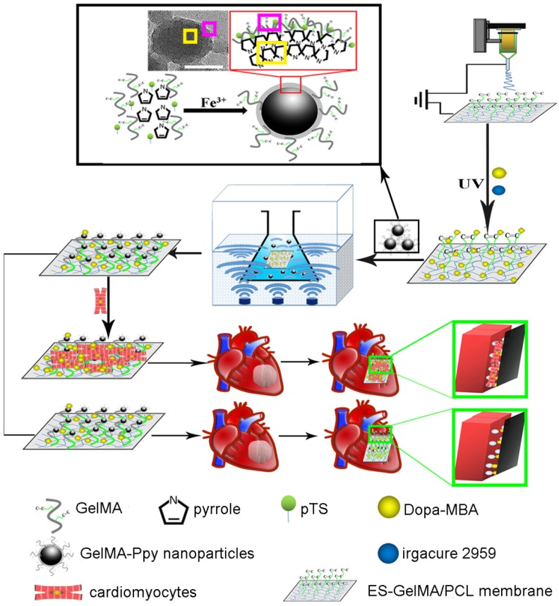
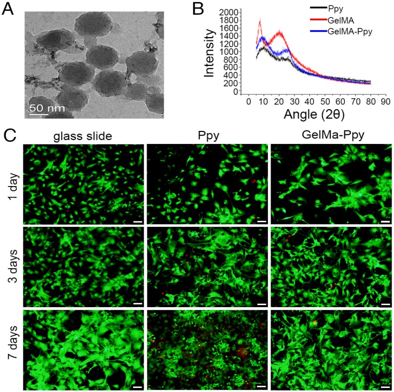
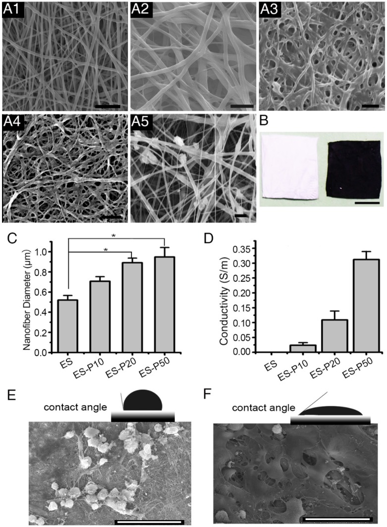
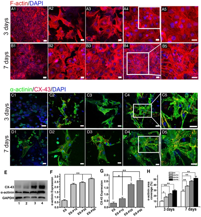
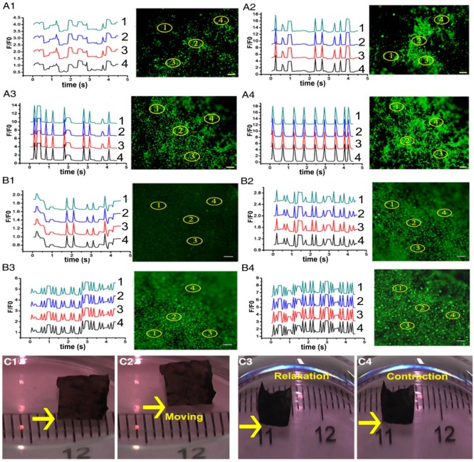
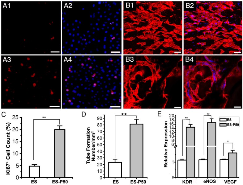
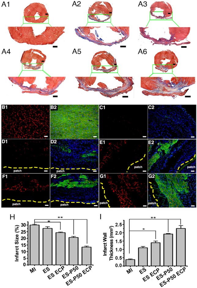
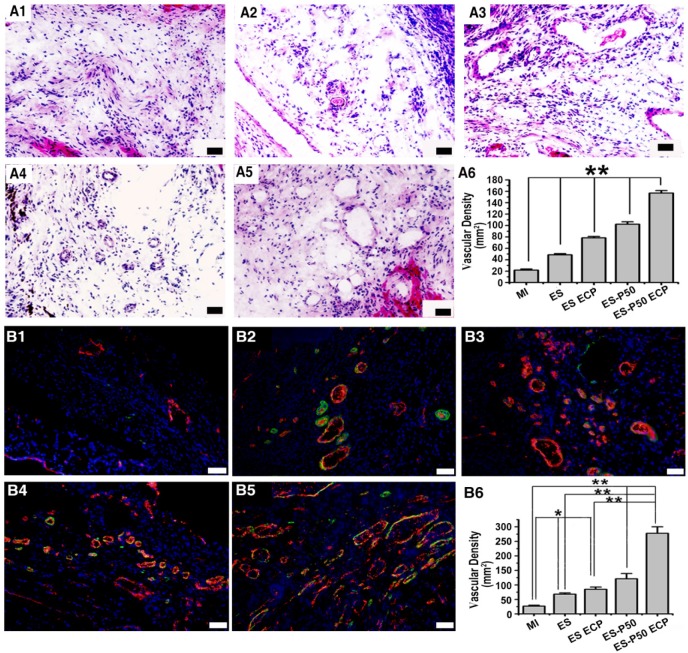
The controversy between polypyrrole's (Ppy) biocompatibility and its aggregation on nanofibers impedes application of conductive Ppy-incorporated nanofibers to create engineered cardiac microenvironments. The purpose of this study was to fabricate a functional scaffold for engineering cardiac patches (ECP) using a high concentration of methyl acrylic anhydride-gelatin (GelMA)-Ppy nanoparticles, mussel-inspired crosslinker, and electrospun (ES)-GelMA/polycaprolactone (PCL) nanofibrous membrane. Methods: First, spherical GelMA-Ppy nanoparticles were obtained when the methacrylate groups of GelMA formed a self-crosslinked network through oxidative polymerization of Ppy. Second, GelMA-Ppy nanoparticles were uniformly crosslinked on the ES-GelMA/PCL membrane through mussel-inspired dopamine-N'N'-methylene-bis-acrylamide (dopamine-MBA) crosslinker. Finally, the feasibility of the dopa-based conductive functional ECP scaffold was investigated in vitro and in vivo. Results: The GelMA-Ppy nanoparticles displayed excellent biocompatibility at a high concentration of 50 mg/mL. The massive GelMA-Ppy nanoparticles could be uniformly distributed on the ES nanofibers through dopamine-MBA crosslinker without obvious aggregation. The high concentration of GelMA-Ppy nanoparticles produced high conductivity of the dopamine-based (dopa-based) conductive membrane, which enhanced the function of cardiomyocytes (CMs) and yielded their synchronous contraction. GelMA-Ppy nanoparticles could also modify the topography of the pristine ES-GelMA/PCL membrane to promote vascularization in vitro. Following transplantation of the conductive membrane-derived ECP on the infarcted heart for 4 weeks, the infarct area was decreased by about 50%, the left ventricular shortening fraction percent (LVFS%) was increased by about 20%, and the neovascular density in the infarct area was significantly increased by about 9 times compared with that in the MI group. Conclusion: Our study reported a facile and effective approach to developing a functional ECP that was based on a mussel-inspired conductive nanofibrous membrane. This functional ECP could repair infarct myocardium through enhancing cardiac function and revascularization.

Keywords: dopamine; electrospun membrane; myocardial infarction; polypyrrole nanoparticles; revascularization.


Conflict of interest statement

Competing Interests: The authors have declared that no competing interest exists.

Figures

Scheme 1
Scheme 1
Schematic illustration of the fabrication of mussel-inspired conductive membrane and its application in engineered cardiac tissue patch (ECP) in a rat MI model. During the oxypolymerization of Ppy, paratoluenesulfonic acid (pTS) served as the dopant, FeCl3 acted as the oxidizing agent, and GelMA self-crosslinked onto the network structure, forming GelMA-Ppy nanoparticles. After the dopa-grafted ES-GelMA/PCL membrane was fabricated under the photoinitiator irgacure 2959, GelMA-Ppy nanoparticles were concussively crosslinked onto the dopa-MAB-grafted ES-GelMA/PCL membrane through the mussel-inspired dopamine crosslinker. Scale bar, 50 nm.
Figure 1
Figure 1
Characterization and biocompatibility of the GelMA-Ppy nanoparticles. (A) TEM image of GelMA-Ppy nanoparticles. (B) XRD analysis of GelMA-Ppy nanoparticles. (C) Live/dead staining of untreated CMs, 25 mg/mL Ppy-treated CMs, and 50 mg/mL GelMA-Ppy nanoparticles-treated CMs for 1, 3 and 7 days. The live cells are green and the dead cells are red. Scale bar, 50 µm.
Figure 2
Figure 2
Characterization of the mussel-inspired conductive membranes. (A) SEM images of ES (A1), ES-P10 (A2), ES-P20 (A3), ES-P50 (A4), and ES-Ppy (A5). Scale bars, 5 µm. (B) Gross photographs of ES (white, left), and ES-P50 (dark, right). Scale bar, 5 mm. (C) Average fiber diameters of different membranes. All data are presented as mean ± SD. *p<0.05, n=3. (D) Conductivities of membranes. (E-F) The contact angles and outspread structure of CMs on ES (E), and on ES-P50 (F). ES: pristine ES-GelMA/PCL membrane; ES-P10: dopa-based ES-P10 membrane; ES-P20: dopa-based ES-P20 membrane; ES-P50: dopa-based ES-P50 membrane. ES-Ppy: directly blending-spun ES-Ppy membrane. Scale bars in (E-F), 50 µm.
Figure 3
Figure 3
Mussel-inspired conductive membranes enhanced the function of CMs. (A-B) Filamentous actin (F-actin) staining for CMs on membranes on day 3 (A) and day 7 (B). (A1, B1) ES. (A2, B2) ES-P10. (A3, B3) ES-P20. (A4, B4) ES-P50. Scale bars, 20 µm. (C-D) α-actinin staining for sarcomere (green) and CX-43 proteins expression (red) of CMs on membranes on day 3 (C) and day 7 (D). (C1, D1) ES. (C2, D2) ES-P10. (C3, D3) ES-P20. (C4, D4) ES-P50. Scale bars, 20 µm. (E) Western blotting for the expressions of α-actinin protein and CX-43 protein in CMs on membranes on day 7 of culture. Line 1: ES. Line 2: ES-P10. Line 3: ES-P20. Line 4: ES-P50. (F-G) Quantitative analysis of α-actinin (F) and CX-43 (G) of CMs on membranes based on Western blotting. (H) α-actinin area coverage of CMs on membranes on day 3 and day 7 based on immunofluorescence. ES: pristine ES-GelMA/PCL membrane; ES-P10: dopa-based ES-P10 membrane; ES-P20: dopa-based ES-P20 membrane; ES-P50: dopa-based ES-P50 membrane. All data are presented as mean ± SD. *p<0.05, **p<0.01.
Figure 4
Figure 4
Dopa-based conductive membranes increase calcium transient's frequency within CMs and spontaneous movement of ECPs. (A-B) Transient and extracted frequency signals of Ca2+ within CMs on membranes on day 3 (A) and day 7 (B). (A1, B1) Pristine ES-GelMA/PCL membranes. (A2, B2) Dopa-based ES-P10 membranes. (A3, B3) Dopa-based ES-P20 membranes. (A4, B4) Dopa-based ES-P50 membranes. Scale bars, 100 µm. (C) Spontaneous movement (C1-C2) and contraction activity (C3-C4) of dopa-based ES-P50 ECPs on day 7.
Figure 5
Figure 5
ES-P50 membrane was beneficial for the proliferation and vascularization of HUVECs in vitro. (A) Ki67-positive cells (red) on ES (A1-A2) and ES-P50 (A3-A4). Scale bars, 50 µm. (B) vWF-positive HUVECs (red) on ES (B1-B2) and ES-P50 (B3-B4). Scale bars, 20 µm. (C) Percentage of Ki67-positive cells on ES and ES-P50. (D) Tube number per mm2 formed by HUVECs on ES and ES-P50. (E) Expression of KDR, eNOS and VEGF genes in HUVECs on ES and ES-P50. ES: pristine ES-GelMA/PCL membrane; ES-P50: dopa-based ES-P50 membrane. All data are presented as mean ± SD. *p<0.05, **p<0.01. n=3. Cell nuclei were stained with DAPI.
Figure 6
Figure 6
Mussel-inspired conductive membranes were effective for infarct myocardium repair. (A) Masson's staining for fibrous tissue (blue) and myocardium (red) of heart sections in rats. (A1) Sham group. (A2) MI group. (A3) ES group. (A4) ES ECP group. (A5) ES-P50 group. (A6) ES-P50 ECP group. Scale bars, 1 mm. (B-G) α-actinin-positive myocardial tissue (green) and CX-43 protein expression (red) in the infarcted area. (B1-B2) Sham group. (C1-C2) MI group. (D1-D2) ES group. (E1-E2) ES ECP group. (F1-F2) ES-P50 group. (G1-G2) ES-P50 ECP group. Cell nuclei were stained with DAPI. Scale bars, 50 µm. (H-I) Statistical analysis of infarct size (H) and infarct wall thickness (I). ES: pristine ES-GelMA/PCL membrane group; ES-ECP: pristine ES-GelMA/PCL ECP group; ES-P50: dopa-based ES-P50 membrane group; ES-P50 ECP: dopa-based ES-P50 ECP group. All data are presented as mean ± SD. *p<0.05, **p<0.01.
Figure 7
Figure 7
Left ventricular function determined by echocardiography 4 weeks after transplantation. (A) Representative echocardiographic images. (B-G) Representative parameters of left ventricular function based on echocardiography. ES: pristine ES-GelMA/PCL membrane group; ES ECP: pristine ES-GelMA/PCL ECP group; ES-P50: dopa-based ES-P50 membrane group; ES-P50 ECP: dopa-based ES-P50 ECP group. All data are presented as mean ± SD. *p<0.05, **p<0.01.
Figure 8
Figure 8
Mussel-inspired conductive ECP enhanced vascularization of infarct myocardium. (A) H&E staining of the infarct area in the MI group (A1), ES group (A2), ES ECP group (A3), ES-P50 group (A4), and ES-P50 ECP group (A5). Scale bars, 50 µm. (A6) Microvessel density within infarcted myocardium based on H&E staining. All data are presented as mean ± SD. **p<0.01. n=3. (B) vWF immunostaining (red) and α-SMA immunostaining (green) of the infarct area in the MI group (B1), ES group (B2), ES ECP group (B3), ES-P50 group (B4), and ES-P50 ECP group (B5). Scale bars, 50 µm. (B6): Microvessel density within infarcted myocardium based on vWF immunostaining. ES: pristine ES-GelMA/PCL membrane group; ES ECP: pristine ES-GelMA/PCL ECP group; ES-P50: dopa-based ES-P50 membrane group; ES-P50 ECP: dopa-based ES-P50 ECP group. All data are presented as mean ± SD. *p<0.05, **p<0.01.
Figure 9
Figure 9
Inflammation in the infarcted area after patch transplantation, and the location of the dopa-based ES-P50 ECP ex vivo 4 weeks after transplantation. (A) F4/80 immunostaining in MI group (A1), ES group (A2), ES ECP group (A3), ES-P50 group (A4) and ES-P50 ECP group (A5). Scale bars, 50 µm. (A6) F4/80-positive cells in different groups. ES: pristine ES-GelMA/PCL membrane group; ES ECP: pristine ES-GelMA/PCL ECP group; ES-P50: dopa-based ES-P50 membrane group; ES-P50 ECP: dopa-based ES-P50 ECP group. All data are presented as mean ± SD. **p<0.01. (B) Fluorescence from FITC-marked scaffold signal (B1) and DiI-positive CMs signal (B2) in different organs.

Similar articles

Cited by

References

    1. Wang W, Tao H, Zhao Y, Sun X, Tang J, Selomulya C. et al. Implantable and biodegradable macroporous iron oxide frameworks for efficient regeneration and repair of infracted heart. Theranostics. 2017;7:1966–75. - PMC - PubMed
    1. Besser RR, Ishahak M, Mayo V, Carbonero D, Claure I, Agarwal A. Engineered microenvironments for maturation of stem cell derived cardiac myocytes. Theranostics. 2018;8:124–40. - PMC - PubMed
    1. Choi YJ, Yi HG, Kim SW, Cho DW. 3D cell printed tissue analogues: a new platform for theranostics. Theranostics. 2017;7:3118–37. - PMC - PubMed
    1. Navaei A, Saini H, Christenson W, Sullivan RT, Ros R, Nikkhah M. Gold nanorod-incorporated gelatin-based conductive hydrogels for engineering cardiac tissue constructs. Acta Biomater. 2016;41:133–46. - PubMed
    1. Tao ZW, Mohamed M, Hogan M, Gutierrez L, Birla RK. Optimizing a spontaneously contracting heart tissue patch with rat neonatal cardiac cells on fibrin gel. J Tissue Eng Regen Med. 2017;11:153–63. - PMC - PubMed

Publication types

MeSH terms