

Overexpression Cathepsin D Contributes to Perineural Invasion of Salivary Adenoid Cystic Carcinoma
- PMID: 30430081
- PMCID: PMC6220369
- DOI: 10.3389/fonc.2018.00492
Overexpression Cathepsin D Contributes to Perineural Invasion of Salivary Adenoid Cystic Carcinoma
Erratum in
-
Corrigendum: Overexpression Cathepsin D Contributes to Perineural Invasion of Salivary Adenoid Cystic Carcinoma.Front Oncol. 2021 Jun 23;11:693392. doi: 10.3389/fonc.2021.693392. eCollection 2021. Front Oncol. 2021. PMID: 34249746 Free PMC article.
Abstract
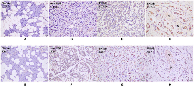
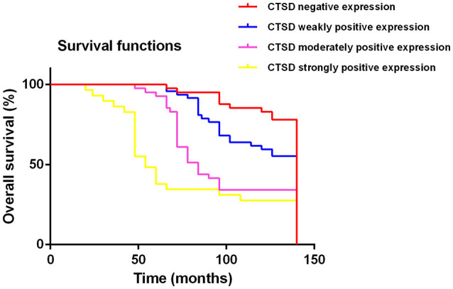
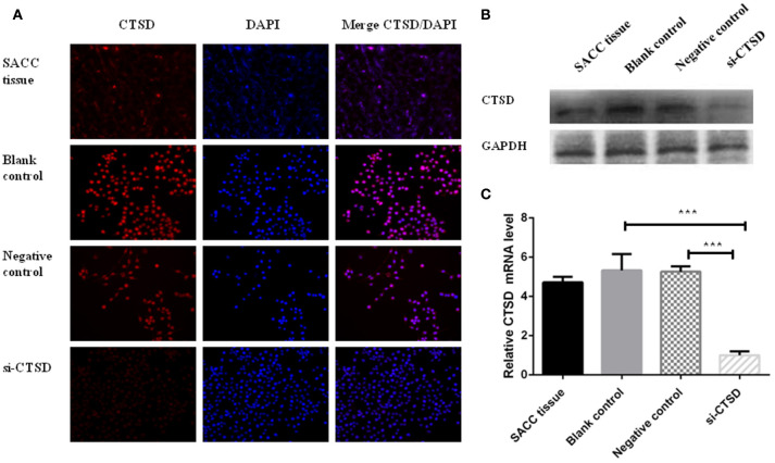
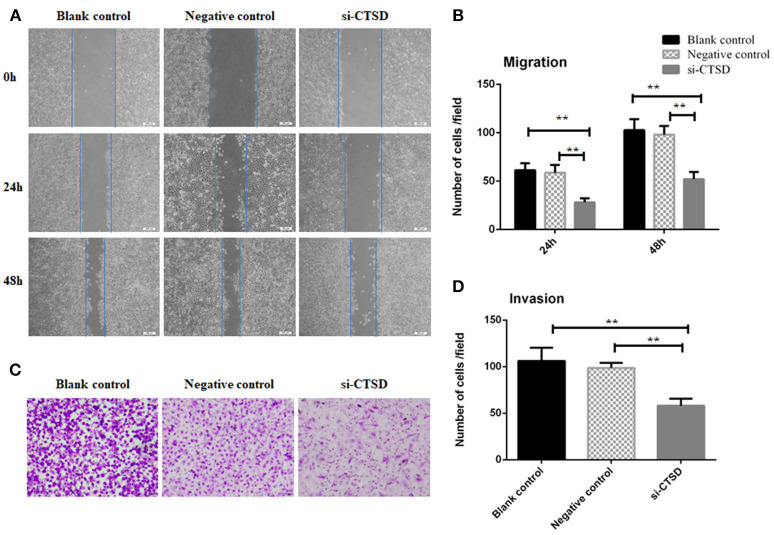
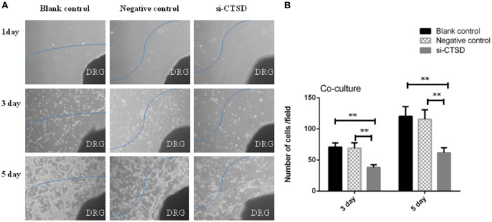
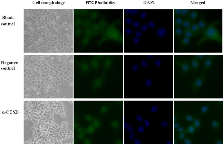
Objective: Cathepsin D (CTSD) is a pivotal orchestrator in the occurrence and development of tumors. Recently, CTSD was detected in salivary adenoid cystic carcinoma (SACC). However, its functional role in perineural invasion (PNI) of SACC remained elusive. We conducted the present study to detect the expression of CTSD in SACC, analyze the correlation between CTSD expression and prognosis of SACC patients and elucidate the role of CTSD in occurrence of PNI in SACC to lay the foundation for further studies. Methods: Immunohistochemical analysis was conducted to assess CTSD and Ki67 expression in 158 SACC samples and 20 normal salivary gland samples adjacent to carcinoma. Meanwhile, the correlation between CTSD and PNI of SACC specimens was analyzed using Wilcoxon test. QRT-PCR, immunofluorescence and western blot analysis were used to examine the levels of CTSD mRNA and protein in SACC-LM cell line. SiRNA-mediated CTSD silence was performed. Scratch wound healing assay, transwell invasion assay and DRG co-culture assay of PNI was used to detect the ability of migration, invasion and PNI. FITC-phalloidin was used to detect cytoskeletal organization. Results: Our data demonstrated that the positive expression of CTSD was observed in 74.1% (117/158) of SACC cases, and the expression of CTSD was significantly correlated with the PNI (p < 0.05). The ability of migration, invasion, and PNI could be inhibited significantly by siRNA-mediated CTSD silence (p < 0.01). Furthermore, siRNA-mediated CTSD silence inhibited cytoskeletal organization and pseudo foot formation in SACC-LM cells. Conclusion: Our results suggested that an association between PNI and expression of CTSD existed. CTSD may promote PNI of SACC accompanied by cytoskeletal organization and pseudo foot formation.
Keywords: cathepsin D (CTSD); cytoskeletal organization; invasion frontier; perineural invasion (PNI); salivary adenoid cystic carcinoma (SACC).
Figures
Similar articles
-
MIF promotes perineural invasion through EMT in salivary adenoid cystic carcinoma.Mol Carcinog. 2019 Jun;58(6):898-912. doi: 10.1002/mc.22979. Epub 2019 Feb 18. Mol Carcinog. 2019. PMID: 30667094
-
Sympathetic innervation contributes to perineural invasion of salivary adenoid cystic carcinoma via the β2-adrenergic receptor.Onco Targets Ther. 2019 Feb 21;12:1475-1495. doi: 10.2147/OTT.S190847. eCollection 2019. Onco Targets Ther. 2019. PMID: 30863115 Free PMC article.
-
Slug silencing inhibited perineural invasion through regulation of EMMPRIN expression in human salivary adenoid cystic carcinoma.Tumour Biol. 2016 Feb;37(2):2161-9. doi: 10.1007/s13277-015-4043-5. Epub 2015 Sep 9. Tumour Biol. 2016. PMID: 26349748
-
Perineural Invasion in Adenoid Cystic Carcinoma of the Salivary Glands: Where We Are and Where We Need to Go.Front Oncol. 2020 Aug 18;10:1493. doi: 10.3389/fonc.2020.01493. eCollection 2020. Front Oncol. 2020. PMID: 33014792 Free PMC article. Review.
-
[Treatment plan and prognosis of salivary adenoid cystic carcinoma with lung metastasis].Hua Xi Kou Qiang Yi Xue Za Zhi. 2019 Apr 1;37(2):214-219. doi: 10.7518/hxkq.2019.02.015. Hua Xi Kou Qiang Yi Xue Za Zhi. 2019. PMID: 31168990 Free PMC article. Chinese.
Cited by
-
Endoplasmic Reticulum Adaptation and Autophagic Competence Shape Response to Fluid Shear Stress in T24 Bladder Cancer Cells.Front Pharmacol. 2021 May 3;12:647350. doi: 10.3389/fphar.2021.647350. eCollection 2021. Front Pharmacol. 2021. PMID: 34012396 Free PMC article.
-
MiR-185-3p regulates epithelial mesenchymal transition via PI3K/Akt signaling pathway by targeting cathepsin D in gastric cancer cells.Transl Cancer Res. 2020 Nov;9(11):6988-7000. doi: 10.21037/tcr-19-2133. Transl Cancer Res. 2020. PMID: 35117305 Free PMC article.
-
Phospholipase A2 Drives Tumorigenesis and Cancer Aggressiveness through Its Interaction with Annexin A1.Cells. 2021 Jun 11;10(6):1472. doi: 10.3390/cells10061472. Cells. 2021. PMID: 34208346 Free PMC article. Review.
-
Cathepsin B defines leader cells during the collective invasion of salivary adenoid cystic carcinoma.Int J Oncol. 2019 Apr;54(4):1233-1244. doi: 10.3892/ijo.2019.4722. Epub 2019 Feb 22. Int J Oncol. 2019. PMID: 30968153 Free PMC article.
-
Transcriptional Profiles of Skeletal Muscle Associated With Increasing Severity of White Striping in Commercial Broilers.Front Physiol. 2020 Jun 16;11:580. doi: 10.3389/fphys.2020.00580. eCollection 2020. Front Physiol. 2020. PMID: 32612536 Free PMC article.
References
-
- Pour PM, Bell RH, Batra SK. Neural invasion in the staging of pancreatic cancer. Pancreas (2003) 26:322–5. - PubMed
-
- Feng FY, Qian Y, Stenmark MH, Halverson S, Blas K, Vance S, et al. . Perineural invasion predicts increased recurrence, metastasis, and death from prostate cancer following treatment with dose-escalated radiation therapy. Int J Radiat Oncol Biol Phys (2011) 81:e361–7. 10.1016/j.ijrobp.2011.04.048 - DOI - PubMed
LinkOut - more resources
Full Text Sources
Miscellaneous