Review
. 2019 Mar;76(5):837-863.
doi: 10.1007/s00018-018-2963-0. Epub 2018 Nov 14.

Mitochondrial functions and melatonin: a tour of the reproductive cancers

Affiliations
Review

Mitochondrial functions and melatonin: a tour of the reproductive cancers

Luiz Gustavo de Almeida Chuffa et al. Cell Mol Life Sci. 2019 Mar.

Abstract

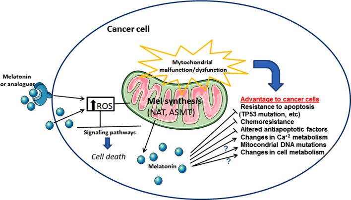
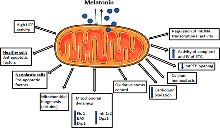
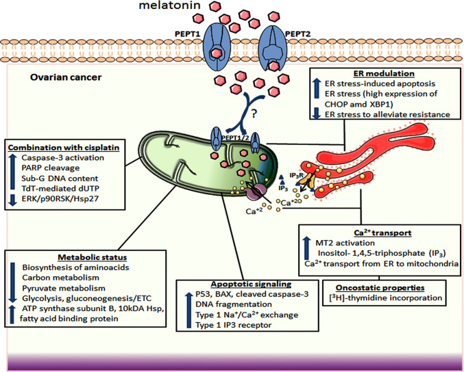
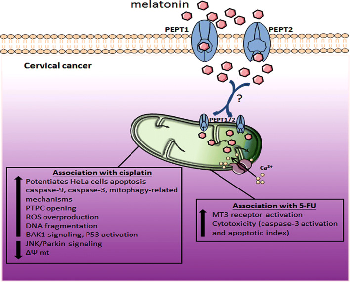
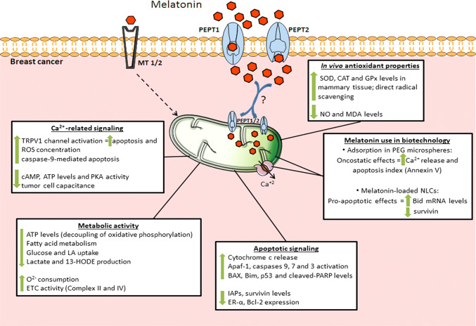
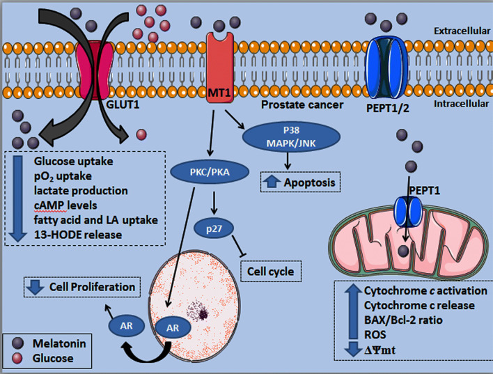
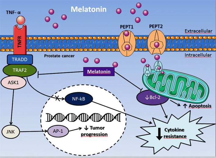
Cancers of the reproductive organs have a strong association with mitochondrial defects, and a deeper understanding of the role of this organelle in preneoplastic-neoplastic changes is important to determine the appropriate therapeutic intervention. Mitochondria are involved in events during cancer development, including metabolic and oxidative status, acquisition of metastatic potential, resistance to chemotherapy, apoptosis, and others. Because of their origin from melatonin-producing bacteria, mitochondria are speculated to produce melatonin and its derivatives at high levels; in addition, exogenously administered melatonin accumulates in the mitochondria against a concentration gradient. Melatonin is transported into tumor cell by GLUT/SLC2A and/or by the PEPT1/2 transporters, and plays beneficial roles in mitochondrial homeostasis, such as influencing oxidative phosphorylation and electron flux, ATP synthesis, bioenergetics, calcium influx, and mitochondrial permeability transition pore. Moreover, melatonin promotes mitochondrial homeostasis by regulating nuclear DNA and mtDNA transcriptional activities. This review focuses on the main functions of melatonin on mitochondrial processes, and reviews from a mechanistic standpoint, how mitochondrial crosstalk evolved in ovarian, endometrial, cervical, breast, and prostate cancers relative to melatonin's known actions. We put emphasis on signaling pathways whereby melatonin interferes within cancer-cell mitochondria after its administration. Depending on subtype and intratumor metabolic heterogeneity, melatonin seems to be helpful in promoting apoptosis, anti-proliferation, pro-oxidation, metabolic shifting, inhibiting neovasculogenesis and controlling inflammation, and restoration of chemosensitivity. This results in attenuation of development, progression, and metastatic potential of reproductive cancers, in addition to lowering the risk of recurrence and improving the life quality of patients.

Keywords: Breast cancer; Cervical cancer; Endometrial cancer; Melatonin; Mitochondrial function; Ovarian cancer; Prostate cancer.


Conflict of interest statement

Authors declare no conflict of interest.

Figures

Fig. 1
Fig. 1
Mitochondrial dysfunction is often present in cancer cells and, as a consequence, it may support cancer development. For instance, exogenous melatonin or its analogs may either activate membrane melatonin receptors or its intrinsic synthesis in mitochondria (via precursors and related enzymes), thus showing important effects in ROS production and cell death. Moreover, high melatonin levels in tumor cells are linked to inhibition of apoptosis resistance and chemoresistance, and stimulation of Ca2+ signaling. Although still not completely defined, other mechanisms by which melatonin contribute to reduce the advantage of cancer cells over normal cells may include mitochondrial DNA mutations and changes in metabolic processes. ROS reactive oxygen species, TP53 gene for tumor suppressor protein p53, Ca2+ calcium, DNA deoxyribonucleic acid, NAT N-acetyltransferase, ASMT acetylserotonin methyltransferase, ? uncertain mechanisms
Fig. 2
Fig. 2
Possible mitochondrial processes by which melatonin influences tumor growth depending on the cell status. Since melatonin can be both taken up and synthesized in mitochondria, a more direct and close relationship is expected to finely orchestrate these functions. UCP uncoupling proteins, Fis1 mitochondrial fission 1 protein, BAX Bcl-2-associated X protein, Drp1 dynamin-related protein 1, mfn1/2 mitofusins 1 and 2, Opa1 optic atrophy 1, mtPTP mitochondrial permeability transition pore, ETC electron transport chain, mtDNA mitochondrial DNA
Fig. 3
Fig. 3
A summary of the effects of melatonin on mitochondria of ovarian cancer cells. In addition to internal production by mitochondria, melatonin can be transported into mitochondria possibly via PEPT1/2, thus promoting effective responses on apoptosis, cellular energy metabolism, ER-stress modulation, and stimulating the action of other chemotherapeutics (e.g., cisplatin). Upon MT2 activation, Ca2+ is transported from ER to mitochondria triggering apoptosis and alleviating chemoresistance. P53 tumor suppressor protein p53, Ca2+ calcium, DNA deoxyribonucleic acid, MT2 melatonin receptor 2, IP3 inositol-1,4,5-triphosphate, IP3R inositol-1,4,5-triphosphate receptor, ERK extracellular signal-regulated kinase, p90RSK dephosphorylation of 90-kDa ribosomal S6 kinase, Hsp27 heat shock protein 27, ER endoplasmic reticulum, BAX bcl-2-like protein 4, ETC electron transport chain, PARP poly(ADP-ribose) polymerase, PEPT1/2 human oligotransporters 1 and 2, CHOP Cruxhalorhodopsin-1, XBP1 X-box binding protein 1, ? uncertain actions for OC
Fig. 4
Fig. 4
The effects of melatonin on cervical cancer mitochondria. Melatonin can be transported into mitochondria possibly via PEPT1/2 and the most ameliorative effects occur in association with other chemotherapies (e.g., cisplatin and 5-fluorouracil) to induce apoptosis and overcome chemoresistance. PTPC mitochondrial permeability transition pore, ROS reactive oxygen species, DNA deoxyribonucleic acid, BAK1 Bcl-2 homologous antagonist/killer gene, P53 tumor suppressor protein p53, 5-FU 5-fluorouracil, Ca2+ calcium, JNK c-Jun N-terminal kinase, ΔΨ mt mitochondrial membrane potential, MT3 melatonin receptor 3, PEPT1/2 human oligotransporters 1 and 2, ? uncertain actions for CC
Fig. 5
Fig. 5
Melatonin effects on mitochondrial function in breast cancer cells may be mediated by MT1 and MT2 receptors or via PEPT1/2 transporters. Melatonin might be transported into the organelle through PEPT1/2, and in addition to the intra-organelle production, triggers apoptosis through different pathways, such as caspases activation, upregulation of pro-apoptotic proteins, and ROS generation. In association with nanoparticles, melatonin stimulates Ca2+ release, which enhances the apoptotic process. Moreover, melatonin efficiently restores aerobic metabolism and promotes alterations in ATP metabolism. Also, melatonin has important protective effects against oxidative stress, by stimulating antioxidant activities and repressing pro-oxidative processes in healthy mammary cells. 13-HODE 13-hydroxyoctadecadienoic acid, Apaf1 apoptotic protease activating factor 1, ATP adenosine triphosphate, BAX bcl-2-like protein 4, Bcl-2 B-cell lymphoma 2, Bid pro-apoptotic member of the Bcl-2 family, Bim Bcl-2-like protein 11, Ca2+ calcium, CAT catalase, cAMP cyclic adenosine monophosphate, ER-α estrogen receptor alpha, ETC electron transport chain, GPx glutathione peroxidase, IAPs inhibitors of apoptosis proteins family, LA linoleic acid, MDA malondialdehyde, MT1/2 melatonin receptors 1 and 2, mRNA messenger ribonucleic acid, NLCs nanostructured lipid carriers, NO nitric oxide, O2− oxygen, p53 tumor suppressor protein p53, PARP poly(ADP-ribose) polymerase, PEG polyethylene glycol, PEPT1/2 human oligotransporters 1 and 2, PKA protein kinase A, ROS reactive oxygen species, SOD superoxide dismutase, TRPV1 transient receptor potential vanilloid 1, ? uncertain actions for BC
Fig. 6
Fig. 6
Melatonin exerts differential effects in prostate cancer cells including signaling via MT1 receptor or after being transported into the cell by GLUT1 and PEPT1/2. Melatonin competes with glucose uptake by the GLUT1, which reduces the PCa metabolic activity associated to the Warburg effect. Upon melatonin binding to the MT1 receptor, PKC and PKA are activated, resulting in AR translocation from nucleus to the cytoplasm while upregulating the p27 protein, a cell cycle inhibitor. In addition, MT1 activation significantly increases the p38MAPK/JNK activity finally resulting in elevation of apoptosis rate. Melatonin transport into PCa cell mitochondria is likely through PEPT1 increasing cytochrome c release to the cytoplasm, in association with higher BAX/Bcl-2 ratio and ROS production; these functional alterations are accompanied by a reduced ΔΨmt, which favor the induction of apoptosis. GLUT1 glucose transporter 1, MT1 melatonin receptor 1, ROS reactive oxygen species, ΔΨmt mitochondrial membrane potential, BAX Bcl-2-associated X protein, Bcl-2 B-cell lymphoma 2, PEPT1/2 human peptide transporter 1 and 2, cAMP cyclic adenosine monophosphate, pO2 oxygen pressure, LA linoleic acid, 13-HODE 13-hydroxyoctadecadienoic acid, AR androgen receptor, p27 p27 cell cycle protein, P38 MAPK P38 mitogen-activated protein kinase, JNK Jun N-terminal kinases, PKC protein kinase C, PKA protein kinase A
Fig. 7
Fig. 7
Melatonin enters the prostate cancer cell cytoplasm and mitochondria through the PEPT1 and PEPT2 transporters and promotes the inhibition of Bcl-2, thereby increasing apoptosis and attenuating cytokine resistance. By inhibiting TRAF2, melatonin also reduces the nuclear translocation of NF-κB, resulting in a decline in cytokine resistance and favoring the ASK1 pathway; the latter activates the JNK and p38 and, as a consequence, they stimulate the nuclear translocation of AP-1 reducing the tumor progression. PEPT1/2 human peptide transporter 1 and 2, Bcl-2 B-cell lymphoma 2, TRAF2 TNF receptor-associated factor 2, TRADD tumor necrosis factor receptor type 1-associated DEATH domain protein, NF-κB nuclear factor kappa B, ASK1 apoptosis signal-regulating kinase 1, JNK Jun N-terminal kinases, AP-1 activating protein, TNFR tumor necrosis factor receptor, TNF-α tumor necrosis factor alpha

Similar articles

Cited by

References

    1. Weiderpass E, Labrèche F. Malignant tumors of the female reproductive system. Saf Health Work. 2012;3:166–180. doi: 10.5491/SHAW.2012.3.3.166. - DOI - PMC - PubMed
    1. Chuffa LG, Lupi-Júnior LA, Costa AB, Amorim JP, Seiva FR. The role of sex hormones and steroid receptors on female reproductive cancers. Steroids. 2017;118:93–108. doi: 10.1016/j.steroids.2016.12.011. - DOI - PubMed
    1. Stangelberger A, Waldert M, Djavan B. Prostate cancer in elderly men. Rev Urol. 2008;10:111–119. - PMC - PubMed
    1. Kong B, Tsuyoshi H, Orisaka M, Shieh DB, Yoshida Y, Tsang BK. Mitochondrial dynamics regulating chemoresistance in gynecological cancers. Ann N Y Acad Sci. 2015;1350:1–16. doi: 10.1111/nyas.12883. - DOI - PubMed
    1. Dan HC, Sun M, Kaneko S, et al. Akt phosphorylation and stabilization of x linked inhibitor of apoptosis protein (XIAP) J Biol Chem. 2004;279:5405–5412. doi: 10.1074/jbc.M312044200. - DOI - PubMed