Clinical Trial
. 2019 May 1;36(9):1451-1460.
doi: 10.1089/neu.2018.5921. Epub 2018 Dec 15.

Electrophysiological Guidance of Epidural Electrode Array Implantation over the Human Lumbosacral Spinal Cord to Enable Motor Function after Chronic Paralysis

Affiliations
Clinical Trial

Electrophysiological Guidance of Epidural Electrode Array Implantation over the Human Lumbosacral Spinal Cord to Enable Motor Function after Chronic Paralysis

Jonathan S Calvert et al. J Neurotrauma. .

Abstract

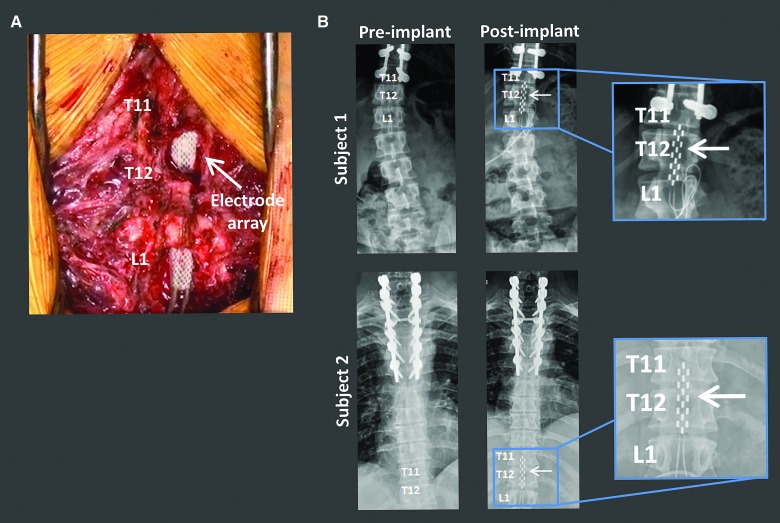
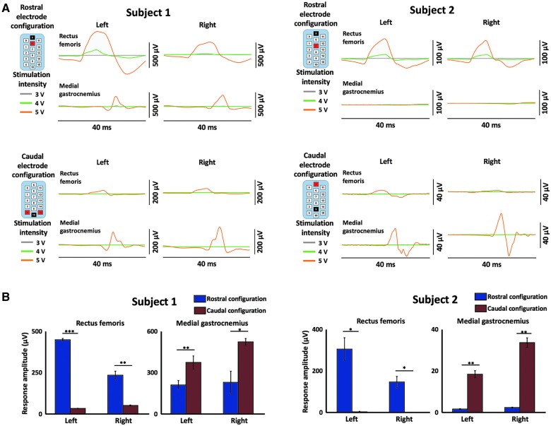
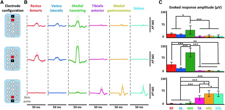
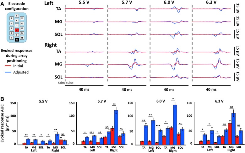
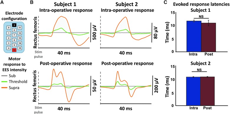
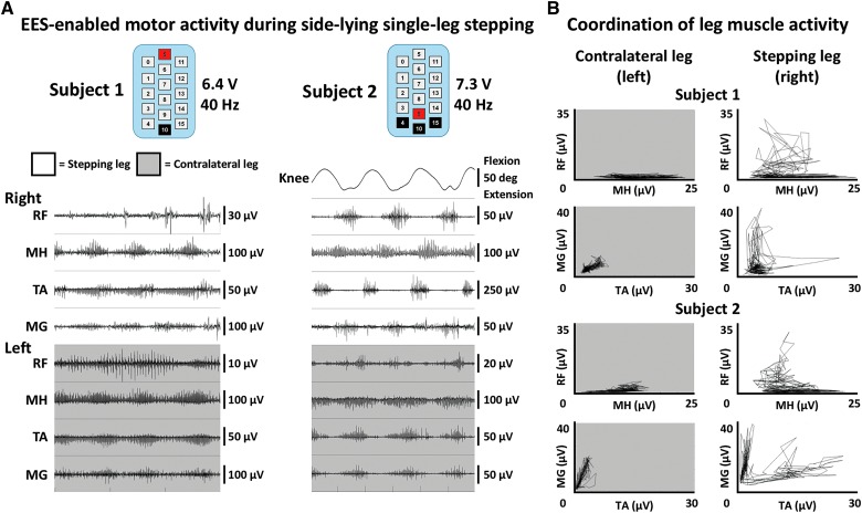
Epidural electrical stimulation (EES) of the spinal cord has been shown to restore function after spinal cord injury (SCI). Characterization of EES-evoked motor responses has provided a basic understanding of spinal sensorimotor network activity related to EES-enabled motor activity of the lower extremities. However, the use of EES-evoked motor responses to guide EES system implantation over the spinal cord and their relation to post-operative EES-enabled function in humans with chronic paralysis attributed to SCI has yet to be described. Herein, we describe the surgical and intraoperative electrophysiological approach used, followed by initial EES-enabled results observed in 2 human subjects with motor complete paralysis who were enrolled in a clinical trial investigating the use of EES to enable motor functions after SCI. The 16-contact electrode array was initially positioned under fluoroscopic guidance. Then, EES-evoked motor responses were recorded from select leg muscles and displayed in real time to determine electrode array proximity to spinal cord regions associated with motor activity of the lower extremities. Acceptable array positioning was determined based on achievement of selective proximal or distal leg muscle activity, as well as bilateral muscle activation. Motor response latencies were not significantly different between intraoperative recordings and post-operative recordings, indicating that array positioning remained stable. Additionally, EES enabled intentional control of step-like activity in both subjects within the first 5 days of testing. These results suggest that the use of EES-evoked motor responses may guide intraoperative positioning of epidural electrodes to target spinal cord circuitry to enable motor functions after SCI.

Keywords: electrically evoked spinal motor potentials; epidural electrical stimulation, spinal cord injury; neuromodulation; spinal cord intraoperative electrophysiology.


Conflict of interest statement

V.R.E. and Y.P.G. are shareholders in NeuroRecovery Technologies. V.R.E. is president and chair of the company's board of directors. V.R.E. and Y.P.G. hold investorship rights on intellectual property licensed by the regents of the University of California to NeuroRecovery Technologies and its subsidiaries.

Figures

<b>FIG. 1.</b>
FIG. 1.
Surgical implantation of the EES electrode array spanning the lumbosacral spinal cord. (A) Intraoperative image of the location of the EES array at the T11–L1 vertebral levels. (B) Anterior-posterior X-ray of each subject before and after EES electrode array implantation. Subject 1 imaging was captured in a seated position. Subject 2 imaging was captured while lying supine. Inserts depict zoomed in view of the EES array. EES, epidural electrical stimulation.
<b>FIG. 2.</b>
FIG. 2.
Intraoperative EES-evoked motor response recordings demonstrates selective activation of rostral and caudal spinal circuitry. (A) EES-evoked responses during rostral electrode array configurations demonstrate proximal muscle activation (rectus femoris) and during caudal electrode array configurations demonstrate distal muscle activation (medial gastrocnemius). Each line represents the average evoked response to stimulation over five stimulations. Gray, green, and orange lines represent stimulation at 3, 4, and 5 volts, respectively. Stimulation occurs at the 0 time point of each plot. Stimulation configuration is shown in the upper left of each figure; black = cathode, red = anode. (B) Bar plots displaying maximum evoked response in given muscles from (A) with amplitude calculated as maximum – minimum response. Blue bars indicate rostral configurations and red bars indicate caudal configurations. * = <0.05; ** = <0.01; *** = <0.001. EES, epidural electrical stimulation; NS, not significant; R, right; L, left.
<b>FIG. 3.</b>
FIG. 3.
EES-evoked motor responses activate specific muscle circuitry intraoperatively. (A) EES-evoked responses during stimulation of the rostral, intermediate, and caudal portions of the EES array are demonstrated in six muscles (rectus femoris, vastus lateralis, medial hamstring, tibialis anterior, medial gastrocnemius, and soleus) from subject 1. Stimulation occurs at the start of each EES-evoked response trace as indicated by the gray dashed line. Dark traces are average of five individual responses that are shown in light traces. Data shown are from motor threshold responses. EES electrode array configuration is shown in the upper left; black = cathode, red = anode. (B) Bar plots displaying maximum EES-evoked responses in given muscles (RF = rectus femoris, VL = vastus lateralis, MH = medial hamstring, TA = tibialis anterior, MG = medial gastrocnemius, and SOL = soleus) from (A) with amplitude calculated as maximum – minimum EES-evoked response. Statistical significance was calculated by a one-way ANOVA followed by a multiple comparisons test and is shown above the bar plots. * = <0.05; ** = <0.01; *** = <0.001; no stars indicates not significant. ANOVA, analysis of variance; EES, epidural electrical stimulation.
<b>FIG. 4.</b>
FIG. 4.
Electrode location adjustment guided by intraoperative EES-evoked motor response recordings. (A) Intraoperative data from subject 2 using a caudal, symmetric (−10/+8) configuration as displayed. Bilateral electromyography (EMG) data from three bilateral distal muscles are shown (TA = tibialis anterior, MG = medial gastrocnemius, and SOL = soleus). Each line is an average of five motor-evoked potentials where stimulation occurs at the start of each trace. Data are shown while increasing the stimulation intensity incrementally from 5.5 to 6.3 V before and after shifting of the array during surgery. (B) Area under the curve of the EES-evoked responses at the four different voltages. * = <0.05; ** = <0.01; *** = <0.001; NS = not significant. Red indicates data before array shift. Blue indicates data after shift. EES, epidural electrical stimulation.
<b>FIG. 5.</b>
FIG. 5.
Comparison of intraoperative and post-operative EES-evoked motor responses. (A) EES electrode array configuration used intraoperatively and post-operatively; black = cathode, red = anode. (B) EMG (electromyography) data are shown from the left rectus femoris of both subjects recorded intraoperatively and post-operatively using the same electrode configuration for each subject after 3 weeks of recovery from surgery. Each trace represents the average of five consecutive evoked motor responses at each EES voltage intensity. Data are shown at subthreshold levels of stimulation when no response was observed, at motor threshold where the first appearance of motor activity was observed, and at the maximum level of stimulation. These voltage values ranged from 3 to 6 V. (C) Latency of the suprathreshold evoked response in the left rectus femoris in both subjects both intraoperatively and at 3 weeks post-operatively. No significant difference was found between intraoperative and post-operative latency for either subject. EES, epidural electrical stimulation; NS, not significant.
<b>FIG. 6.</b>
FIG. 6.
Intentional control of rhythmic movement while side-lying. (A) EMG (electromyography) and goniometer data from both subjects are shown while each subject intentionally attempted to generate EES-enabled step-like movements of their right leg. EMG data were recorded bilaterally from rectus femoris (RF), medial hamstring (MH), tibialis anterior (TA), and medial gastrocnemius (MG). Subject 2 wore sagittal knee goniometers during testing to quantify leg motion. Stimulation configurations were chosen that allowed for optimal movement. Pulse width and frequency were held constant at 210 μs and 40 Hz, respectively. Voltage was incrementally increased until subjects displayed ability to intentionally control leg movements. White background indicates the leg that was supported in a gravity-neutral position in order to move freely. Gray background indicates the leg that was resting on a table and limited with respect to movement capability. (B) Muscle coordination plots from the same data as part (A). Root mean square (RMS) envelopes of the EMG data were calculated and antagonistic muscles are plotted against one another to demonstrate patterns of coordination. Note that a more normal L-shaped (reciprocal) coordination patterned was generated when the leg was suspended and freed of surface tension. EES, epidural electrical stimulation.

References

    1. Harvey L.A. (2016). Physiotherapy rehabilitation for people with spinal cord injuries. J. Physiother. 62, 4–11 - PubMed
    1. Sekhon L.H., and Fehlings M.G. (2001). Epidemiology, demographics, and pathophysiology of acute spinal cord injury. Spine (Phila Pa 1976) 26, 24 Suppl., S2–S12 - PubMed
    1. Bydon M., Lin J., Macki M., Gokaslan Z.L., and Bydon A. (2014). The current role of steroids in acute spinal cord injury. World Neurosurg. 82, 848–854 - PubMed
    1. Chen B.K., Madigan N.N., Hakim J.S., Dadsetan M., McMahon S.S., Yaszemski M.J., and Windebank A.J. (2018). GDNF Schwann cells in hydrogel scaffolds promote regional axon regeneration, remyelination and functional improvement after spinal cord transection in rats. J. Tissue Eng. Regen. Med. 12, e398–e407 - PubMed
    1. Anderson M.A., Burda J.E., Ren Y., Ao Y., Coppola G., Khakh B.S., Deming T.J., Michael V., and Angeles L. (2016). Astrocyte scar formation aids CNS axon regeneration. Nature 532, 195–200 - PMC - PubMed

Publication types