Review
. 2018 Nov;15(Suppl 3):S164-S170.
doi: 10.1513/AnnalsATS.201806-371AW.

Airway Mucin Secretion

Affiliations
Review

Airway Mucin Secretion

Ana M Jaramillo et al. Ann Am Thorac Soc. 2018 Nov.

Abstract

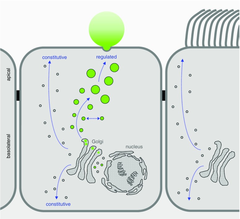
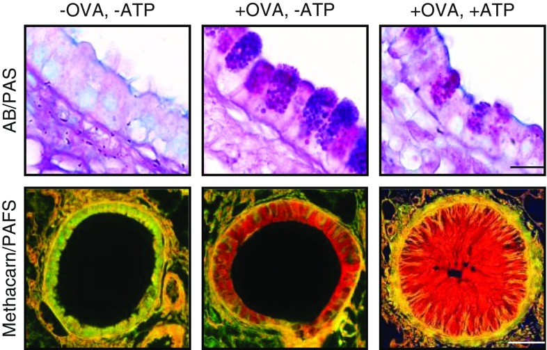
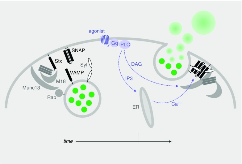
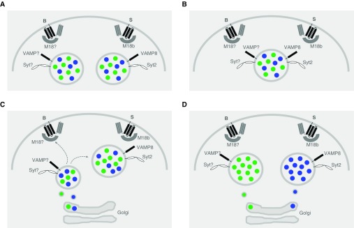
Exocytosis of secreted mucins is the final step in their intracellular processing, resulting in their release into the airway lumen to interact with water and ions to form mucus. Mucins are secreted at a low baseline rate and a high stimulated rate, and both rates are regulated by second messengers acting on components of the exocytic machinery. The principal physiologic function of the low baseline rate is to support steady-state mucociliary clearance of inhaled particles and pathogens that enter the airways during normal breathing. Even in the setting of mucin hyperproduction, baseline secretion generally does not induce mucus occlusion. The principal physiologic function of the high stimulated rate of secretion from both submucosal glands and surface goblet cells in proximal airways appears to be to sweep away larger particles, whereas in distal airways it appears to act in concert with mucin hyperproduction to induce mucus occlusion to trap migrating helminths. Pathophysiologically, stimulated mucin secretion in the setting of mucin hyperproduction from allergic or other types of airway inflammation in the absence of helminth infection causes airflow obstruction and infection. Molecular components of the mucin exocytic machinery are increasingly being identified, and surprisingly, many components are not shared between baseline and stimulated machines. The physiologic significance of the presence of two distinct molecular machines is not yet known, such as whether these interact selectively with secretory granules of different sizes or contents. A full understanding of the mechanism and regulation of airway mucin secretion will provide further insight into pathophysiologic processes and may identify therapeutic strategies to alleviate obstructive airway diseases.

Keywords: exocytosis; mucin; mucus; secretion.


Figures

Figure 1.
Figure 1.
Regulated versus constitutive secretory pathways of airway epithelial cells. A simplified model of airway surface epithelium is illustrated, showing a secretory (club) cell in the center and a ciliated cell on the right. Proteins destined for secretion, including the secreted mucins (green) that are produced exclusively in club cells and not in ciliated cells, are synthesized in rough endoplasmic reticulum surrounding the nucleus and extending into cytoplasm at the basal pole of the cell. Newly synthesized proteins are transported from the endoplasmic reticulum to the cis-Golgi in coated vesicles and become fully glycosylated as they progress through the Golgi stacks. In the trans-Golgi, mucins are likely segregated from other secreted proteins and packaged into large carrier vesicles as described for other large proteins such as collagen (37). New mucin granules then likely fuse homotypically (laterally) to generate large secretory granules, as occurs in other regulated secretory cells, but this has not yet been directly studied in club cells. Mucin secretory granules can then be released in response to extracellular signals and intracellular second messengers in a classic regulated secretory pathway (see text for details). Distinct apical and basolateral constitutive secretory pathways responsible for the replacement of lipids and proteins in the plasma membrane and secretion of proteins such as cytokines are likely to exist in both club cells and ciliated cells as they do in other polarized cell types, though the constitutive secretory pathways of airway epithelial cells have not yet been delineated at a molecular level. It is important to recognize that the baseline regulated pathway for mucin secretion is not synonymous with the apical constitutive secretory pathway and probably shares few or no molecular components (see text for details).
Figure 2.
Figure 2.
Increased mucin production followed by stimulated secretion, resulting in airway occlusion. Mouse bronchi fixed with formalin, sectioned longitudinally, and stained with Alcian blue and periodic acid–Schiff stain (AB/PAS, top row) or fixed with methacarn to preserve mucus volume, sectioned transversely, and stained with periodic acid–fluorescent Schiff (PAFS, bottom row). Left: In the naive (uninflamed) state, there is minimal mucin staining inside epithelial cells or in the airway lumen. Center: Mucin hyperproduction (“mucous metaplasia”) was produced by allergic inflammation using an ovalbumin (+OVA) model. Intracellular mucin is visible within numerous purple granules in secretory cells (top) and extensive red staining (bottom) 3 days after OVA aerosol challenge. Right: Sudden mucin release from metaplastic cells was induced by an adenosine triphosphate aerosol (+ATP). Ten minutes later, mucus occludes the airway lumen. Not shown is ATP stimulation of naive airways (OVA−ATP+), because there is no observable difference from unstimulated naive airways (OVA−ATP−). Scale bars = 10 μm in the top row and 50 μm in the bottom row.
Figure 3.
Figure 3.
Generic model of mucin granule exocytosis progressing over time as illustrated from left to right. This model is based on structural and functional information obtained from the study of neurons and other regulated exocytic cells, as well as functional information obtained from the study of airway secretory cells, as referenced in the text. Left: In the resting state, mucin granules (green) become tethered to the plasma membrane by Rab proteins interacting with effectors (not yet identified or illustrated) and the priming protein Munc13. Center: Agonist binding to heptahelical receptors (blue), such as those for adenosine triphosphate (P2Y2R), adenosine (A3R), and serine proteases (protease-activated receptors 1 and 2), leads to activation of a trimeric G protein (Gq), then phospholipase-Cβ (PLC), resulting in generation of the second messengers diacylglycerol (DAG) and inositol trisphosphate (IP3). DAG activates Munc13 to open syntaxin (Stx) in collaboration with the scaffolding protein Munc18 and promote Stx interactions with the other SNARE proteins (SNAP [synaptosomal-associated protein] and VAMP [vesicle-associated membrane protein], black bars). IP3 induces the release of Ca2+ from apical endoplasmic reticulum (ER) to activate the exocytic calcium sensor synaptotagmin (Syt), which promotes further coiling of the SNARE complex. (Note that in electrically excitable cells such as neurons, Ca2+ instead enters the cytoplasm through plasma membrane channels.) Right: Complete coiling of the SNARE complex induces fusion of the granule and plasma membranes, releasing mucins into the extracellular space, where they absorb water and swell.
Figure 4.
Figure 4.
Models illustrating the possible interactions of the two regulated exocytic machines of airway secretory cells with mucin granules. In each model, B designates the baseline exocytic machine and S designates the stimulated machine. Munc18 has been shortened to M18 to avoid clutter, and “?” is used to designate an unknown isoform of Munc18, VAMP (vesicle-associated membrane protein), and Syt (synaptotagmin) proteins. Blue dots within the secretory granules designate mucin 5ac (Muc5ac), and green dots designate Muc5b. (A) Two different populations of granules, each containing both Muc5ac and Muc5b, but differentially containing only one of the VAMP and Syt isoforms, are each capable of only interacting with either a baseline or a stimulated plasma membrane machine. (B) A single population of granules containing both Muc5ac and Muc5b also contains both VAMP and Syt isoforms, rendering the granules capable of interacting with both baseline and stimulated plasma membrane machines. (C) A single population of granules containing both Muc5ac and Muc5b derives from small post-Golgi vesicles that contain only the baseline VAMP and Syt isoforms that make them competent for interactions only with the baseline plasma membrane machine. These granules then enlarge by homotypic fusion and mature by losing the VAMP and Syt baseline isoforms and acquiring the VAMP and Syt stimulated isoforms, making them increasingly incompetent for fusion with the baseline plasma membrane machine but increasingly competent for the stimulated plasma membrane machine. (D) Two populations of granules containing only Muc5ac or Muc5b, and differentially containing only one of the VAMP and Syt isoforms, are each capable of only interacting with either a baseline or a stimulated plasma membrane machine.

Similar articles

Cited by

References

    1. Roy MG, Livraghi-Butrico A, Fletcher AA, McElwee MM, Evans SE, Boerner RM, et al. Muc5b is required for airway defence. Nature. 2014;505:412–416. - PMC - PubMed
    1. Ren B, Azzegagh Z, Jaramillo AM, Zhu Y, Pardo-Saganta A, Bagirzadeh R, et al. SNAP23 is selectively expressed in airway secretory cells and mediates baseline and stimulated mucin secretion. Biosci Rep. 2015;35:e00220. - PMC - PubMed
    1. Kaul S, Mittal SK, Feigenbaum L, Kruhlak MJ, Roche PA. Expression of the SNARE protein SNAP-23 is essential for cell survival. PLoS One. 2015;10:e0118311. - PMC - PubMed
    1. Zhu Y, Abdullah LH, Doyle SP, Nguyen K, Ribeiro CM, Vasquez PA, et al. Baseline goblet cell mucin secretion in the airways exceeds stimulated secretion over extended time periods, and is sensitive to shear stress and intracellular mucin stores. PLoS One. 2015;10:e0127267. - PMC - PubMed
    1. Davis CW, Dickey BF. Regulated airway goblet cell mucin secretion. Annu Rev Physiol. 2008;70:487–512. - PubMed

Publication types