Review
. 2018 Nov;15(Suppl 3):S216-S226.
doi: 10.1513/AnnalsATS.201806-368AW.

Emerging Concepts and Therapies for Mucoobstructive Lung Disease

Affiliations
Review

Emerging Concepts and Therapies for Mucoobstructive Lung Disease

Marcus A Mall et al. Ann Am Thorac Soc. 2018 Nov.

Abstract

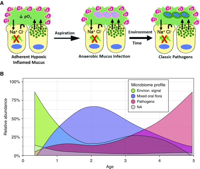
A spectrum of intrapulmonary airway diseases, for example, cigarette smoke-induced bronchitis, cystic fibrosis, primary ciliary dyskinesia, and non-cystic fibrosis bronchiectasis, can be categorized as "mucoobstructive" airway diseases. A common theme for these diseases appears to be the failure to properly regulate mucus concentration, producing mucus hyperconcentration that slows mucus transport and, importantly, generates plaque/plug adhesion to airway surfaces. These mucus plaques/plugs generate long diffusion distances for oxygen, producing hypoxic niches within adherent airway mucus and subjacent epithelia. Data suggest that concentrated mucus plaques/plugs are proinflammatory, in part mediated by release of IL-1α from hypoxic cells. The infectious component of mucoobstructive diseases may be initiated by anaerobic bacteria that proliferate within the nutrient-rich hypoxic mucus environment. Anaerobes ultimately may condition mucus to provide the environment for a succession to classic airway pathogens, including Staphylococcus aureus, Haemophilus influenzae, and ultimately Pseudomonas aeruginosa. Novel therapies to treat mucoobstructive diseases focus on restoring mucus concentration. Strategies to rehydrate mucus range from the inhalation of osmotically active solutes, designed to draw water into airway surfaces, to strategies designed to manipulate the relative rates of sodium absorption versus chloride secretion to endogenously restore epithelial hydration. Similarly, strategies designed to reduce the mucin burden in the airways, either by reducing mucin production/secretion or by clearing accumulated mucus (e.g., reducing agents), are under development. Thus, the new insights into a unifying process, that is, mucus hyperconcentration, that drives a significant component of the pathogenesis of mucoobstructive diseases promise multiple new therapeutic strategies to aid patients with this syndrome.

Keywords: IL-1α; anaerobes; hydration therapies; mucoobstruction; mucus hyperconcentration.


Figures

Figure 1.
Figure 1.
Mucus plugging causes hypoxic epithelial necrosis that triggers sterile inflammation in mucoobstructive lung disease. Mucus plugging produces regional hypoxia and necrosis of a subset of epithelial cells lining the airway surfaces. Dying epithelial cells release the alarmin IL-1α into the airway lumen. Binding of IL-1α to IL-1 receptors (IL-1Rs) on neighboring cells results in activation of the IL-1R/MyD88 signaling pathway, inducing neutrophilic airway inflammation in the absence of bacterial infection. Image courtesy of Joshua Bird. MyD88 = myeloid differentiation primary response 88; NF-κB = nuclear factor-κB; pO2 = partial pressure of oxygen.
Figure 2.
Figure 2.
Early infection in the cystic fibrosis (CF) lung. (A) Predicted sequence of bacterial pathogen acquisition in early CF. The CF airway early in life (left) has heterogeneous areas characterized by a hypoxic mucoinflammatory environment dominated by adherent mucus (green). Aspiration (middle) introduces oral anaerobic pathogens into the lung. Oral anaerobes survive and proliferate in lower airway hypoxic mucus, in part by fermenting sugars cleaved from mucins. With time and environmental exposure (right), classic CF pathogens (Staphylococcus and Pseudomonas) infect adherent CF mucus. Fermentation products of mucins may promote classic pathogen infection. (B) Relative abundance of bacteria in the bronchoalveolar lavage of infants/children with CF, as a function of age. Bacteria were binned into three classes: 1) environmental bacteria (green); 2) oral anaerobes (purple); and 3) classic CF pathogens (pink). Image courtesy of Bryan Zorn and Matthew Wolfgang.
Figure 3.
Figure 3.
Response of normal versus cystic fibrosis (CF) airway epithelia to bacterial infection. (A, part I) Response of normal (white columns) versus CF (red columns) human bronchial epithelial cultures to luminal application of supernatant of mucopurulent material (SMM) harvested from lumens of CF airways. Shown are total mucin secretion rates in response to SMM. (A, part II) Short-circuit current (Isc) profiles of Na+ absorption (amiloride-sensitive Isc) and cystic fibrosis transmembrane conductance regulator (CFTR) Cl secretion (forskolin-induced Isc) responses to SMM. (A, part III) Change in airway surface liquid height in response to SMM. (A, part IV) Change in mucus concentration (% solids) in response to SMM (38). (B, part I) Normal epithelia: Airway epithelia in basal state (left) coordinate mucin secretion rates and epithelial Na+ channel–mediated Na+/liquid absorption versus cystic fibrosis transmembrane/Ca2+-activated Cl channel (CaCC, i.e., TMEM16a) Cl/liquid secretion to maintain mucus at an approximately 2% solids hydration state commensurate with robust mucociliary clearance (MCC) rates. The balance of liquid transport and mucin secretion is maintained by ATP and adenosine interaction with apical P2Y2 and A2b purinoceptors, respectively. Epithelial responses to bacterial/host products include mucin secretion, which via corelease with mucins of adenosine (and AMP) stimulates a disproportionate increase in CFTR-mediated Cl/fluid secretion, “super”-hydration of mucus (1.5% solids), and accelerated MCC. The net effect is to flush bacteria off airway surfaces. (B, part II) CF: Under basal, that is, the “nondiseased” but vulnerable, state (left), CF airway epithelia manage to maintain quasi-normal mucus hydration via upregulation of CaCC activity to offset missing CFTR Cl transport and unregulated Na+ absorption. In response to a bacterial/host product challenge, mucin secretion is upregulated, but the absence of CFTR negates a coupled adenosine-mediated fluid secretory response. The net effect is to increase CF mucus concentration, slow MCC, and lead to spread/worsening of CF airway disease. Image 3B courtesy of Joshua Bird. A2BR = A2B adenosine receptor; ADO = adenosine; ASL = airway surface liquid; ENaC = epithelial Na+ channel; P2Y2R = purinergic receptor P2Y2; PBS = phosphate-buffered saline; PCL = perciliary layer.

References

    1. Boucher RC. Airway surface dehydration in cystic fibrosis: pathogenesis and therapy. Annu Rev Med. 2007;58:157–170. - PubMed
    1. Accurso FJ, Rowe SM, Clancy JP, Boyle MP, Dunitz JM, Durie PR, et al. Effect of VX-770 in persons with cystic fibrosis and the G551D-CFTR mutation. N Engl J Med. 2010;363:1991–2003. - PMC - PubMed
    1. Ramsey BW, Davies J, McElvaney NG, Tullis E, Bell SC, Drevinek P, et al. A CFTR potentiator in patients with cystic fibrosis and the G551D mutation. N Engl J Med. 2011;365:1663–1672. - PMC - PubMed
    1. Van Goor F, Hadida S, Grootenhuis PD, Burton B, Cao D, Neuberger T, et al. Rescue of CF airway epithelial cell function in vitro by a CFTR potentiator, VX-770. Proc Natl Acad Sci USA. 2009;106:18825–18830. - PMC - PubMed
    1. Wainwright CE, Elborn JS, Ramsey BW, Marigowda G, Huang X, Cipolli M, et al. Lumacaftor–ivacaftor in patients with cystic fibrosis homozygous for Phe508del CFTR. N Engl J Med. 2015;373:220–231. - PMC - PubMed

Publication types

MeSH terms

LinkOut - more resources