

Fungi in Mucoobstructive Airway Diseases
- PMID: 30431347
- PMCID: PMC6322025
- DOI: 10.1513/AnnalsATS.201803-154AW
Fungi in Mucoobstructive Airway Diseases
Abstract
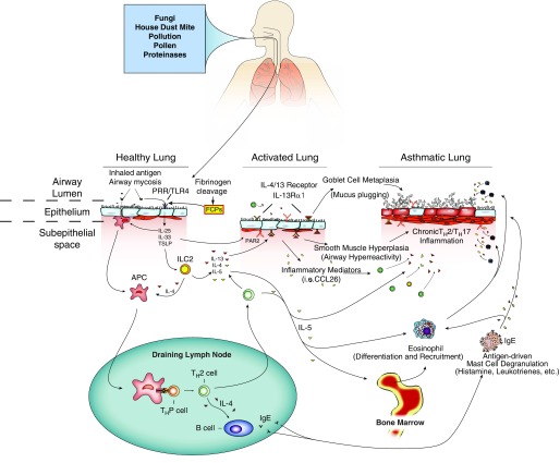
Asthma, chronic rhinosinusitis, and related incurable allergic afflictions of the upper and lower airways are medically important because of their association with the disabling symptom of dyspnea and, at least for asthma, the potential to cause fatal asphyxiation. Extensive research over the past two decades has uncovered both the physiological basis of airway obstruction in asthma and key governing molecular pathways. Exaggerated airway constriction in response to diverse provocative stimuli, termed airway hyperresponsiveness, is mediated through the cytokines interleukin 4 (IL-4) and IL-13 and the transcription factor signal transducer and activator of transcription 6 (STAT6). Overproduction of mucus has long been known to be an essential second component of airway obstruction and is also mediated in part through the IL-4/IL-13/STAT6 pathway. In this review, we discuss a second major signaling pathway which underlies mucus production that is mediated through proteinase-cleaved fibrinogen signaling through Toll-like receptor 4. Unexpectedly, our analysis of human sputum and paranasal sinus fluid indicates that in most cases of severe allergic airway disease, a unique type of airway fungal infection, termed airway mycosis, is pathogenically linked to these conditions. We further discuss how fungal and endogenous proteinases mediate the fibrinogenolysis that is essential to both Toll-like receptor 4 signaling and fibrin deposition that, together with mucus, contribute to airway obstruction.
Keywords: airway mycosis; asthma; chronic rhinosinusitis; fibrinogen; fungi.
Figures
References
-
- Molfino NA, Nannini LJ, Martelli AN, Slutsky AS. Respiratory arrest in near-fatal asthma. N Engl J Med. 1991;324:285–288. - PubMed
-
- Bresciani M, Paradis L, Des Roches A, Vernhet H, Vachier I, Godard P, et al. Rhinosinusitis in severe asthma. J Allergy Clin Immunol. 2001;107:73–80. - PubMed
-
- Bachert C, Claeys SE, Tomassen P, van Zele T, Zhang N. Rhinosinusitis and asthma: a link for asthma severity. Curr Allergy Asthma Rep. 2010;10:194–201. - PubMed
-
- Pakdaman MN, Luong A. The links between chronic rhinosinusitis and asthma. Curr Opin Otolaryngol Head Neck Surg. 2011;19:218–223. - PubMed
Publication types
MeSH terms
Substances
Grants and funding
LinkOut - more resources
Full Text Sources
Medical
Research Materials
Miscellaneous
