

Common atrium and the associated malformations: Evaluation by low-dose dual-source computed tomography
- PMID: 30431572
- PMCID: PMC6257481
- DOI: 10.1097/MD.0000000000012983
Common atrium and the associated malformations: Evaluation by low-dose dual-source computed tomography
Abstract
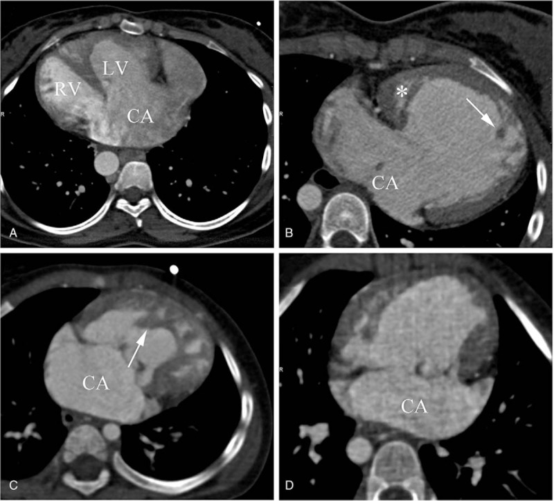
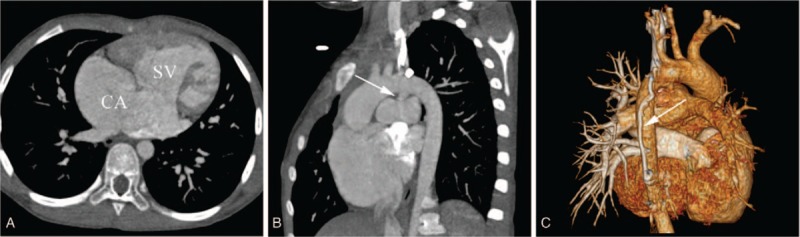
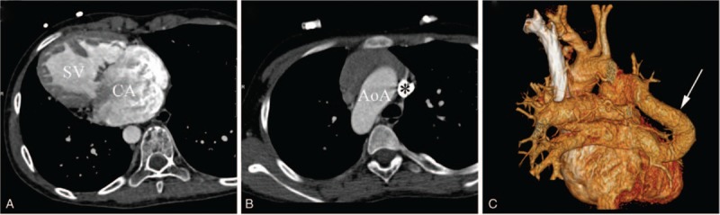
Common atrium (CA) is a rare complex congenital heart disease. The studies of CA are mostly case reports, while few have been done regarding its morphological characteristics. We aimed to determine CA characteristics and diagnostic accuracy in assessing associated malformations in these patients with low-dose dual-source computed tomography (DSCT).Twenty-one pediatric and adolescent CA patients underwent low-dose DSCT. Different ventricular types and associated malformations were assessed. The diagnostic accuracy of DSCT and transthoracic echocardiography (TTE) in evaluating associated malformations were assessed. The effective doses of DSCT were calculated.Patients (n = 21) were divided into CA with biventricular physiology (n = 7) and CA with single ventricle (SV) (n = 14). There were 3 types of SV morphology: single left ventricle (n = 5), single right ventricle (n = 6), and undifferentiated ventricle (n = 3). In all, 22 associated malformations were seen in CA and 56 in CA with SV. DSCT was superior to TTE for detecting intracardiac anomalies (sensitivity: DSCT, 92.31% vs TTE, 76.92%), great vessels anomalies (sensitivity: DSCT, 100.00% vs TTE, 77.50%), and of collateral vessels (sensitivity: DSCT, 100% vs TTE, 20.00%). The estimated mean effective dose was 0.95 ± 0.44 mSv (<1 mSv).This study indicated that low-dose DSCT is an ideal alternative for pediatric and adolescent patients with CA, providing morphological details of CA and associated malformations with high accuracy.
Conflict of interest statement
The authors declare that they have no competing interests.
Figures
Similar articles
-
Assessment of Double Outlet Right Ventricle Associated with Multiple Malformations in Pediatric Patients Using Retrospective ECG-Gated Dual-Source Computed Tomography.PLoS One. 2015 Jun 26;10(6):e0130987. doi: 10.1371/journal.pone.0130987. eCollection 2015. PLoS One. 2015. PMID: 26115034 Free PMC article.
-
Assessment of transposition of the great arteries associated with multiple malformations using dual-source computed tomography.PLoS One. 2017 Nov 20;12(11):e0187578. doi: 10.1371/journal.pone.0187578. eCollection 2017. PLoS One. 2017. PMID: 29155835 Free PMC article.
-
Assessment of tetralogy of Fallot-associated congenital extracardiac vascular anomalies in pediatric patients using low-dose dual-source computed tomography.BMC Cardiovasc Disord. 2017 Dec 4;17(1):285. doi: 10.1186/s12872-017-0718-8. BMC Cardiovasc Disord. 2017. PMID: 29202750 Free PMC article.
-
Dual-source Computed Tomography for Evaluating Pulmonary Artery and Aorta in Pediatric Patients with Single Ventricle.Sci Rep. 2017 Oct 17;7(1):13398. doi: 10.1038/s41598-017-11809-6. Sci Rep. 2017. PMID: 29042577 Free PMC article.
-
Preoperative evaluation of anomalous pulmonary venous connection using dual-source computed tomography: Comparison with echocardiography.Eur J Radiol. 2017 Sep;94:107-114. doi: 10.1016/j.ejrad.2017.06.015. Epub 2017 Jun 21. Eur J Radiol. 2017. PMID: 28669428
Cited by
-
Situs Inversus Partialis With a Wandering Spleen Having a Single Atrium: A Rare Tale of Survival.Cureus. 2023 Jul 14;15(7):e41860. doi: 10.7759/cureus.41860. eCollection 2023 Jul. Cureus. 2023. PMID: 37581126 Free PMC article.
-
A 78-year-old man diagnosed with single atrium as congenital heart disease.SAGE Open Med Case Rep. 2023 Jul 29;11:2050313X231189772. doi: 10.1177/2050313X231189772. eCollection 2023. SAGE Open Med Case Rep. 2023. PMID: 37529079 Free PMC article.
References
-
- Jiang H, Wang H, Wang Z, et al. Surgical correction of common atrium without noncardiac congenital anomalies. J Card Surg 2013;28:580–6. - PubMed
-
- Nabati M, Bagheri B, Habibi V. Coincidence of total anomalous pulmonary venous drainage to the superior vena cava, common atrium, and single ventricle: a very rare condition. Echocardiography 2013;30:E98–101. - PubMed
-
- Xiao Y, Qiao W, Zhan Y, et al. Single ventricle and single atrium with anomalous hepatic venous drainage and azygos continuation of inferior vena cava in an adult patient. J Clin Ultrasound 2015;43:458–61. - PubMed
-
- Ferdman DJ, Brady D, Rosenzweig EB. Common atrium and pulmonary vascular disease. Pediatr Cardiol 2011;32:595–8. - PubMed
-
- Stern KW, McElhinney DB, Gauvreau K, et al. Echocardiographic evaluation before bidirectional Glenn operation in functional single-ventricle heart disease: comparison to catheter angiography. Circ Cardiovasc Imaging 2011;4:498–505. - PubMed
