

A case report on cystic meningioma in cerebellopontine angle and recommendations for management
- PMID: 30431593
- PMCID: PMC6257460
- DOI: 10.1097/MD.0000000000013179
A case report on cystic meningioma in cerebellopontine angle and recommendations for management
Abstract
Rationale: Cystic meningioma located at the cerebellopontine angle (CPA) is an extremely rare occurrence. It is frequently misdiagnosed preoperatively. Little is known about the clinical features and outcome of this rare disease.
Patient concerns: A 70-year-old male presenting with progressive headache and gait disturbance.
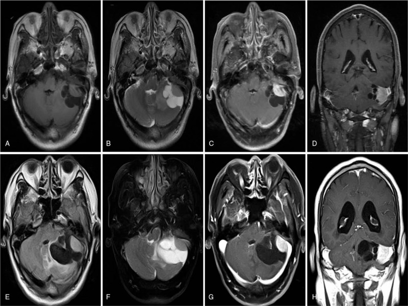
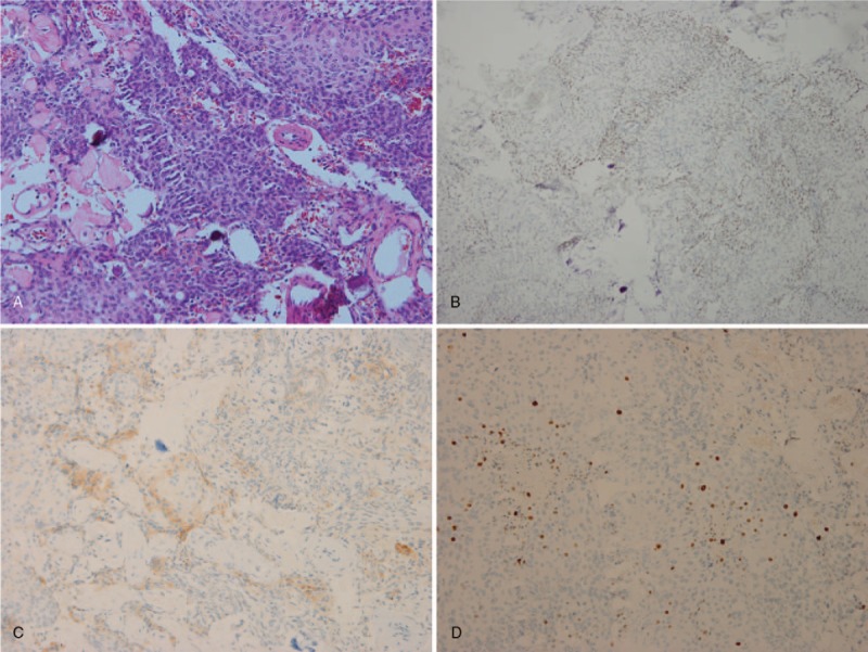
Diagnosis: According to the symptoms, signs, and Gd-enhanced magnetic resonance images (MRI), a preoperative diagnosis of hemangioblastoma located in left CPA was made. Finally, the histological examination revealed a meningioma.
Interventions: A complete resection, including the part of the solid mass together with cyst, was performed.
Outcomes: The postoperative course of the patient was uneventful, and no residual or recurrent tumor was found during the 24-month follow-up period.
Lessons: Cystic meningioma should be included in the differential diagnosis of a CPA mass with atypical radiologic features, such as a large cyst and enhanced mural nodule. By summarizing the related literature, we found that the most common pathological subtype of CPA cystic meningioma is the clear cell subtype, which belongs to WHO grade II. Gross total resection including the enhanced cyst wall is extremely important. A close follow-up is necessary because of the high recurrence rate in this subset of meningioma.
Conflict of interest statement
The authors have no conflicts of interest to disclose.
Figures
References
-
- Campbell BA, Jhamb A, Maguire JA, et al. Meningiomas in 2009: controversies and future challenges. Am J Clin Oncol 2009;32:73–85. - PubMed
-
- Buetow MP, Buetow PC, Smirniotopoulos JG. Typical, atypical, and misleading features in meningioma. Radiographics 1991;11:1087–106. - PubMed
-
- Ercan Ö, Yücesoy K, Çitak G, et al. Cystic meningioma imitating vestibular schwannoma at the cerebellopontine angle: case report. J Neurol Sci (Turkish) 2007;24:84–7.
-
- Deb P, Sahni H, Bhatoe HS. Cystic angiomatous meningioma in the cerebellopontine angle mimicking hemangioblastoma. J Cancer Res Ther 2010;6:560–3. - PubMed
Publication types
MeSH terms
LinkOut - more resources
Full Text Sources
