Case Reports
. 2018 Jun;57(2):366-371.
doi: 10.20471/acc.2018.57.02.19.

Brachial Plexus Schwannoma - Case Report and Literature Review

Affiliations
Case Reports

Brachial Plexus Schwannoma - Case Report and Literature Review

Luka Vučemilo et al. Acta Clin Croat. 2018 Jun.

Abstract

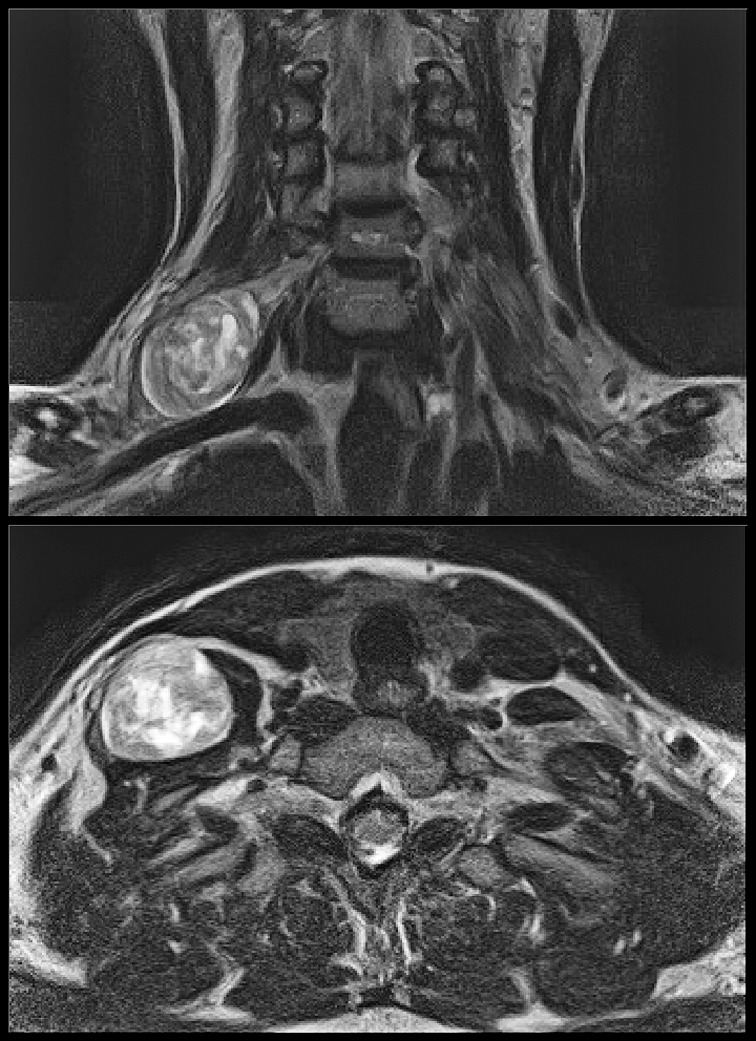
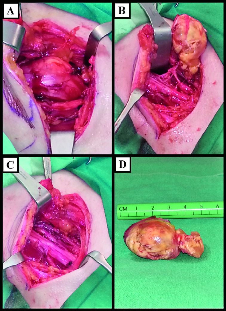
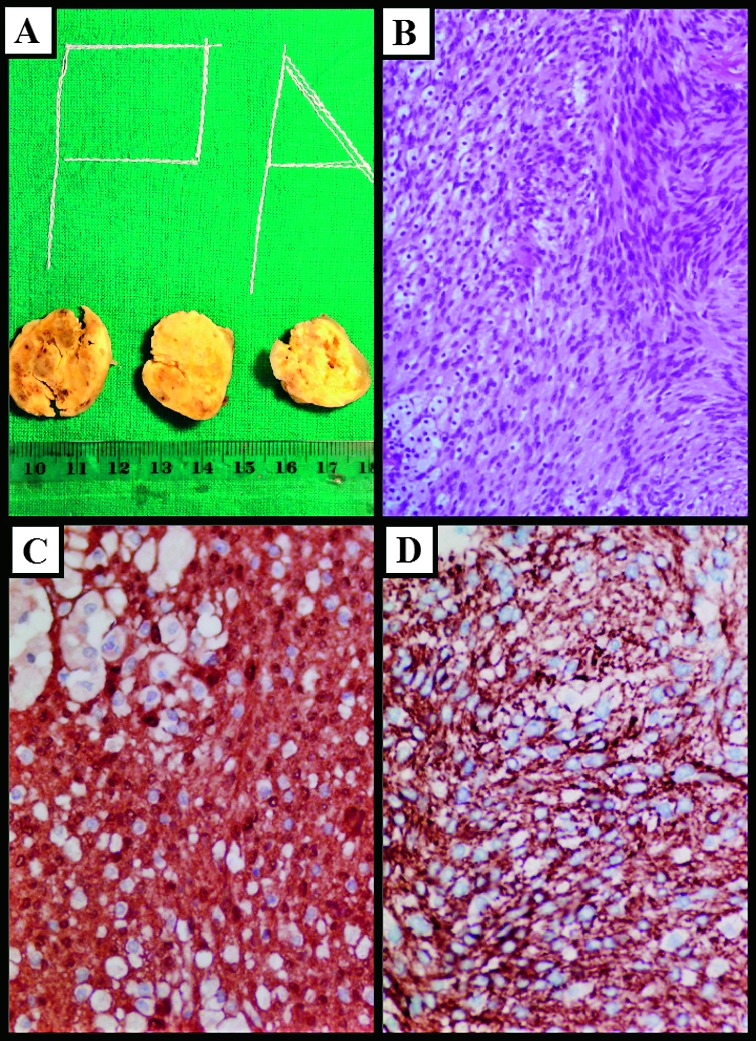
Schwannoma as an extracranial nerve sheath tumor rarely affects brachial plexus. Due to the fact that brachial plexus schwannomas are a rare entity and due to the brachial plexus anatomic complexity, schwannomas in this region present a challenge for surgeons. We present a case of a 49-year-old female patient with a slow growing painless mass in the right supraclavicular region that was diagnosed as schwannoma and operated at our department. The case is described to remind that in case of supraclavicular tumors, differential diagnosis should take brachial plexus tumors, i.e. schwannomas, in consideration. Extra caution is also required on fine needle aspiration procedures or biopsies of schwannomas due to the possible iatrogenic injury of the nerve and adjacent structures. On operative treatment of schwannoma, complete tumor resection should be achieved while preserving the nerve.

Keywords: Biopsy, Fine-Needle; Brachial Plexus; Case Reports; Nerve Sheath Neoplasms; Neurilemmoma.


Figures

Fig. 1
Fig. 1
Radiological findings. Magnetic resonance imaging, coronal and axial scans showing a large schwannoma arising from the right brachial plexus.
Fig. 2
Fig. 2
Intraoperative findings. (A) Encapsulated mass splitting the fasciscles of the right brachial plexus; (B) schwannoma after separation from fascicles of the brachial plexus; (C) brachial plexus after enucleation of schwannoma; (D) enucleated schwannoma, macroscopic view.
Fig. 3
Fig. 3
Histologic findings. (A) Tumor cut surface was yellowish with focal areas of hemorrhage; (B) fascicles of monomorphic spindle shaped cells and foamy histiocytes (hematoxylin and eosin staining, X20); (C) intense cytoplasmic expression of S100 in tumor cells (X40); (D) intense cytoplasmic expression of GFAP in tumor cells (X40).

References

    1. Knight DM, Birch R, Pringle J. Benign solitary schwannomas: a review of 234 cases. J Bone Joint Surg Br. 2007;89:382–7. 10.1302/0301-620X.89B3.18123 - DOI - PubMed
    1. Yun DH, Kim HS, Chon J, Lee J, Jung PK. Thoracic outlet syndrome caused by schwannoma of brachial plexus. Ann Rehabil Med. 2013;37:896–900. 10.5535/arm.2013.37.6.896 - DOI - PMC - PubMed
    1. Tang CY, Fung B, Fok M, Zhu J. Schwannoma in the upper limbs. BioMed Res Int. 2013;2013:167196. 10.1155/2013/167196 - DOI - PMC - PubMed
    1. Kohyama S, Hara Y, Nishiura Y, Hara T, Nakagawa T, Ochiai N. A giant plexiform schwannoma of the brachial plexus: case report. J Brachial Plex Peripher Nerve Inj. 2011;6:9. 10.1186/1749-7221-6-9 - DOI - PMC - PubMed
    1. Lusk MD, Kline DG, Garcia CA. Tumors of the brachial plexus. Neurosurgery. 1987;21:439–53. 10.1227/00006123-198710000-00001 - DOI - PubMed

Publication types

LinkOut - more resources