. 2018 Dec;33(12):1928-1937.
doi: 10.1002/mds.27541. Epub 2018 Nov 15.

Progressively disrupted somatodendritic morphology in dopamine neurons in a mouse Parkinson's model

Affiliations

Progressively disrupted somatodendritic morphology in dopamine neurons in a mouse Parkinson's model

William B Lynch et al. Mov Disord. 2018 Dec.

Abstract

Background: Parkinson's disease is characterized by the progressive loss of dopamine neurons in the substantia nigra, leading to severe motor deficits. Although the disease likely begins to develop years before observable motor symptoms, the specific morphological and functional alterations involved are poorly understood.

Objectives: MitoPark mice lack the gene coding for mitochondrial transcription factor A specifically in dopamine neurons, which over time produces a progressive decline of neuronal function and related behavior that phenotypically mirrors human parkinsonism. Our previous work identified a progressive decrease in cell capacitance in dopamine neurons from MitoPark mice, possibly suggesting reduced membrane surface area. We therefore sought to identify and quantify somatodendritic parameters in this model across age.

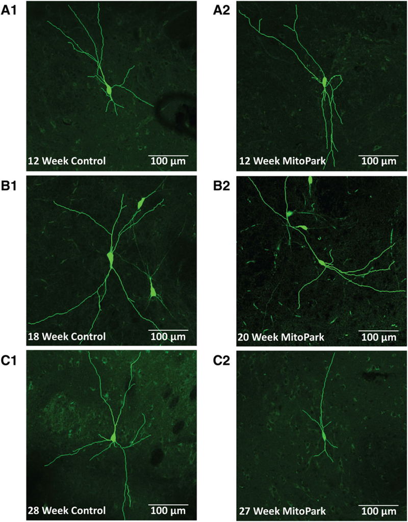
Methods: We used whole-cell patch clamp and fluorescent labeling to quantify somatodendritic morphology of single, neurobiotin-filled dopamine neurons in acutely isolated brain slices from MitoPark mice.

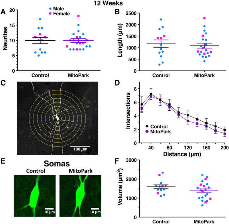
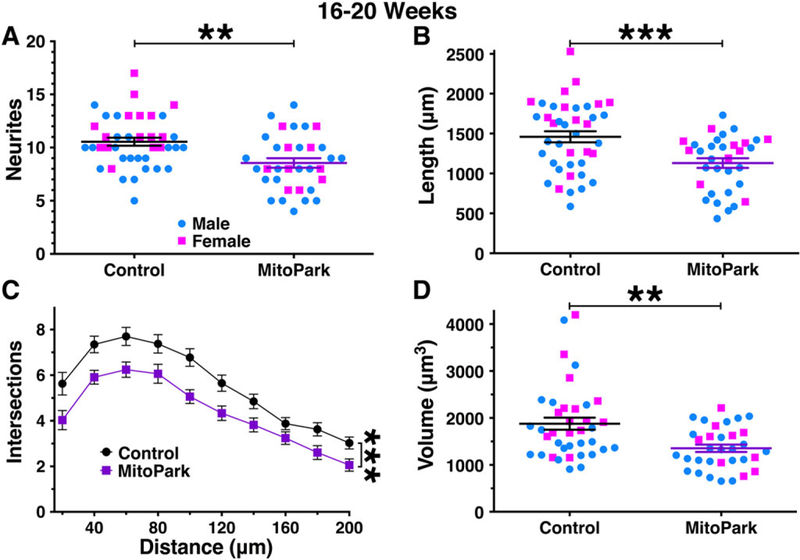
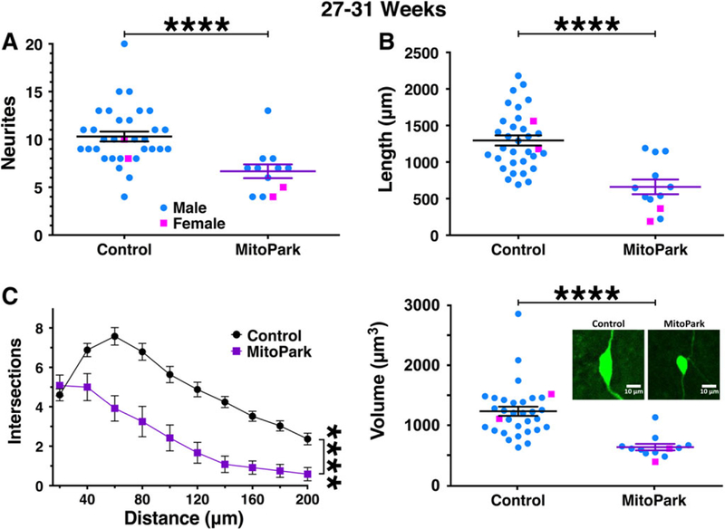
Results: We found that MitoPark mice exhibit an adult-onset, age-dependent reduction of neuritic branching and soma size in dopamine neurons. This decline proceeds similarly in MitoPark mice of both sexes, but does not begin until after the age that early decrements in ion channel physiology and behavior have previously been observed.

Conclusions: A progressive and severe decline in somatodendritic morphology occurs prior to cell death, but is not responsible for the subtle decrements observable in the earliest stages of neurodegeneration. This work could help identify the ideal time window for specific treatments to halt disease progression and avert debilitating motor deficits in Parkinson's patients. © 2018 International Parkinson and Movement Disorder Society.

Keywords: MitoPark; branching; dendrites; in vivo; soma.


Conflict of interest statement

Relevant conflicts of interests/financial disclosures: Nothing to report.

Figures

FIG. 1.
FIG. 1.
As age increases, somatodendritic morphology in dopamine neurons from MitoPark mice becomes disrupted. Substantia nigra dopamine neurons from 12-week-old MitoPark mice (A2) exhibited a similar appearance to neurons from littermate controls (A1). In contrast, the number of branches appeared to be reduced in 16–20-week old MitoPark mice (B2 vs B1). Furthermore, branch number and length as well as soma size were dramatically reduced in 27- to 31-week-old MitoPark mice (C2 vs C1). Neurons from control mice exhibited consistent morphology throughout all age groups.
FIG. 2.
FIG. 2.
Dopamine neurons from MitoPark mice exhibit normal morphology at 12 weeks of age. Morphological parameters were quantified for individual dopamine neurons from 12-week-old male (blue circles) and female (pink squares) MitoPark mice and littermate controls. Neurite number (A) and cumulative neurite length (B) were not different between genotypes. Sholl analysis was performed by digitally drawing concentric circles at 20-μm intervals around the soma and counting neurite intersections (C). The number of intersections did not vary between genotypes (D). Soma size (E) was estimated by measuring the two-dimensional area through multiple z-planes. Soma volumes were not significantly different in MitoPark mice at this age (F).
FIG. 3.
FIG. 3.
Morphological parameters are mildly reduced in dopamine neurons from 16- to 20-week-old MitoPark mice. By 16 to 20 weeks of age, dopamine neurons from male (blue circles) and female (pink squares) MitoPark mice exhibited a measurable impairment in somatodendritic morphology. This was observed for neurite count (A), cumulative neurite length (B), number of intersections/Sholl analysis (C), and soma volume (D). **P < .01; ***P < .001 between genotypes.
FIG. 4.
FIG. 4.
Morphological parameters are severely reduced in dopamine neurons from 27- to 31-week-old MitoPark mice. By 27 to 31 weeks of age, dopamine neurons from male (blue circles) and female (pink squares) MitoPark mice exhibited a severe impairment in somatodendritic morphology. This was observed for all parameters tested including neurite count (A), cumulative neurite length (B), and number of intersections/Sholl analysis (C). Soma volume was also severely reduced (D) with the inset showing representative 2-dimensional images from this age group. ****P < .0001 between genotypes.

Similar articles

Cited by

References

    1. Goetz CG. The history of Parkinson’s disease: early clinical descriptions and neurological therapies. Cold Spring Harb Perspect Med 2011;1:a008862. - PMC - PubMed
    1. Kalia LV, Lang AE. Parkinson’s disease. The Lancet 2015;386: 896–912. - PubMed
    1. Dauer W, Przedborski S. Parkinson’s disease: mechanisms and models. Neuron 2003;39:889–909. - PubMed
    1. Hornykiewicz O Parkinson’s disease and its chemotherapy. Bio-chem Pharmacol 1975;24:1061–1065. - PubMed
    1. Kordower JH, Olanow CW, Dodiya HB, et al. Disease duration and the integrity of the nigrostriatal system in Parkinson’s disease. Brain 2013;136:2419–2431. - PMC - PubMed

Publication types

MeSH terms