. 2019 Apr;16(2):360-369.
doi: 10.1111/iwj.13041. Epub 2018 Nov 15.

The clinical dynamic changes of macrophage phenotype and function in different stages of human wound healing and hypertrophic scar formation

Affiliations

The clinical dynamic changes of macrophage phenotype and function in different stages of human wound healing and hypertrophic scar formation

Lin Chen et al. Int Wound J. 2019 Apr.

Abstract

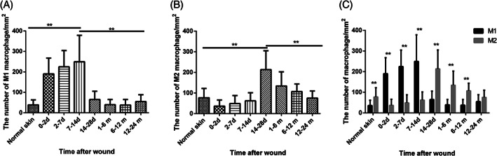
The pathogenesis of hypertrophic scar (HS) is still poorly understood. Macrophages, especially the polarisation of that to M1 or M2, play a pivotal role in control of the degree of scar formation. Profiling of macrophage phenotypes in human specimens during long-term period of wound healing and HS formation may provide valuable clinical evidence for understanding the pathology of human scars. Human wound and HS specimens were collected, the macrophage phenotype was identified by immunofluorescence, and biomarkers and cytokines associated with M1 and M2 macrophages were detected by RT-PCR. The correlation between the macrophage phenotype and HS characteristics was analysed by linear regression analyses. We found excessive and persistent infiltration by M1 macrophages around the blood vessels in the superficial layer of the dermis at early wound tissues, whereas M2 macrophages predominated in later wound tissues and the proliferative phase of HS and were scattered throughout the dermis. The density of M1 macrophages was positively correlated with mRNA expression levels of tumour necrosis factor-alpha (TNF-α) and IL-6. The density of M2 macrophages was positively correlated with ARG1 and negatively correlated with the duration of HS. The sequential infiltration by M1 macrophage and M2 macrophages in human wound and HS tissues was confirmed.

Keywords: cytokines; hypertrophic scar; macrophage phenotype; wound healing.


Figures

Figure 1
Figure 1
The dynamic changes of the macrophage M1/M2 phenotype during wound healing and hypertrophic scar (HS) development. A, B, C, D, I, J, K, and L showed the M1 macrophages of normal skin, wounds of the 0 to 2 days group, the 2 to 7 days group, the 7 to 14 days group, the 14 to 28 days group, HS of the 1 to 6 months group, the 6 to 12 months group, and the 12 to 24 months group. M1 macrophages were identified as double positive for membrane staining of CD68 (green) and nuclear transcription factor staining of RBP‐J (red). Similarly, M2 macrophages were identified as double positive for membrane staining of CD68 (green) and nuclear transcription factors staining of CMAF (red). E, F, G, H, M, N, O, and P showed the M2 macrophages of normal skin, wounds of the 0 to 2 days group, the 2 to 7 days group, the 7 to 14 days group, the 14 to 28 days group, HS of the 1 to 6 m group, the 6 to 12 m group, and the 12 to 24 m group. Original magnification: 40×. The arrows indicate the positive cells. d, days; m, months
Figure 2
Figure 2
The density changes of M1 and M2 macrophages in wound‐healing process and hypertrophic scar (HS) development and the ratio of M1 and M2 macrophages in each group. The density was calculated as the ratio of the number of double positive cells in the dermal region using 400‐fold magnification and the area. A showed the density changes of M1 macrophages and B showed the density changes of M2 macrophages. C showed the ratio of the density of M1 macrophage and M2 macrophage. Error bars represent the mean ± standard deviation (n = 5; independent experiments). Significantly different at **P < 0.01 as determined using one‐way analysis of variance with post hoc LSD test
Figure 3
Figure 3
Histologic analysis of M1 macrophage infiltration in the human tissues (green: CD68, red: RBP‐J, blue: DAPI). The dotted lines indicate the boundaries of the epidermis and the dermis. A and C showed the histological distribution of M1 macrophage in the 0 to 2 and 2 to 7 days wounds with the original magnification of 20×. B and D showed the local amplification of A and C with the original magnification of 40× and a double positive cell was showed in the lower right box of B and the top right box of D
Figure 4
Figure 4
Histologic analysis of M2 macrophage infiltration in the human tissues. (green: CD68, red: CMAF, blue: DAPI). The dotted lines indicate the boundaries of the epidermis and the dermis. A and C showed the histological distribution of M2 macrophage in the 1 to 6 and 12 to 24 months hypertrophic scar tissues with the original magnification of 20×. B and D showed the local amplification of a and C with the original magnification of 40× and a double positive cell was shown in the lower right box of B and D
Figure 5
Figure 5
Relative mRNA expression of biomarkers and cytokines associated with M1 macrophages and M2 macrophages normalised to GAPDH and normal skin expressed as 2−(ΔΔCt) in human specimens during wound‐healing process and hypertrophic scar development. A–C showed the relative mRNA expression of M1‐associated genes NOS2, TNF‐α, and IL‐6. D–F showed the relative mRNA expression of M2‐associated genes ARG1, TGF‐β1, and CD206. Error bars represent the mean ± standard deviation (n = 5; independent experiments). Significantly different at *P < 0.05, **P < 0.01 as determined using one‐way analysis of variance with post hoc LSD test. NS, normal skin; d, days; m, months

Similar articles

Cited by

References

    1. Finnerty CC, Jeschke MG, Branski LK, Barret JP, Dziewulski P, Herndon DN. Hypertrophic scarring: the greatest unmet challenge after burn injury. The Lancet. 2016;388(10052):1427‐1436. - PMC - PubMed
    1. Sen CK, Gordillo GM, Roy S, et al. Human skin wounds: a major and snowballing threat to public health and the economy. Wound Repair Regen. 2009;17(6):763‐771. - PMC - PubMed
    1. Singer AJ, Clark RA. Cutaneous wound healing. N Engl J Med. 1999;341(10):738‐746. - PubMed
    1. Lucas T, Waisman A, Ranjan R, et al. Differential roles of macrophages in diverse phases of skin repair. J Immunol. 2010;184(7):3964‐3977. - PubMed
    1. Zhu Z, Ding J, Ma Z, Iwashina T, Tredget EE. Systemic depletion of macrophages in the subacute phase of wound healing reduces hypertrophic scar formation. Wound Repair Regen. 2016;24(4):644‐656. - PubMed