Review
. 2018 Nov 14;9(11):594.
doi: 10.3390/mi9110594.

Ultrasonic Based Tissue Modelling and Engineering

Affiliations
Review

Ultrasonic Based Tissue Modelling and Engineering

Karl Olofsson et al. Micromachines (Basel). .

Abstract

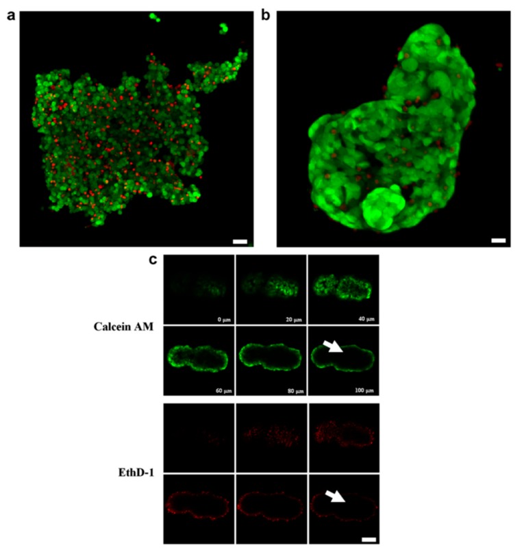
Systems and devices for in vitro tissue modelling and engineering are valuable tools, which combine the strength between the controlled laboratory environment and the complex tissue organization and environment in vivo. Device-based tissue engineering is also a possible avenue for future explant culture in regenerative medicine. The most fundamental requirements on platforms intended for tissue modelling and engineering are their ability to shape and maintain cell aggregates over long-term culture. An emerging technology for tissue shaping and culture is ultrasonic standing wave (USW) particle manipulation, which offers label-free and gentle positioning and aggregation of cells. The pressure nodes defined by the USW, where cells are trapped in most cases, are stable over time and can be both static and dynamic depending on actuation schemes. In this review article, we highlight the potential of USW cell manipulation as a tool for tissue modelling and engineering.

Keywords: acoustic trapping; acoustofluidics; microfluidics; tissue engineering; tissue modelling; ultrasonic manipulation.


Conflict of interest statement

The authors declare no conflicts of interest.

Figures

Figure 1
Figure 1
Red blood cell manipulation by ultrasound in a chick embryo. The image shows a chick embryo vasculosa where red blood cells are trapped in clusters separated by half the wavelength at 3 MHz continuous ultrasonic irradiation. The image was acquired through a microscope with 26× magnification. Reprinted with permission from [10].
Figure 2
Figure 2
Ultrasound-formed HepG2 cell aggregates embedded in alginate retains viability and forms spheroids over time. Individual HepG2 cells are distinguishable in the initial aggregate (a) but after three days in culture membrane spreading and cell–cell connections forms a stable spheroid (b). Calcein AM and EthD-1 staining after 10 days in culture shows retained cell viability and poor dye penetration which is typical for larger 3D aggregates (c,d). Scale bars are 30 µm in (a), 10 µm in (b) and 20 µm in (c). Reprinted with permission from [15].
Figure 3
Figure 3
Multiple acoustic pressure nodes are an effective way to parallelize tissue formation. Numerous pressure nodes can be obtained by using multiple wavelengths in a single resonator chamber (a) (scale bar: 1 mm) [29], producing pressure nodes arrays through orthogonal one-dimensional pressure fields (b) [30] or using multiple microwells where the half-wavelength (λ/2) criterion is met in two dimensions (scale bar: 100 µm) (c). Reprinted with permission from (a) [29], (b) [30], and in (c) experiment by Karl Olofsson.
Figure 4
Figure 4
Acoustic holography can be used to form arbitrary pressure field images. The target image (a) is used to backward calculate the required phase distribution (b). The 3D printed transmission hologram, as a height topography in a phase plate (c), is placed in front of the transducer (d) to acquire the sound pressure image (e). Scale bar: 10 mm. Reprinted with permission from [49].
Figure 5
Figure 5
Patterned and in situ differentiated neurons can model the cerebral cortex. USW-based arrangement of neural progenitor cells during fibrin hydrogel cross-linking formed stable cell layers embedded in the hydrogel suitable for long-term culture without USW. This allowed for differentiation of the neural progenitor cells into neurons in situ which interacted between layers after 8 days (a). Fourteen days after culture, mature neuron maker MAP2 was co-expressed together with Tuj1 (b). The excitatory marker CaMKII and glial cell marker GFAP are detected after 30 days of culture (c,d). All scale bars indicate 30 µm. Reprinted with permission from [53].
Figure 6
Figure 6
On-chip confocal imaging illustrates the strength with USW-based multi-cellular tumor spheroid (MCTS) culture platforms. Nuclear dye DAPI-stained A498 MCTSs mounted in a refractive-index-matching solution allowed for 3D reconstruction (a) from a stack of optical sections (b) acquired by on-chip confocal microscopy. The whole MCTS volume could be imaged in detail. Figure adapted from [58].
Figure 7
Figure 7
Implantation of an acoustically generated cartilage graft in healthy human tissue with a drilled defect (a). Close-ups are shown in (b,d), and in particular, (d) shows the fusion between the native tissue and the graft with formation of hyaline cartilage in the gap. A defect before implantation is shown in (e). The scale bars in (a,e) correspond to 500 µm; whereas the scale bars in (b,d) correspond to 50 µm. Reprinted from [63] published by the Royal Society of Chemistry.
Figure 8
Figure 8
Actuating the acoustic trap with a frequency sweep will move a levitated chondrocyte cell aggregate laterally. The displacement can be controlled by the sweep rate, providing a means to control the mechanical stimulation during tissue formation. In the figure, the aggregate position along x and y directions over time is measured at the sweep rates 1 Hz (a), 2 Hz (b), 5 Hz (c) and 20 Hz (d) Reprinted with permission from [23] published by the Royal Society of Chemistry.

Similar articles

Cited by

References

    1. Laurell T., Lenshof A. Microscale Acoustofluidics. Royal Society of Chemistry; London, UK: 2014.
    1. Laurell T., Petersson F., Nilsson A. Chip integrated strategies for acoustic separation and manipulation of cells and particles. Chem. Soc. Rev. 2007;36:492–506. doi: 10.1039/B601326K. - DOI - PubMed
    1. Shi J., Ahmed D., Mao X., Lin S.C., Lawit A., Huang T.J. Acoustic tweezers: Patterning cells and microparticles using standing surface acoustic waves (SSAW) Lab Chip. 2009;9:2890–2895. doi: 10.1039/b910595f. - DOI - PubMed
    1. Brandt E.H. Acoustic physics. Suspended by sound. Nature. 2001;413:474–475. doi: 10.1038/35097192. - DOI - PubMed
    1. Evander M., Nilsson J. Acoustofluidics 20: Applications in acoustic trapping. Lab Chip. 2012;12:4667–4676. doi: 10.1039/c2lc40999b. - DOI - PubMed

LinkOut - more resources