

Mesenchymal Stem Cells and Transforming Growth Factor-β₃ (TGF-β₃) to Enhance the Regenerative Ability of an Albumin Scaffold in Full Thickness Wound Healing
- PMID: 30441760
- PMCID: PMC6306712
- DOI: 10.3390/jfb9040065
Mesenchymal Stem Cells and Transforming Growth Factor-β₃ (TGF-β₃) to Enhance the Regenerative Ability of an Albumin Scaffold in Full Thickness Wound Healing
Abstract
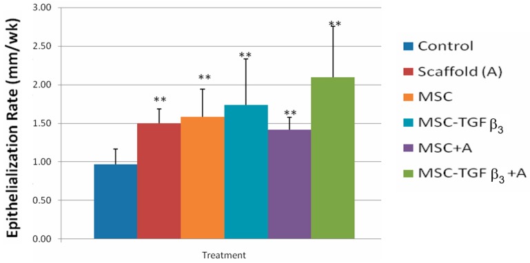
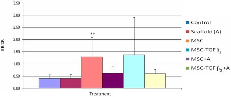
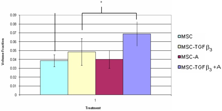
Pressure ulcers are one of the most common forms of skin injury, particularly in the spinal cord injured (SCI). Pressure ulcers are difficult to heal in this population requiring at least six months of bed rest. Surgical treatment (grafting) is the fastest recovery time, but it still requires six weeks of bed rest plus significant additional costs and a high recurrence rate. A significant clinical benefit would be obtained by speeding the healing rate of a non-surgical treatment to close to that of surgical treatment (approximately doubling of healing rate). Current non-surgical treatment is mostly inactive wound coverings. The goal of this project was to look at the feasibility of doubling the healing rate of a full-thickness defect using combinations of three treatments, for the first time, each shown to increase healing rate: application of transforming growth factor-β₃ (TGF-β₃), an albumin based scaffold, and mesenchymal stem cells (MSCs). At one week following surgery, the combined treatment showed the greatest increase in healing rate, particularly for the epithelialization rate. Although the target level of a 100% increase in healing rate over the control was not quite achieved, it is anticipated that the goal would be met with further optimization of the treatment.
Keywords: TGF-β3; albumin scaffold; mesenchymal stem cells; pressure ulcers; wound healing.
Conflict of interest statement
The authors declare no conflicts of interest.
Figures


Similar articles
-
Stromal Tissue Rigidity Promotes Mesenchymal Stem Cell-Mediated Corneal Wound Healing Through the Transforming Growth Factor β Signaling Pathway.Stem Cells. 2016 Oct;34(10):2525-2535. doi: 10.1002/stem.2405. Epub 2016 Jun 25. Stem Cells. 2016. PMID: 27250866
-
Genetically-modified bone mesenchymal stem cells with TGF-β3 improve wound healing and reduce scar tissue formation in a rabbit model.Exp Cell Res. 2018 Jun 1;367(1):24-29. doi: 10.1016/j.yexcr.2018.02.006. Epub 2018 Feb 15. Exp Cell Res. 2018. PMID: 29453974
-
Effects of different transforming growth factor beta (TGF-β) isomers on wound closure of bone cell monolayers.Cytokine. 2014 Sep;69(1):75-86. doi: 10.1016/j.cyto.2014.05.010. Epub 2014 Jun 11. Cytokine. 2014. PMID: 25022965
-
[The modern approach to wound treatment].Med Pregl. 2000 Jul-Aug;53(7-8):363-8. Med Pregl. 2000. PMID: 11214479 Review. Croatian.
-
Role of TGF beta-mediated inflammation in cutaneous wound healing.J Investig Dermatol Symp Proc. 2006 Sep;11(1):112-7. doi: 10.1038/sj.jidsymp.5650004. J Investig Dermatol Symp Proc. 2006. PMID: 17069018 Review.
Cited by
-
Mesenchymal Stem Cells for Regenerative Medicine.Cells. 2019 Aug 13;8(8):886. doi: 10.3390/cells8080886. Cells. 2019. PMID: 31412678 Free PMC article. Review.
-
Mesenchymal Stem Cell-Derived Extracellular Vesicles as Non-Coding RNA Therapeutic Vehicles in Autoimmune Diseases.Pharmaceutics. 2022 Mar 29;14(4):733. doi: 10.3390/pharmaceutics14040733. Pharmaceutics. 2022. PMID: 35456567 Free PMC article. Review.
-
Stem cell and tissue engineering approaches in pressure ulcer treatment.J Spinal Cord Med. 2023 Mar;46(2):194-203. doi: 10.1080/10790268.2021.1916155. Epub 2021 Apr 27. J Spinal Cord Med. 2023. PMID: 33905315 Free PMC article. Review.
-
Using biomaterials to improve mesenchymal stem cell therapies for chronic, nonhealing wounds.Bioeng Transl Med. 2023 Sep 13;9(1):e10598. doi: 10.1002/btm2.10598. eCollection 2024 Jan. Bioeng Transl Med. 2023. PMID: 38193114 Free PMC article. Review.
-
Topical hADSCs-HA Gel Promotes Skin Regeneration and Angiogenesis in Pressure Ulcers by Paracrine Activating PPARβ/δ Pathway.Drug Des Devel Ther. 2024 Oct 26;18:4799-4824. doi: 10.2147/DDDT.S474628. eCollection 2024. Drug Des Devel Ther. 2024. PMID: 39478872 Free PMC article.
References
-
- [(accessed on 7 January 2018)]; Available online: http://www.ihi,org/IHI/Programs/Campaign/Pressure Ulcers.htm.
-
- Makelbust J., Sieggreen M. Pressure Ulcers: Guidelines for Prevention and Management. Springhouse Corporation; Springhouse, PA, USA: 2001. pp. 110–111.
Grants and funding
LinkOut - more resources
Full Text Sources
Miscellaneous
