

Metabolomic Signature in Sera of Multiple Sclerosis Patients during Pregnancy
- PMID: 30441762
- PMCID: PMC6274842
- DOI: 10.3390/ijms19113589
Metabolomic Signature in Sera of Multiple Sclerosis Patients during Pregnancy
Abstract
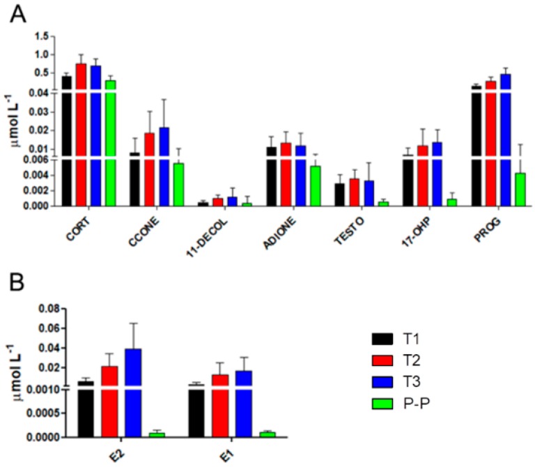
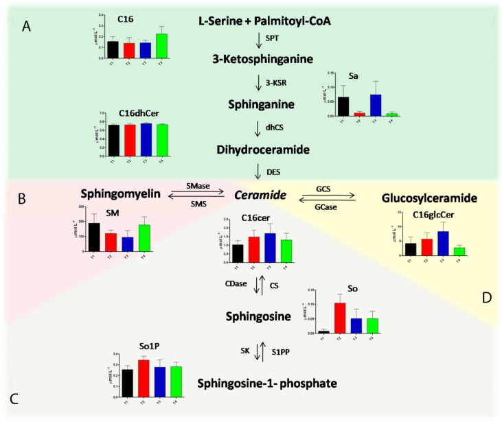
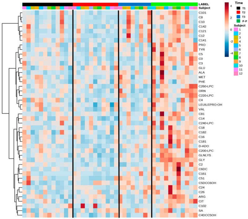
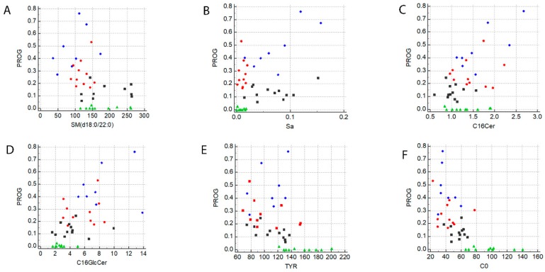
Multiple sclerosis (MuS) is an autoimmune disease of the central nervous system characterized by neuroinflammation, neurodegeneration, and degradation of the myelin sheath. Epidemiological studies have shown that the female gender is more susceptible than the male gender to MuS development, with a female-to-male ratio of 2:1. Despite this high onset, women have a better prognosis than men, and the frequency of the relapsing phase decreases during pregnancy, while it increases soon after birth. Therefore, it is interesting to investigate hormonal fluctuations during pregnancy and whether they correlate with metabolic signatures. To gain a deeper inside into the biochemical mechanism of such a multifactorial disease, we adopted targeted metabolomics approaches for the determination of many serum metabolites in 12 pregnant women affected by MuS by mass spectrometry analysis. Our data show a characteristic hormonal fluctuation for estrogens and progesterone, as expected. They also highlight other interesting hormonal alterations for cortisol, corticosterone, 11-deoxycortisol, 4-androstene-3,17-dione, testosterone, and 17α-hydroxyprogesterone. Furthermore, a negative correlation with progesterone levels was observed for amino acids and for acylcarnitines, while an imbalance of different sphingolipids pathways was found during pregnancy. In conclusion, these data are in agreement with the characteristic clinical signs of MuS patients during pregnancy and, if confirmed, they may add an important tessera in the complex mosaic of maternal neuroprotection.
Keywords: acylcarnitines; amino acids; biomarkers; ceramides; estrogens; mass spectrometry; metabolomics; multiple sclerosis; pregnancy; steroids.
Conflict of interest statement
The authors declare no conflict of interest.
Figures
Similar articles
-
Integrated Lipidomics and Metabolomics Analysis of Tears in Multiple Sclerosis: An Insight into Diagnostic Potential of Lacrimal Fluid.Int J Mol Sci. 2019 Mar 13;20(6):1265. doi: 10.3390/ijms20061265. Int J Mol Sci. 2019. PMID: 30871169 Free PMC article.
-
A Plasma Metabolomic Signature of the Exfoliation Syndrome Involves Amino Acids, Acylcarnitines, and Polyamines.Invest Ophthalmol Vis Sci. 2018 Feb 1;59(2):1025-1032. doi: 10.1167/iovs.17-23055. Invest Ophthalmol Vis Sci. 2018. PMID: 29450546
-
An integrated metabolomics approach for the research of new cerebrospinal fluid biomarkers of multiple sclerosis.Mol Biosyst. 2015 Jun;11(6):1563-72. doi: 10.1039/c4mb00700j. Mol Biosyst. 2015. PMID: 25690641
-
Integration of metabolomics and proteomics in multiple sclerosis: From biomarkers discovery to personalized medicine.Proteomics Clin Appl. 2016 Apr;10(4):470-84. doi: 10.1002/prca.201500083. Epub 2016 Mar 11. Proteomics Clin Appl. 2016. PMID: 27061322 Review.
-
Hormonal influences in multiple sclerosis: new therapeutic benefits for steroids.Maturitas. 2011 Jan;68(1):47-51. doi: 10.1016/j.maturitas.2010.09.014. Epub 2010 Oct 29. Maturitas. 2011. PMID: 21035281 Review.
Cited by
-
Multi-Omics Approach for Studying Tears in Treatment-Naïve Glaucoma Patients.Int J Mol Sci. 2019 Aug 18;20(16):4029. doi: 10.3390/ijms20164029. Int J Mol Sci. 2019. PMID: 31426571 Free PMC article.
-
Contribution of Metabolomics to Multiple Sclerosis Diagnosis, Prognosis and Treatment.Int J Mol Sci. 2021 Oct 15;22(20):11112. doi: 10.3390/ijms222011112. Int J Mol Sci. 2021. PMID: 34681773 Free PMC article. Review.
-
Chronic Oleoylethanolamide Treatment Decreases Hepatic Triacylglycerol Level in Rat Liver by a PPARγ/SREBP-Mediated Suppression of Fatty Acid and Triacylglycerol Synthesis.Nutrients. 2021 Jan 27;13(2):394. doi: 10.3390/nu13020394. Nutrients. 2021. PMID: 33513874 Free PMC article.
-
Influence of hormones in multiple sclerosis: focus on the most important hormones.Metab Brain Dis. 2023 Mar;38(3):739-747. doi: 10.1007/s11011-022-01138-7. Epub 2023 Jan 3. Metab Brain Dis. 2023. PMID: 36595158 Review.
-
Inhibition of de novo ceramide biosynthesis affects aging phenotype in an in vitro model of neuronal senescence.Aging (Albany NY). 2019 Aug 29;11(16):6336-6357. doi: 10.18632/aging.102191. Epub 2019 Aug 29. Aging (Albany NY). 2019. PMID: 31467258 Free PMC article.
References
MeSH terms
Substances
Grants and funding
LinkOut - more resources
Full Text Sources
Medical
Molecular Biology Databases
