. 2018 Nov 14;19(11):3592.
doi: 10.3390/ijms19113592.

Breast Cancer: An Examination of the Potential of ACKR3 to Modify the Response of CXCR4 to CXCL12

Affiliations

Breast Cancer: An Examination of the Potential of ACKR3 to Modify the Response of CXCR4 to CXCL12

Irene Del Molino Del Barrio et al. Int J Mol Sci. .

Erratum in

Abstract

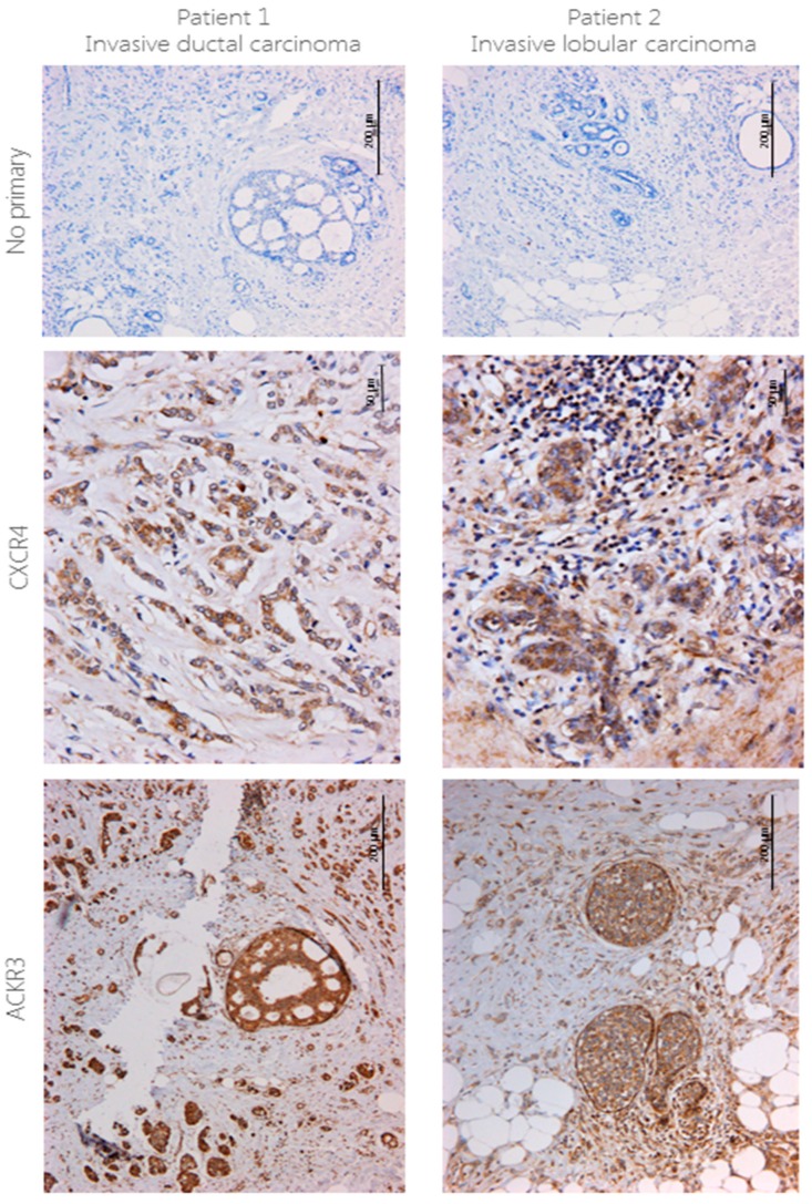
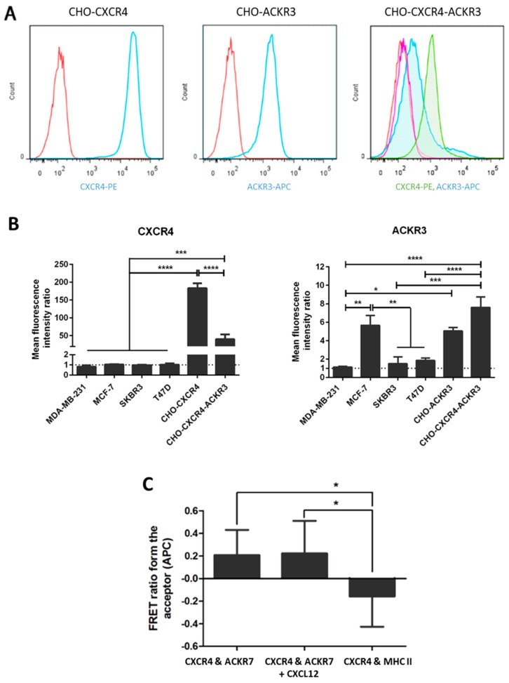
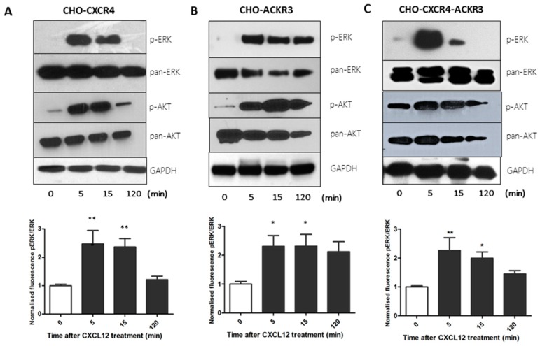
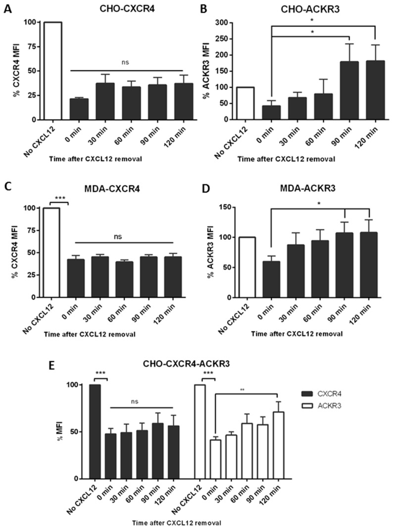
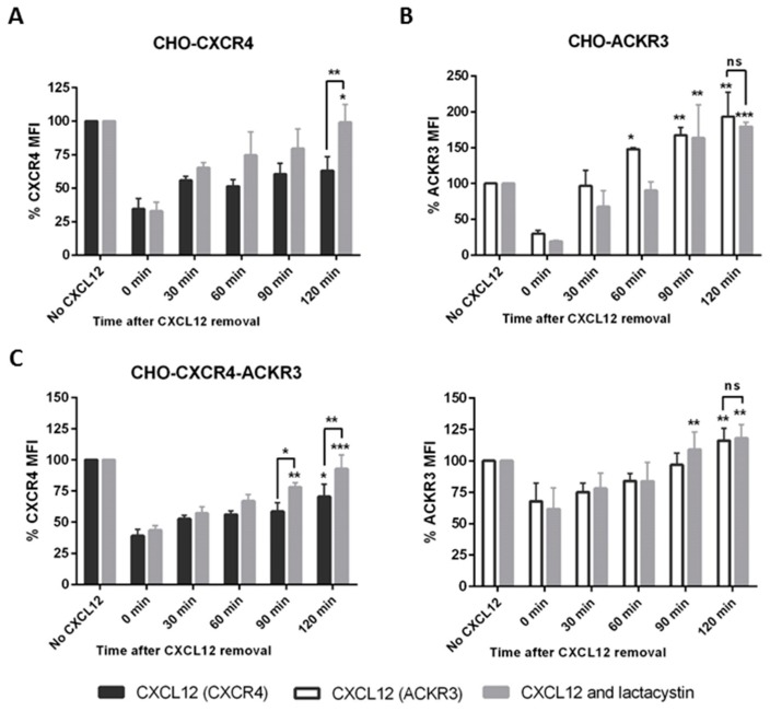
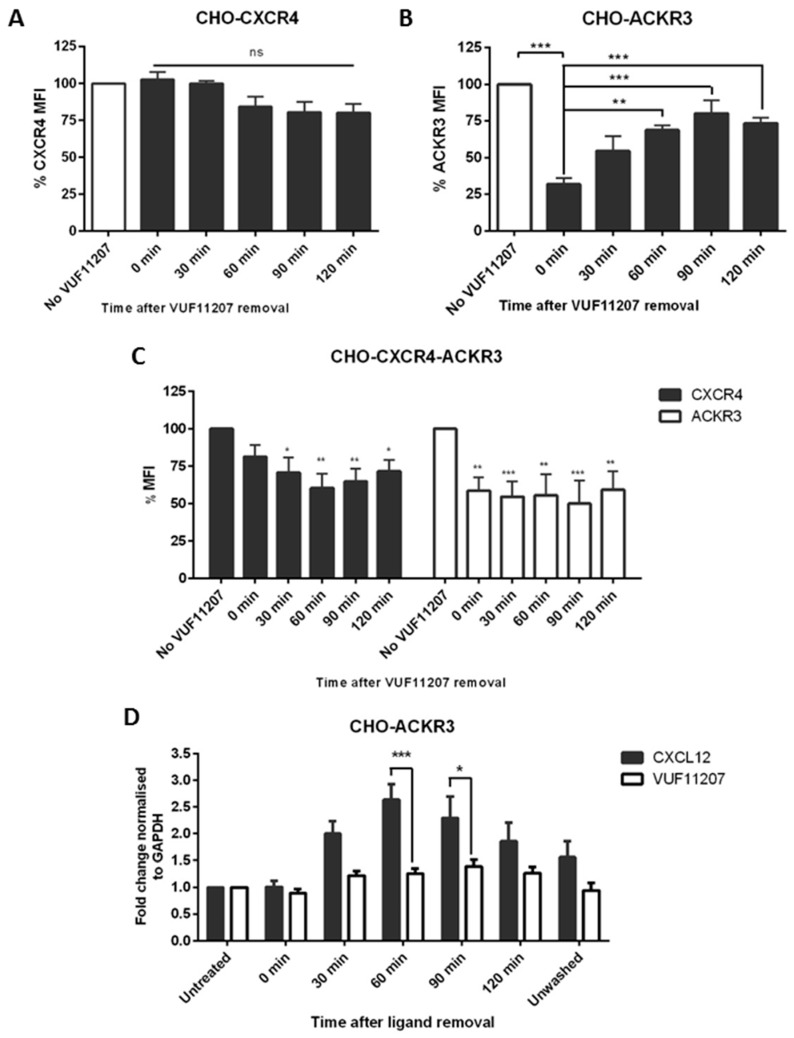
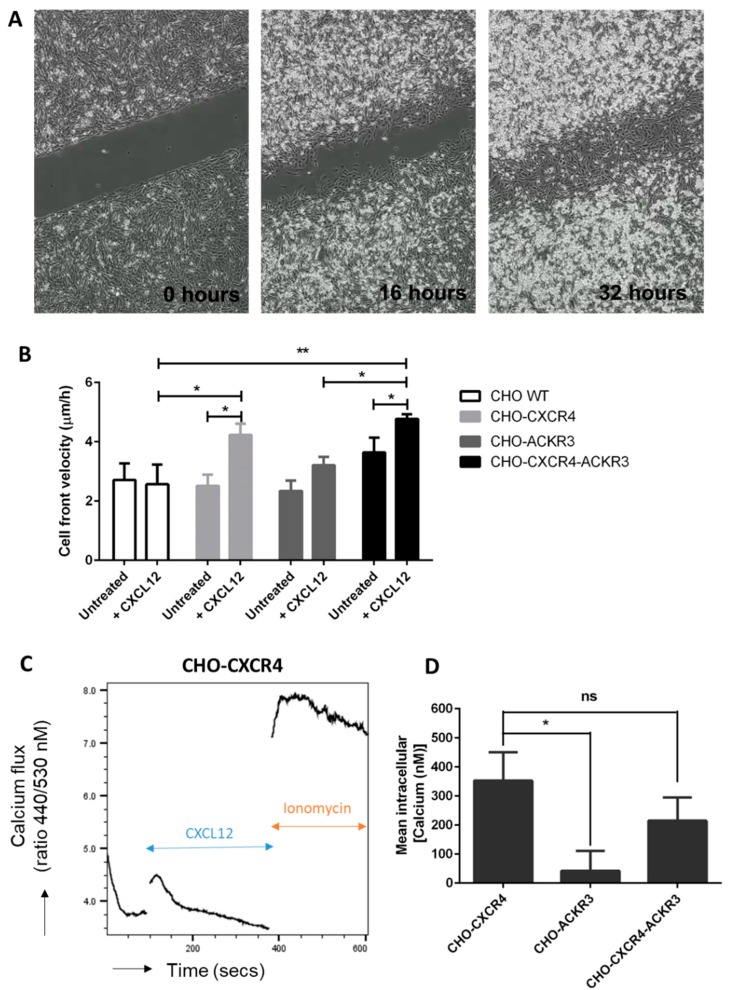
Upon binding with the chemokine CXCL12, the chemokine receptor CXCR4 has been shown to promote breast cancer progression. This process, however, can be affected by the expression of the atypical chemokine receptor ACKR3. Given ACKR3's ability to form heterodimers with CXCR4, we investigated how dual expression of both receptors differed from their lone expression in terms of their signalling pathways. We created single and double CXCR4 and/or ACKR3 Chinese hamster ovary (CHO) cell transfectants. ERK and Akt phosphorylation after CXCL12 stimulation was assessed and correlated with receptor internalization. Functional consequences in cell migration and proliferation were determined through wound healing assays and calcium flux. Initial experiments showed that CXCR4 and ACKR3 were upregulated in primary breast cancer and that CXCR4 and ACKR3 could form heterodimers in transfected CHO cells. This co-expression modified CXCR4's Akt activation after CXCL12's stimulation but not ERK phosphorylation (p < 0.05). To assess this signalling disparity, receptor internalization was assessed and it was observed that ACKR3 was recycled to the surface whilst CXCR4 was degraded (p < 0.01), a process that could be partially inhibited with a proteasome inhibitor (p < 0.01). Internalization was also assessed with the ACKR3 agonist VUF11207, which caused both CXCR4 and ACKR3 to be degraded after internalization (p < 0.05 and p < 0.001), highlighting its potential as a dual targeting drug. Interestingly, we observed that CXCR4 but not ACKR3, activated calcium flux after CXCL12 stimulation (p < 0.05) and its co-expression could increase cellular migration (p < 0.01). These findings suggest that both receptors can signal through ERK and Akt pathways but co-expression can alter their kinetics and internalization pathways.

Keywords: ACKR3; CXCR4; CXCR7; chemokines; heterodimerization; metastasis.


Conflict of interest statement

The authors declare no conflict of interest.

Figures

Figure 1
Figure 1
CXCR4 and ACKR3 staining using IHC in breast cancer tissue. 4 μm sections from human breast cancer were stained for CXCR4 (1:40) and ACKR3 (1:100) using immunohistochemistry following no pre-treatment or EDTA antigen retrieval pre-treatment, respectively. Briefly, protocol from the VECTASTAIN ABC HRP kit was followed, signal was developed using DAB and counterstained with haematoxylin. No primary antibody was used as a control. n = 2.
Figure 2
Figure 2
Expression of CXCR4 and/or ACKR3 in transfected CHO cells. CHO cells were transfected with pcDNA3.1/Zeo-CXCR4 and/or pcDNA3-ACKR3 using Effectene and selected with antibiotics. (A) Histograms showing CXCR4 or ACKR3 expression in the final selected colonies. Cells were stained with PE or APC-conjugated antibodies and expression was assessed using flow cytometry (Red = Isotype, blue = antibody) on CHO-CXCR4 (left) and CHO-ACKR3 (middle); and red = isotype for CXCR4, purple = isotype for ACKR3, green = CXCR4, blue = ACKR3 on CHO-CXCR4-ACKR3 (right) cells. (B) Mean fluorescence levels of the transfected CHO cells were determined and compared to several breast cancer cell lines to assess CXCR4 (left) and ACKR3 (right) receptor levels. Data represents the mean ± SEM of three independent experiments and statistical significance was calculated using a one way ANOVA (* p < 0.05). (C) Formation of heterodimers was assessed using fluorescence resonance energy transfer (FRET) and quantified through the FRET ratio from APC (the acceptor fluorochrome). Data represents the mean ± SEM of three independent experiments and statistical significance was calculated using a one way ANOVA (* p < 0.05, ** p < 0.01, *** p < 0.001, **** p < 0.0001).
Figure 3
Figure 3
Western blot and cell-based ELISA show that CXCL12 treatment of transfected CHO cells differentially activates the ERK and AKT pathways. Serum starved (A) CHO-CXCR4, (B) CHO-ACKR3 and (C) CHO-CXCR4-ACKR3 cells were stimulated with 10 nM CXCL12 for 5, 15 and 120 min. (Top) Cells were lysed and immunoblots probed with p-ERK or p-Akt, stripped and re-probed for pan-ERK or pan-AKT and GAPDH as a loading control. Images are representative of three independent experiments. (Bottom) Cells were fixed with methanol and cell-based ELISA was performed as per protocol using p-ERK and total-ERK antibodies with fluorescence intensity readings at 600 and 450 nm. Data represents the mean ± SEM of three independent experiments and statistical significance was calculated using a one way ANOVA (* p < 0.05, ** p < 0.01).
Figure 4
Figure 4
CXCR4 and ACKR3 follow different internalization pathways after CXCL12 stimulation. (A) CHO-CXCR4, (B) CHO-ACKR3, (C) MDA-MB-231-CXCR4, (D) MDA-MB-231-ACKR3 and (E) CHO-CXCR4-ACKR3 cells were treated with 10–50 nM CXCL12 for 15–30 min, then washed and incubated with chemokine-free media for up to 2 h to assess receptor recycling. Cells were labelled with CXCR4-PE and/or ACKR3-APC antibody and receptor’s mean fluorescence intensity (MFI) was measured using flow cytometry. Data represents the mean ± SEM of three independent experiments and statistical significance was calculated using a one way ANOVA (ns: not significant, * p < 0.05, ** p < 0.01, *** p < 0.001).
Figure 5
Figure 5
Preventing receptor degradation after CXCL12 stimulation using lactacystin, a proteasome inhibitor. (A) CHO-CXCR4, (B) CHO-ACKR3 and (C) CHO-CXCR4-ACKR3 cells were pre-treated for 1 h with 10 μM lactacystin (a proteosome inhibitor) before incubating with 10–50 nM CXCL12 for 15–30 min. Cells were then washed and incubated with chemokine-free media for up to 2 h before being labelled and analysed using a flow cytometer. Data represents the mean ± SEM of three independent experiments and statistical significance was calculated using a one way ANOVA (ns: not significant, * p < 0.05, ** p < 0.01, *** p < 0.001).
Figure 6
Figure 6
CXCR4 and ACKR3 follow different internalization pathways after VUF11207 stimulation. (A) CHO-CXCR4, (B) CHO-ACKR3 and (C) CHO-CXCR4-ACKR3 cells were stimulated with 1 nM VUF11207 for 30 min and then washed and incubated with agonist-free media for up to 2 h to assess receptor recycling. Cells were then labelled and receptor expression was measured using flow cytometry. Data represents the mean ± SEM of three independent experiments and statistical significance was calculated using a one way ANOVA (ns: not significant, * p < 0.05, ** p < 0.01, *** p < 0.001). (D) CXCL12 and VUF11207 have a different effect in ACKR3 transcription. CHO-ACKR3 cells were stimulated with 1 nM VUF11207 or 10 nM CXCL12 and recycling was assessed as described above. RNA was then extracted at each time point and ACKR3 expression was assessed using qPCR and normalized to GAPDH. Data represents the mean ± SEM of three independent experiments and statistical significance was calculated using a one-way ANOVA (* p < 0.05, *** p < 0.001).
Figure 7
Figure 7
CXCR4 but not ACKR3, has an effect in wound healing after CXCL12 stimulation. CHO-WT, CHO-CXCR4, CHO-ACKR3 and CHO-CXCR4-ACKR3 cells were seeded into Ibidi inserts to create a “wound” 24 h later. Wells were then filled with serum-reduced media with or without 10 nM CXCL12 and wound closure was monitored for 48 h using the Nikon Biostation. (A) Example of images captured during a wound healing assay, magnification 4× (B) Cell front velocity was calculated from the wound area. Data represents the mean ± SEM of nine independent experiments and statistical significance was calculated using a two-way ANOVA with Bonferroni post-tests (* p < 0.05, ** p < 0.01). (C) CXCR4- but not ACKR3-expressing cells show calcium flux in response to CXCL12. Indo-1AM stained cells were stimulated with 10 nM CXCL12 followed by 4 μM ionomycin as a positive control and intracellular calcium flux was assessed. A representative plot showing calcium release in CHO-CXCR4 cells can be seen. (D) Ratio between free Indo-1 AM at 510 nm and calcium-bound Indo-1AM at 420 nm was calculated and concentration of calcium released was determined. Data is representative of three independent experiments and statistical significance was calculated using a one-way ANOVA (ns: not significant, * p < 0.05).

Similar articles

Cited by

References

    1. Siegel R.L., Miller K.D., Jemal A. Cancer statistics, 2016. CA Cancer J. Clin. 2016;66:7–30. doi: 10.3322/caac.21332. - DOI - PubMed
    1. Berry D.A., Cronin K.A., Plevritis S.K., Fryback D.G., Clarke L., Zelen M., Mandelblatt J.S., Yakovlev A.Y., Habbema J.D.F., Feuer E.J. Effect of screening and adjuvant therapy on mortality from breast cancer. N. Engl. J. Med. 2005;353:1784–1792. doi: 10.1056/NEJMoa050518. - DOI - PubMed
    1. Olsen O., Gøtzsche P.C. Cochrane review on screening for breast cancer with mammography. Lancet. 2001;358:1340–1342. doi: 10.1016/S0140-6736(01)06449-2. - DOI - PubMed
    1. Greene F.L., Page D.L., Fleming I.D., Fritz A.G., Balch C.M., Haller D.G., Morrow M. AJCC Cancer Staging Manual. 6th ed. Springer; New York, NY, USA: 2002. Breast; pp. 223–240.
    1. Baggiolini M. Chemokines and leukocyte traffic. Nature. 1998;392:565–568. doi: 10.1038/33340. - DOI - PubMed