Review
. 2018 Nov 14;19(11):3601.
doi: 10.3390/ijms19113601.

Small Molecules Enhance Scaffold-Based Bone Grafts via Purinergic Receptor Signaling in Stem Cells

Affiliations
Review

Small Molecules Enhance Scaffold-Based Bone Grafts via Purinergic Receptor Signaling in Stem Cells

Patrick Frank Ottensmeyer et al. Int J Mol Sci. .

Abstract

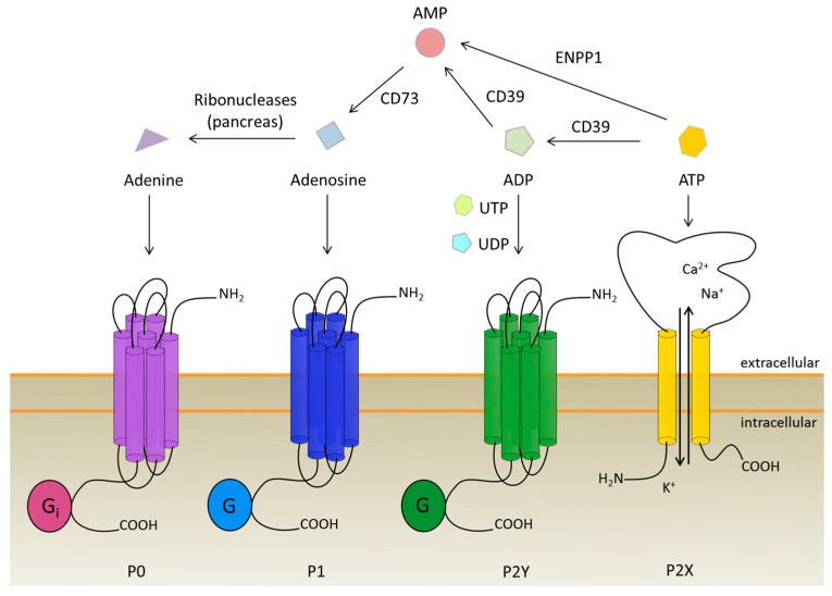
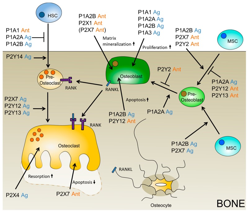
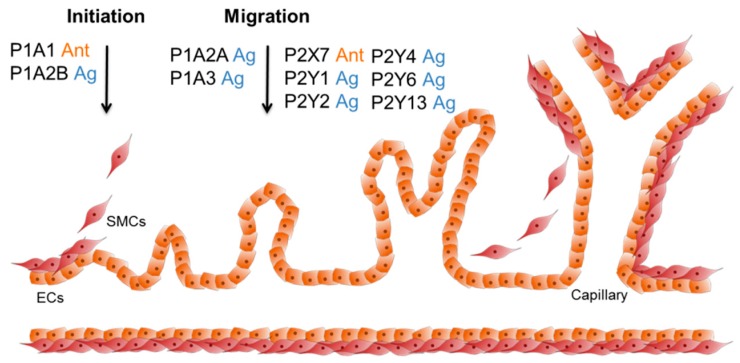
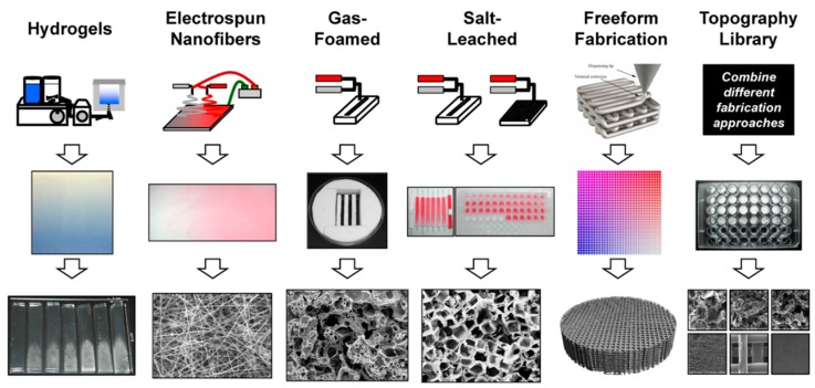
The need for bone grafts is high, due to age-related diseases, such as tumor resections, but also accidents, risky sports, and military conflicts. The gold standard for bone grafting is the use of autografts from the iliac crest, but the limited amount of accessible material demands new sources of bone replacement. The use of mesenchymal stem cells or their descendant cells, namely osteoblast, the bone-building cells and endothelial cells for angiogenesis, combined with artificial scaffolds, is a new approach. Mesenchymal stem cells (MSCs) can be obtained from the patient themselves, or from donors, as they barely cause an immune response in the recipient. However, MSCs never fully differentiate in vitro which might lead to unwanted effects in vivo. Interestingly, purinergic receptors can positively influence the differentiation of both osteoblasts and endothelial cells, using specific artificial ligands. An overview is given on purinergic receptor signaling in the most-needed cell types involved in bone metabolism-namely osteoblasts, osteoclasts, and endothelial cells. Furthermore, different types of scaffolds and their production methods will be elucidated. Finally, recent patents on scaffold materials, as wells as purinergic receptor-influencing molecules which might impact bone grafting, are discussed.

Keywords: angiogenesis; bone; drug release; mesenchymal stem cells; osteoblast; osteoclast; patent; purinergic receptors; scaffold.


Conflict of interest statement

The authors declare no conflict of interest.

Figures

Figure 1
Figure 1
Purinergic receptors and their ligands with respect to ATP conversion to adenine. P0 receptors are activated by adenine, whereas P1 receptors are stimulated via adenosine binding. The P2 receptors are divided into ATP-dependent P2X ion channels and G protein-coupled P2Y receptors which are stimulated by ATP, ADP, UTP, UDP, and UDP-glucose. Degradation of ATP to adenosine is done by ectonucleotidases, namely CD39, CD73, and NPP1. Adenine is produced during the nucleotide recovery in the pancreas.
Figure 2
Figure 2
Putative purinergic receptor ligands which might positively influence bone grafting. The beneficial ligand is depicted, agonist (Ag) is shown in blue, and antagonist (Ant) in orange. If only the data on the negatively influencing ligand is available, then the putative positively influencing ligand is given in brackets.
Figure 3
Figure 3
Purinergic receptor ligands which can influence angiogenesis. During the initiation phase of angiogenesis, the smooth muscle cells (SMCs) are released from the vascular tube. Next, the endothelial cells (ECs) start to migrate into the free space, and create new tubes and branches (sprouting). In the end, new SMCs cover the ECs and the process stops. P1A1 and P1A2B stimulate VEGF expression, which increases angiogenesis. The activation of P1A2A, P1A3, and P2Y1 enhances the migration of hEPCs and ECs. Additionally, P1A3 increases the expression of pro-angiogenic factors. The P2X7 receptor is accountable for EC survival and proliferation. P2Y2, P2Y4, and P2Y6 showed a pro-angiogenic effect upon Up4A signaling, and P2Y13 can increase angiogenesis by activation of Akt and ERK1/2.
Figure 4
Figure 4
Different techniques for additive scaffold manufacturing (printed with permission of C. Simon Jr. [142]). First line: manufactured scaffolds include hydrogels, electrospun nanofibers, foams, and 3D structures obtained from salt leaching, freeform fabrication, and lithography. Second and third line: for each technique, the sample in macroscale is shown, together with a figure of the scaffold microstructure.
Figure 5
Figure 5
Publications between 01/2015 and 08/2018, partitioned by fabrication method of bone scaffold. Search parameters were publications with the terms “bone tissue engineering”, “scaffold”, and [fabrication method]. Search was performed on 21 August 2018, using NCBI PubMed. Reviews are excluded from the results. Possible differences can be due to rounding.
Figure 6
Figure 6
Patents between 01/2015 and 08/2018, partitioned by materials. Search was performed on Espacenet with the keywords “bone tissue engineering” and “scaffold” on August 21st, 2018. Composite materials had the most filed patents in the given period, followed by ceramics and bioglass, and polymers.

References

    1. Tomar K., Sahoo N.K. Evaluation of graft uptake from the iliac crest in secondary alveolar bone grafting: Bergland’s criteria revisited. J. Oral Biol. Craniofac. Res. 2018;8:171–176. doi: 10.1016/j.jobcr.2017.03.005. - DOI - PMC - PubMed
    1. Egol K.A., Nauth A., Lee M., Pape H.C., Watson J.T., Borrelli J., Jr. Bone Grafting: Sourcing, Timing, Strategies, and Alternatives. J. Orthop. Trauma. 2015;29(Suppl. 12):S510–S514. doi: 10.1097/BOT.0000000000000460. - DOI - PubMed
    1. Batista J.D., Justino Oliveira Limirio P.H., Rocha F.S., Gomes Moura C.C., Zanetta-Barbosa D., Dechichi P. Influence of Platelet-Poor Plasma on Angiogenesis and Maintenance of Volume in Autogenous Bone Grafts. J. Oral Maxillofac. Surg. 2018 doi: 10.1016/j.joms.2018.06.024. - DOI - PubMed
    1. Burk T., Del Valle J., Finn R.A., Phillips C. Maximum Quantity of Bone Available for Harvest From the Anterior Iliac Crest, Posterior Iliac Crest, and Proximal Tibia Using a Standardized Surgical Approach: A Cadaveric Study. J. Oral Maxillofac. Surg. 2016;74:2532–2548. doi: 10.1016/j.joms.2016.06.191. - DOI - PubMed
    1. Kilinc A., Korkmaz I.H., Kaymaz I., Kilinc Z., Dayi E., Kantarci A. Comprehensive analysis of the volume of bone for grafting that can be harvested from iliac crest donor sites. Br. J. Oral Maxillofac. Surg. 2017;55:803–808. doi: 10.1016/j.bjoms.2017.07.011. - DOI - PubMed

LinkOut - more resources