. 2018 Dec 3;215(12):3136-3150.
doi: 10.1084/jem.20181031. Epub 2018 Nov 15.

CNBP controls IL-12 gene transcription and Th1 immunity

Affiliations

CNBP controls IL-12 gene transcription and Th1 immunity

Yongzhi Chen et al. J Exp Med. .

Abstract

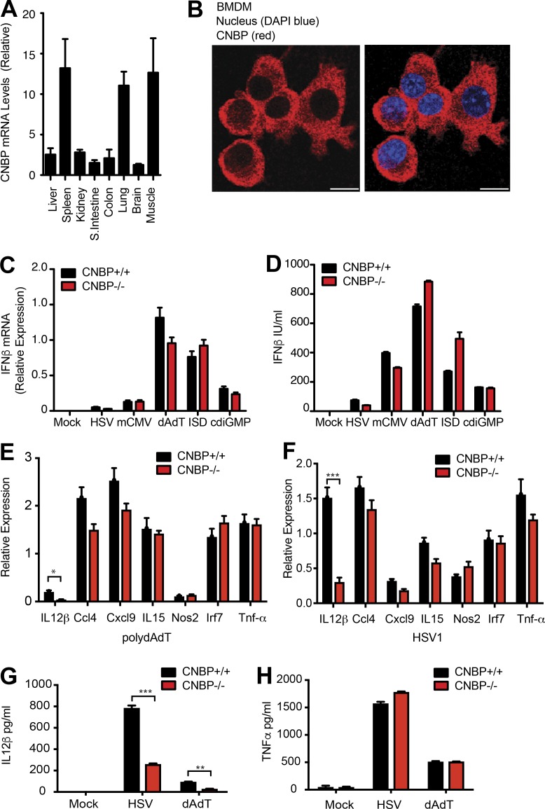
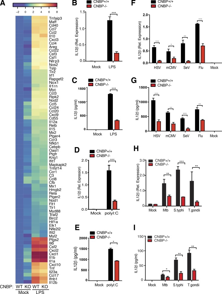
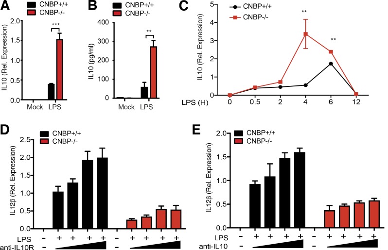
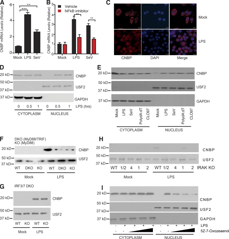
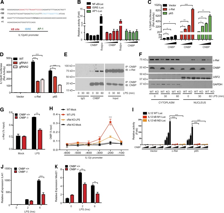
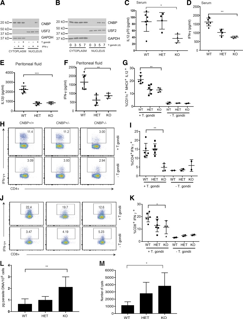
An inducible program of inflammatory gene expression is a hallmark of antimicrobial defenses. Recently, cellular nucleic acid-binding protein (CNBP) was identified as a regulator of nuclear factor-kappaB (NF-κB)-dependent proinflammatory cytokine gene expression. Here, we generated mice lacking CNBP and found that CNBP regulates a very restricted gene signature that includes IL-12β. CNBP resides in the cytosol of macrophages and translocates to the nucleus in response to diverse microbial pathogens and pathogen-derived products. Cnbp-deficient macrophages induced canonical NF-κB/Rel signaling normally but were impaired in their ability to control the activation of c-Rel, a key driver of IL-12β gene transcription. The nuclear translocation and DNA-binding activity of c-Rel required CNBP. Lastly, Cnbp-deficient mice were more susceptible to acute toxoplasmosis associated with reduced production of IL-12β, as well as a reduced T helper type 1 (Th1) cell IFN-γ response essential to controlling parasite replication. Collectively, these findings identify CNBP as important regulator of c-Rel-dependent IL-12β gene transcription and Th1 immunity.


Figures

Figure 1.
Figure 1.
Identification of CNBP, a cytosolic dsDNA–binding protein regulating the cytosolic dsDNA–induced IL-12β response. (A) Tissue-based expression of Cnbp in mouse (normalized to Gapdh); n = 3. (B) Confocal microscopy of primary BMDMs probed with anti-CNBP. Bars, 10 μm. (C and D) IFN-β mRNA and protein levels in Cnbp+/+ and Cnbp−/− BMDMs treated with DNA ligands, including HSV, mCMV, dAdT, ISD, or c-di-GMP, were detected through qRT-PCR (C) and ELISA (D). (E and F) qRT-PCR analysis of Il12b, Ccl4, Cxcl9, Il15, Nos2, Irf7, or Tnf-α mRNA in Cnbp+/+ and Cnbp−/− BMDMs left unstimulated or transfected with poly(dA:dT) (E) or HSV1 infection (F). (G and H) IL-12β (G) or TNF-α (H) protein levels in Cnbp+/+ and Cnbp−/− BMDMs treated with HSV or dA:dT were detected through ELISA. Error bars represent SD of triplicate technical replicates (C, E, and F) or SEM of triplicate biological replicates (A, D, G, and H). All data are representative of three independent experiments. *, P < 0.05; **, P < 0.01; ***, P < 0.001.
Figure 2.
Figure 2.
CNBP is a broad regulator of the IL-12β response in the TLR and RIG-I–like receptor pathway. (A) Heat map of gene expression in WT and KO BMDMs treated or not treated with LPS and analyzed by NanoString; n = 3. (B and C) Analysis of IL-12β by qRT-PCR (B) and ELISA (C) in WT and Cnbp−/− BMDMs left unstimulated or stimulated with LPS. (D and E) Analysis of IL-12β by qRT-PCR (D) and ELISA (E) in WT and Cnbp−/− BMDMs left unstimulated or stimulated with poly(I:C). (F and G) IL-12β mRNA and protein levels in WT and Cnbp−/− BMDMs uninfected (0 h) or infected with HSV, mCMV, SeV, or flu virus were detected through qRT-PCR (F) and ELISA (G). (H and I) IL-12β mRNA and protein levels in WT and Cnbp−/− BMDMs uninfected (0 h) or infected with M. tuberculosis, Salmonella, or T. gondii were detected through qRT-PCR (H) and ELISA (I). Error bars represent SD of triplicate technical replicates (B–I). All data are representative of three independent experiments with similar results. *, P < 0.05; **, P < 0.01; ***, P < 0.001.
Figure 3.
Figure 3.
CNBP negatively regulates IL-10. (A and B) Analysis of IL-10 by qRT-PCR (A) and ELISA (B) in Cnbp WT or KO BMDMs left unstimulated or stimulated with LPS. (C) qRT-PCR analysis of IL-10 mRNA in Cnbp WT or KO BMDMs left unstimulated or stimulated with LPS at different time courses. (D and E) WT and Cnbp−/− macrophages were incubated with anti–IL-10R blocking antibody (D) or anti–IL-10 neutralizing antibody (E) before LPS stimulation, and IL-12β mRNA expression levels were determined by qPCR. Error bars represent SD of triplicate technical replicates. All data are representative of three independent experiments with similar results. **, P < 0.01; ***, P < 0.001.
Figure 4.
Figure 4.
CNBP translocates to the nucleus depending on TLR–MyD88–IRAK–TAK1 signaling. (A) qRT-PCR analysis of CNBP expression in BMDMs stimulated with LPS or SeV. (B) WT BMDMs were pretreated with the NF-κB inhibitor BAY11-7082 (10 μM) or DMSO, followed by LPS stimulation or SeV infection. (C) Confocal analysis of CNBP distribution in primary macrophage stimulated with LPS. Bars, 10 μm. (D) The cytosolic and nuclear extracts were analyzed for CNBP by Western blotting in WT BMDMs treated with LPS. Anti-GAPDH or anti-USF2 was used for the cytosolic or nuclear loading control, respectively. (E) The cytosolic and nuclear extracts were analyzed for CNBP by Western blotting in WT BMDMs treated with LPS, SeV, poly(dA:dT), or CL097. (F–H) Nuclear extracts were analyzed for CNBP by Western blotting in MyD88−/− and MyD88−/−TRIF−/− (F), IRF3/7−/− (G), or IRAK1/2−/−, IRAK4−/−, IRAK1−/−, and IRAK2−/− (H) BMDMs treated with LPS. (I) The cytosolic and nuclear extracts were analyzed for CNBP by Western blotting in WT BMDMs treated with the TAK1 kinase inhibitor 5Z-7-Oxozeaenol in the presence of LPS. Error bars represent SD of triplicate technical replicates (A and B). All data are representative of three independent experiments with similar results. **, P < 0.01; ***, P < 0.001.
Figure 5.
Figure 5.
CNBP regulates IL-12β via the NF-κB/Rel family member c-Rel. (A) IL-12β promoter analysis. (B) Luciferase activity of NF-κB, ISRE, or AP1 in Hek293 cells after 36-h transfection with increasing amounts of plasmids encoding Cnbp or mSting. (C) Luciferase activity of IL12p40 in Hek293 cells after 36-h cotransfection of c-Rel or p65 with increasing amounts of CNBP. (D) Luciferase activity of IL12p40 in Hek293 Cnbp WT or KO cells after 36-h transfection of c-Rel or p65. Plasmids containing different guide RNAs targeting CNBP or scramble control guide RNAs were cotransfected with Cas9 plasmid into Hek293 cells using Lipofectamine 2000, and cells were selected with puromycin resistance after 36 h. Cells were passaged for 1–2 wk before luciferase experimental use. (E) Coimmunoprecipitation of CNBP and c-Rel in LPS-stimulated BMDMs. (F) c-Rel nuclear translocation analysis in CNBP WT and KO BMDMs stimulated with LPS. (G) ChIP followed by qPCR (ChIP-qPCR) of c-Rel at the Il12p40 promoter in CNBP WT and KO BMDMs stimulated with LPS. (H) ChIP-qPCR of CNBP at the Il12p40 promoter in c-Rel WT and KO BMDMs stimulated with LPS. Il-12β regions (horizontal axis) are as follows: −600, positions −600 to −489; −500, positions −500 to −394; −400, positions −406 to −297; −300, positions −300 to −192; −200, positions −200 to −78; and −100, positions −100 to +10. (I) Luciferase activity of IL12p40 or IL12p40 containing different κB site mutations in Hek293 cells after 36-h cotransfection of c-Rel or p65 with increasing amounts of CNBP. (J and K) qRT-PCR analysis of Il4i1 (J) and Med21 (K) in CNBP WT or KO BMDMs unstimulated or stimulated with LPS. Error bars represent SEM of triplicate biological replicates. All data are representative of three independent experiments. *, P < 0.05; **, P < 0.01; ***, P < 0.001.
Figure 6.
Figure 6.
CNBP protects mice against infection with T. gondii. (A) CD11c+ splenocytes isolated by magnetic purification from mice injected with Flt3L were infected with T. gondii. The cytosolic and nuclear extracts were analyzed for CNBP by Western blotting. (B) Splenocytes from WT mice infected with T. gondii at different times after infection (days 0, 3, 5, and 7) were analyzed for CNBP translocation by Western blotting. (C and D) ELISA quantification of IL-12p70 (C) and IFN-γ (D) levels in the serum 7 d after T. gondii infection in WT, Cnbp+/− (HET), and Cnbp−/− (KO) mice. (E and F) ELISA quantification of IL-12β (E) and IFN-γ (F) levels in the peritoneal fluid 7 d after T. gondii infection in WT, Cnbp+/− (HET), and Cnbp−/− (KO) mice. (G) Frequency of IL-12p40–producing DCs (CD11c+MHC II+) in CNBP WT, HET, and KO mice. (H–K) Splenocytes were stimulated for 4 h with PMA/ionomycin in the presence of brefeldin A. The frequency of IFN-γ–producing CD4+ (H and I) and CD8+ (J and K) T cells was measured by flow cytometry. Flow plots are gated on CD3+CD4+ (H) or CD3+CD8+ (J) T cells. (L) Parasite burden in PECs at day 7 after infection was determined by qRT-PCR. (M) Brain cysts in long-term infection with T. gondii. WT, HET, and KO mice were infected with T. gondii, and brains were collected at day 44 after infection. Each symbol represents an individual mouse; small horizontal lines indicate the mean. All data are representative of at least two to three independent experiments with similar results. *, P < 0.05; **, P < 0.01; ***, P < 0.001.

Comment in

  • Sensing danger through a "finger".
    Ma X. Ma X. J Exp Med. 2018 Dec 3;215(12):2969-2971. doi: 10.1084/jem.20182034. Epub 2018 Nov 20. J Exp Med. 2018. PMID: 30459157 Free PMC article.

Similar articles

Cited by

References

    1. Andrade W.A., Souza M.C., Ramos-Martinez E., Nagpal K., Dutra M.S., Melo M.B., Bartholomeu D.C., Ghosh S., Golenbock D.T., and Gazzinelli R.T.. 2013. Combined action of nucleic acid-sensing Toll-like receptors and TLR11/TLR12 heterodimers imparts resistance to Toxoplasma gondii in mice. Cell Host Microbe. 13:42–53. 10.1016/j.chom.2012.12.003 - DOI - PMC - PubMed
    1. Aste-Amezaga M., Ma X., Sartori A., and Trinchieri G.. 1998. Molecular mechanisms of the induction of IL-12 and its inhibition by IL-10. J. Immunol. 160:5936–5944. - PubMed
    1. Atianand M.K., Hu W., Satpathy A.T., Shen Y., Ricci E.P., Alvarez-Dominguez J.R., Bhatta A., Schattgen S.A., McGowan J.D., Blin J., et al. . 2016. A Long Noncoding RNA lincRNA-EPS Acts as a Transcriptional Brake to Restrain Inflammation. Cell. 165:1672–1685. 10.1016/j.cell.2016.05.075 - DOI - PMC - PubMed
    1. Bafica A., Scanga C.A., Feng C.G., Leifer C., Cheever A., and Sher A.. 2005. TLR9 regulates Th1 responses and cooperates with TLR2 in mediating optimal resistance to Mycobacterium tuberculosis. J. Exp. Med. 202:1715–1724. 10.1084/jem.20051782 - DOI - PMC - PubMed
    1. Benhalevy D., Gupta S.K., Danan C.H., Ghosal S., Sun H.W., Kazemier H.G., Paeschke K., Hafner M., and Juranek S.A.. 2017. The Human CCHC-type Zinc Finger Nucleic Acid-Binding Protein Binds G-Rich Elements in Target mRNA Coding Sequences and Promotes Translation. Cell Reports. 18:2979–2990. 10.1016/j.celrep.2017.02.080 - DOI - PMC - PubMed

Publication types

MeSH terms