. 2018 Dec 4;115(49):E11485-E11494.
doi: 10.1073/pnas.1811997115. Epub 2018 Nov 15.

Ubiquilin 2 modulates ALS/FTD-linked FUS-RNA complex dynamics and stress granule formation

Affiliations

Ubiquilin 2 modulates ALS/FTD-linked FUS-RNA complex dynamics and stress granule formation

Elizabeth J Alexander et al. Proc Natl Acad Sci U S A. .

Abstract

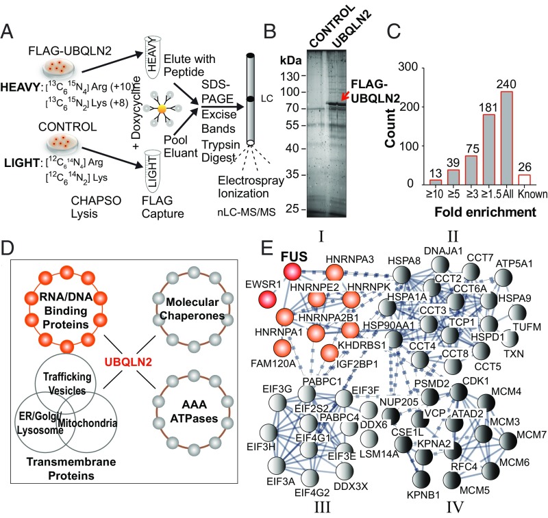
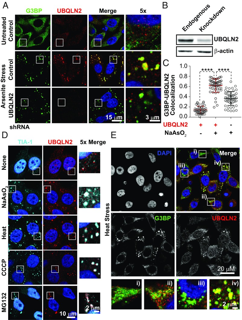
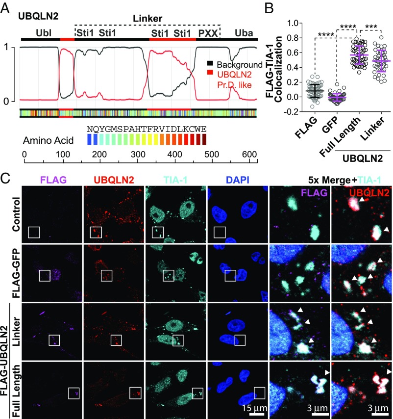
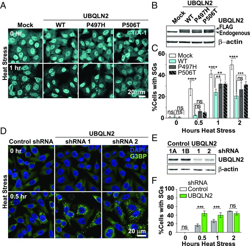
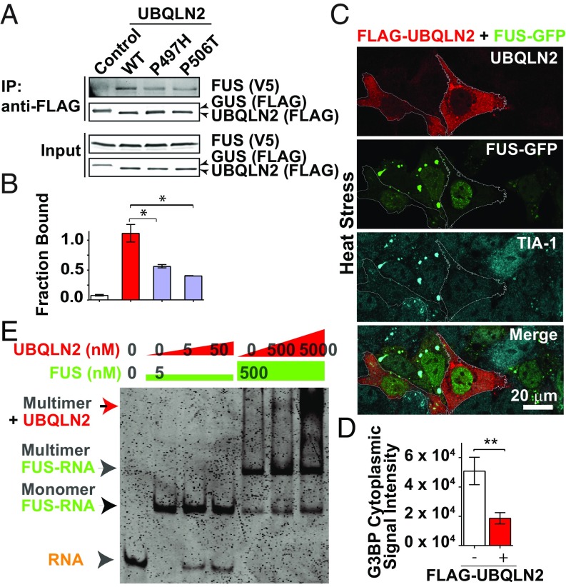
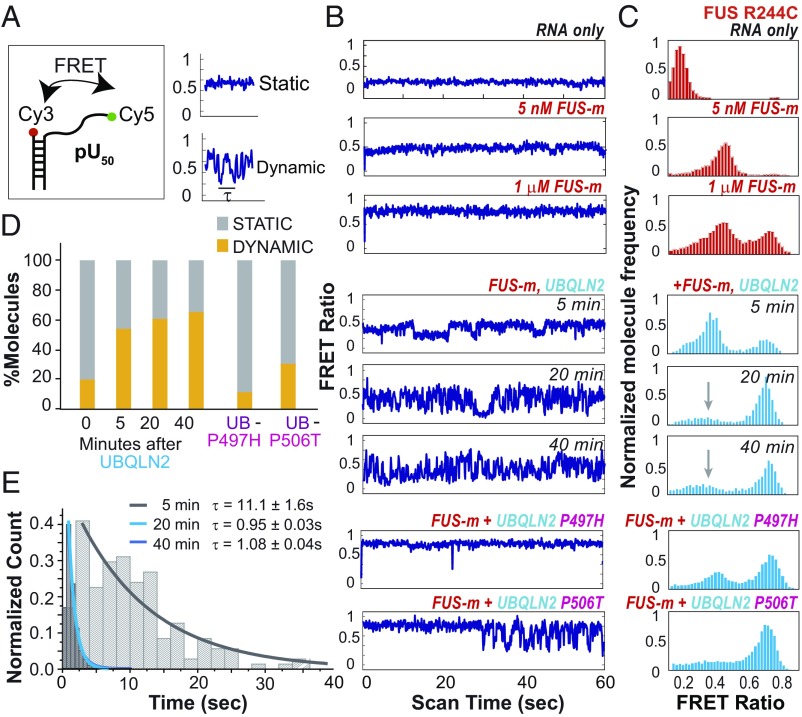
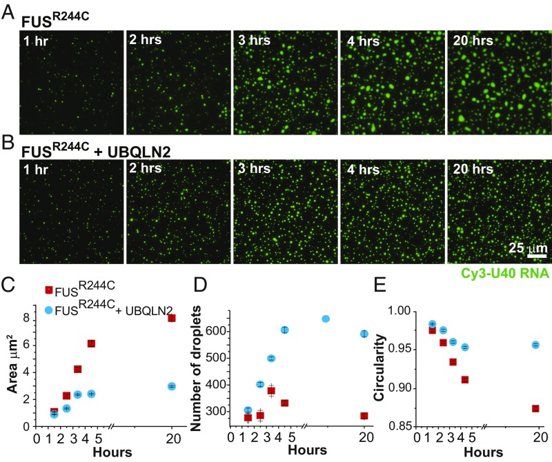
The ubiquitin-like protein ubiquilin 2 (UBQLN2) has been genetically and pathologically linked to the neurodegenerative diseases amyotrophic lateral sclerosis (ALS) and frontotemporal dementia (FTD), but its normal cellular functions are not well understood. In a search for UBQLN2-interacting proteins, we found an enrichment of stress granule (SG) components, including ALS/FTD-linked heterogeneous ribonucleoprotein fused in sarcoma (FUS). Through the use of an optimized SG detection method, we observed UBQLN2 and its interactors at SGs. A low complexity, Sti1-like repeat region in UBQLN2 was sufficient for its localization to SGs. Functionally, UBQLN2 negatively regulated SG formation. UBQLN2 increased the dynamics of FUS-RNA interaction and promoted the fluidity of FUS-RNA complexes at a single-molecule level. This solubilizing effect corresponded to a dispersal of FUS liquid droplets in vitro and a suppression of FUS SG formation in cells. ALS-linked mutations in UBQLN2 reduced its association with FUS and impaired its function in regulating FUS-RNA complex dynamics and SG formation. These results reveal a previously unrecognized role for UBQLN2 in regulating the early stages of liquid-liquid phase separation by directly modulating the fluidity of protein-RNA complexes and the dynamics of SG formation.

Keywords: ALS; FTD; FUS; stress granule; ubiquilin 2.


Conflict of interest statement

The authors declare no conflict of interest.

Figures

Fig. 1.
Fig. 1.
SG components immunoprecipitate with UBQLN2. (A) Schematic of coimmunoprecipitation and SILAC nano-LC (nLC)-MS/MS analysis. Heavy-isotope-labeled stable HEK293T cell lines that inducibly express amino-terminal FLAG-UBQLN2 were treated with doxycycline and lysed in buffer containing the detergent CHAPSO. Light-isotope-labeled HEK293T cells treated with doxycycline were used as a control. Lysates were incubated with FLAG (M2) magnetic beads and eluted with FLAG peptide. Eluants were pooled at a 1:1 ratio and separated by SDS/PAGE. Bands were cut out, and proteins were digested with trypsin. Peptides were then extracted, separated via nLC, and injected via electrospray ionization into an LTQ Orbitrap Elite mass spectrometer for analysis. (B) Representative silver-stained gel of UBQLN2 CHAPSO immunoprecipitation. The red arrow points to exogenously expressed FLAG-UBQLN2 protein. (C) Cumulative frequency distribution of SILAC heavy (UBQLN2)/light (control) ratios from LC-MS/MS analysis of proteins that coimmunoprecipitated with FLAG-UBQLN2. A total of 240 putative interactors were identified, many of which cluster into SG component complexes shown in E. (D) Classes of UBQLN2 interactors grouped by domain structure. (E) STRING network of UBQLN2 interactors found in the G3BP-dependent SG proteome (56). Dotted and solid lines represent lower confidence and higher confidence connections, respectively. Members represented in the four clusters include hnRNPs (I), molecular chaperones (II), translation factors (III), and RNA trafficking proteins (IV). We focused further work on the class I hnRNP FUS.
Fig. 2.
Fig. 2.
UBQLN2 associates with SGs. (A) Immunofluorescence (IF) images of endogenous UBQLN2 at SGs in response to sodium arsenite stress (30 min, 0.5 mM NaAsO2) in HeLa cells. G3BP marks SGs. Regions of UBQLN2-G3BP overlap appear yellow. Specific depletion of UBQLN2 eliminates the UBQLN2 signal and regions of overlap. Control shRNA is a scrambled nontargeting shRNA. (B) Western blot showing average endogenous UBQLN2 protein depletion 96 h after shRNA transfection. β-Actin is a loading control. (C) Scatter plot showing the Pearson’s R coefficient of overlap between the G3BP (green) and UBQLN2 (red) signals shown in A for 50 individual cells (○) chosen at random. Error bars are SD. ****P < 0.0001 by Dunnett’s multiple comparison test done with one-way ANOVA (P < 0.0001). UBQLN2 (−) represents the partial shRNA depletion of UBQLN2 shown in B. UBQLN2 appears to colocalize with SGs. (D) IF images of UBQLN2 localization under different stress conditions. HeLa cells were fixed and permeabilized simultaneously as shown in A. Stress conditions include: 0.5 h, 0.5 mM NaAsO2; 1 h, 43.7 °C heat stress; 1.5 h, 1 μM CCCP in glucose-free media; 1 h, 10 μM MG132. (E) IF images showing localization of UBQLN2 to SGs with differing morphologies and localization after 30 min of heat stress at 43.7 °C. G3BP SG distribution is diffuse (i), concentrated but not punctate (ii), perinuclear punctate (iii), or large cytoplasmic punctate (iv).
Fig. 3.
Fig. 3.
Sti1-like linker alone is sufficient for UBQLN2 SG localization. (A) PrD analysis of UBQLN2. The dotted line highlights the bounds of the linker region tested in B. Identified PrD 1 is amino acids 105–143. PrD 2 is amino acids 338–460 of human UBQLN2. (B) Scatter plots showing the Pearson’s R coefficient of overlap between FLAG-tagged proteins (magenta) with the SG marker TIA-1 (cyan) for 50 individual cells (○) chosen at random. FLAG-tagged proteins were expressed from an integrated short flippase recognition target (FRT) site in HeLa Flp-In TRex cells and exposed to 60 min of heat stress at 43.7 °C. Error bars are 1 SD from the mean. ****P < 0.0001, ***P = 0.0006; Dunnett’s multiple comparison test. (C) Representative immunofluorescence images of HeLa Flp-In TRex cells expressing FLAG-tagged proteins as quantitated in B. The linker region alone drives UBQLN2 into SGs.
Fig. 4.
Fig. 4.
UBQLN2 levels negatively regulate SG assembly. (A) Immunofluorescence (IF) maximum intensity projections (MIPs) of SGs in mock, FLAG-UBQLN2 wild-type (WT), FLAG-UBQLN2 P497H, and FLAG-UBQLN2 P506T transfected HeLa cells fixed and stained before and after heat stress. TIA-1 was used as an SG marker. (B) Western blot showing the level of UBQLN2 WT and mutant overexpression. β-Actin is used as a loading control. (C) Quantitation of percentage of cells with SGs larger than 1 μm shown in A. ****P < 0.0001, ***P < 0.001, **P < 0.01, *P < 0.05; Sidak’s multiple comparison test. ns, not significant. (D) IF MIPs of SGs in control and UBQLN2-specific shRNA-treated HeLa cells before and after heat stress. G3BP was used as an SG marker, with DAPI marking the nuclei of individual cells. (E) Western blot showing the level of UBQLN2 depletion by the two UBQLN2-specific shRNAs. Approximately double the number of UBQLN2 shRNA-depleted cells show large cytoplasmic SGs after 30 min of heat stress. (F) Quantitation of percentage of cells with SG larger than 1 μm shown in D. ***P < 0.001, Sidak’s multiple comparison test. ns, not significant. A representative dataset is shown. More than 400 cells from four fields of view were imaged and averaged at each time point. Error bars are SD. The experiments were repeated using both G3BP and TIA-1 markers. UBQLN2 appears to negatively regulate SG formation in all cases.
Fig. 5.
Fig. 5.
UBQLN2 forms a complex with FUS and suppresses its SG formation. (A) FUS-V5 immunoprecipitates (IP) with FLAG-UBQLN2 in HEK293T cells. UBQLN2 ALS-linked missense mutations P497H and P506T partially disrupt UBQLN2’s interaction with FUS. The FLAG-tagged plant reporter protein β-glucuronidase (GUS) was used as a control. (B) Quantitation of Western blot shown in A. The experiment was repeated twice, and the average results are presented here. Error bars are SD. *P < 0.01, Dunnett’s multiple comparison test. (C) Immunofluorescence images of fixed HeLa cells cotransfected with FLAG-UBQLN2 and FUS-GFP. Cells overexpressing FLAG-UBQLN2 are outlined. TIA-1 marks SGs. (D) Quantitation of G3BP cytoplasmic signal in 13 pairs of cells expressing just FUS-GFP or FUS-GFP in the presence of FLAG-UBQLN2. **P < 0.01, two-tailed student’s t test. UBQLN2 overexpression suppresses FUS-GFP SG formation in response to heat stress (30 min at 43.7 °C). (E) Coomassie blue-stained native PAGE gel of FUS-RNA and FUS-RNA-UBQLN2 by EMSA. The RNA probe is Cy3-labeled pU40. Five nanomolar FUS results in a monomer FUS–RNA complex, whereas 500 nM FUS results in a shifted mobility multimer FUS–RNA complex. Addition of UBQLN2 to this preformed complex supershifts the multimer, but not the monomer, FUS–RNA complex.
Fig. 6.
Fig. 6.
UBQLN2 increases FUSR244C–RNA interaction dynamics. (A) pU50 probe design and sample static and dynamic single-molecule traces showing the dwell time constant (τ). (B) Representative traces showing the fluctuation of the FRET ratio for single molecules over time (panels 1–8). FUS-m, FUS mutant FUSR244C. (C) Histograms of smFRET ratios for FUS-m mixed with pU50 RNA (red; panels 1–3), FUS-m mixed with pU50 in the presence of wild-type UBQLN2 (cyan; panels 4–6), and mutant UBQLN2 P497H and P506T (cyan; panels 7 and 8). The gray arrows point to the FRET peak broadened and flattened by UBQLN2 addition. (D) Percentage of single molecules with dynamic vs. static smFRET ratios. More than 1,000 traces were surveyed for this analysis. UBQLN2 mutant traces were collected between 20 and 40 min. Wild-type UBQLN2 addition alters FUSR244C–RNA complex dynamics, while mutant UBQLN2 does so to a lesser extent. (E) τ of FRET fluctuation taken at 5, 20, and 40 min after addition of wild-type UBQLN2 to FUSR244C. At 5–20 min after UBQLN2 addition, the FRET fluctuation rate dramatically increases for single molecules that are dynamic.
Fig. 7.
Fig. 7.
UBQLN2 suppresses mutant FUS recruitment into phase-separated droplets. (A) Phase-separated droplets of FUSR244C mutants formed over 20 h. (B) Droplets of FUSR244C with UBQLN2 formed over 20 h. (C) Area of droplets taken over 20 h. Red and light blue indicate FUSR244C without and with UBQLN2 added, respectively. UBQLN2 addition leads to an increase in average liquid droplet area. (D) Number of droplets per imaging area. UBQLN2 addition leads to a decrease in liquid droplet number. (E) Circularity of droplets over 20 h. UBQLN2 addition leads to maintenance of liquid droplet circularity. More than 400 droplets in three to four fields of view were used for this analysis. All error bars shown are SEM. The experiment was repeated twice.

References

    1. Ling SC, Polymenidou M, Cleveland DW. Converging mechanisms in ALS and FTD: Disrupted RNA and protein homeostasis. Neuron. 2013;79:416–438. - PMC - PubMed
    1. Ratnavalli E, Brayne C, Dawson K, Hodges JR. The prevalence of frontotemporal dementia. Neurology. 2002;58:1615–1621. - PubMed
    1. van den Berg-Vos RM, et al. Sporadic lower motor neuron disease with adult onset: Classification of subtypes. Brain. 2003;126:1036–1047. - PubMed
    1. Van Langenhove T, van der Zee J, Van Broeckhoven C. The molecular basis of the frontotemporal lobar degeneration-amyotrophic lateral sclerosis spectrum. Ann Med. 2012;44:817–828. - PMC - PubMed
    1. Mackenzie IR, Rademakers R, Neumann M. TDP-43 and FUS in amyotrophic lateral sclerosis and frontotemporal dementia. Lancet Neurol. 2010;9:995–1007. - PubMed

Publication types