. 2018 Dec 4;115(49):E11532-E11541.
doi: 10.1073/pnas.1800886115. Epub 2018 Nov 15.

Genetic deletion of vesicular glutamate transporter in dopamine neurons increases vulnerability to MPTP-induced neurotoxicity in mice

Affiliations

Genetic deletion of vesicular glutamate transporter in dopamine neurons increases vulnerability to MPTP-induced neurotoxicity in mice

Hui Shen et al. Proc Natl Acad Sci U S A. .

Erratum in

Abstract

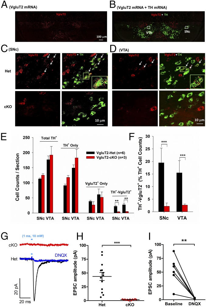
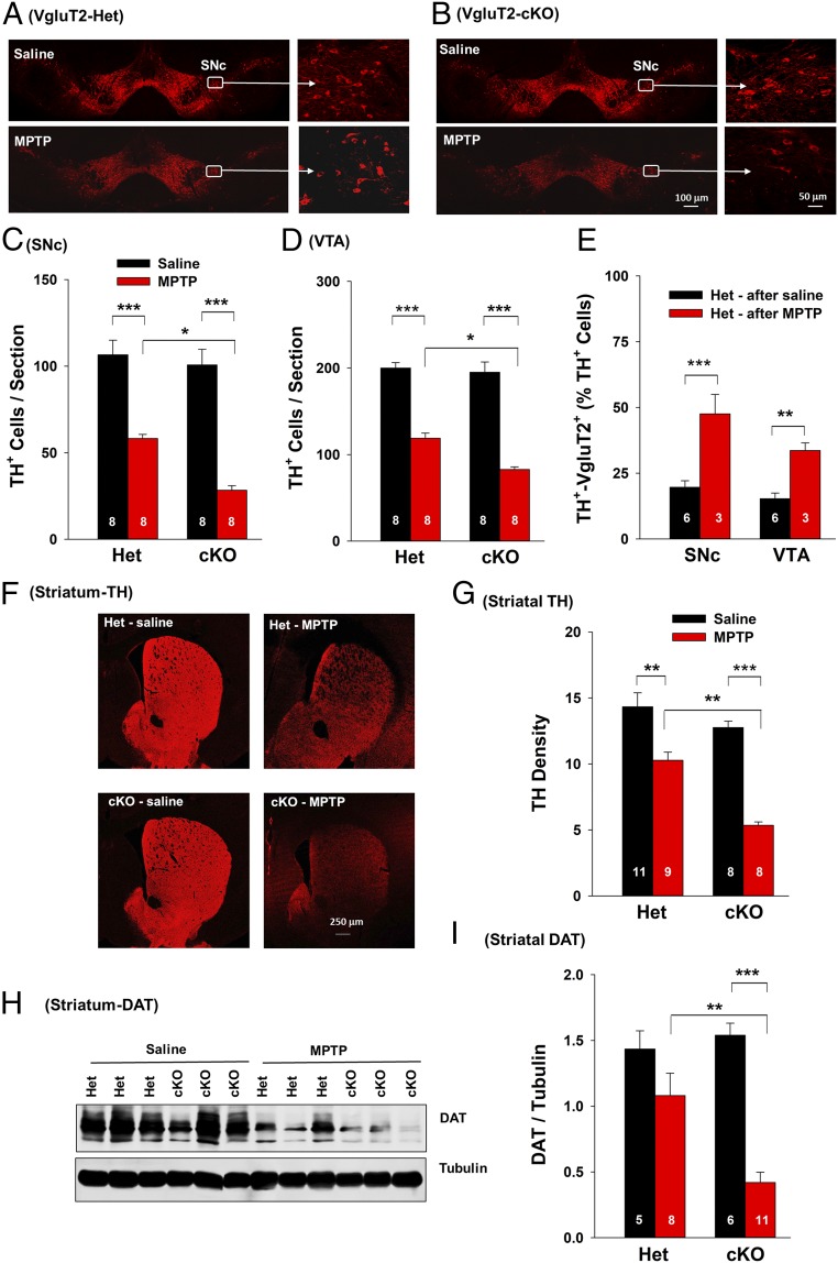
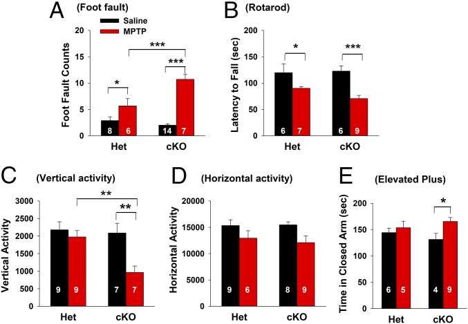
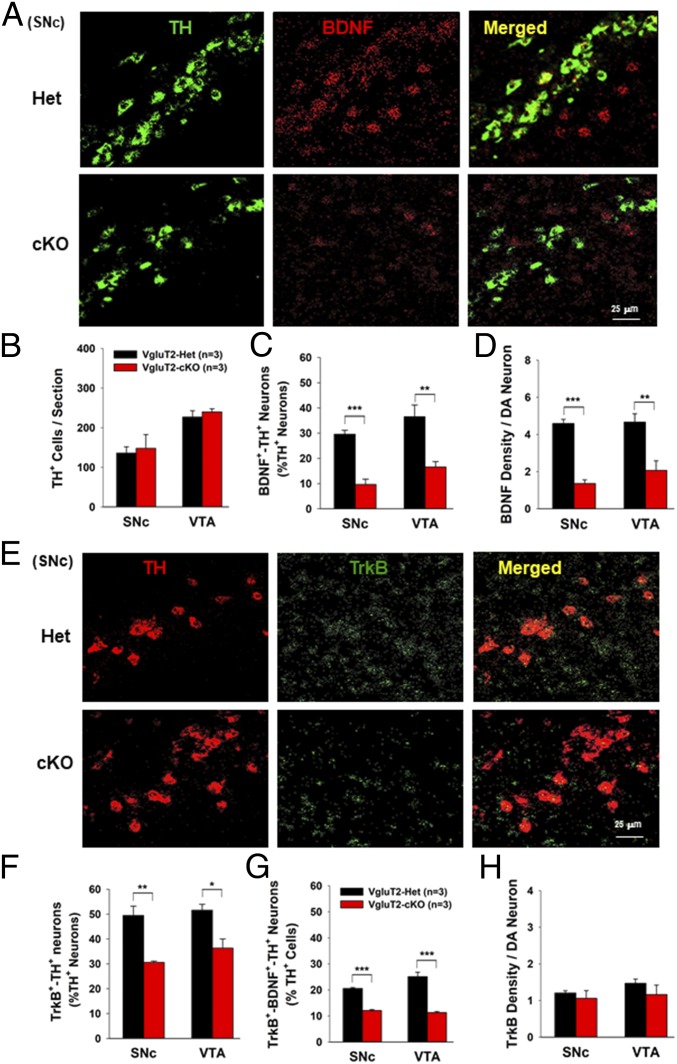
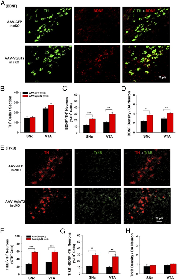
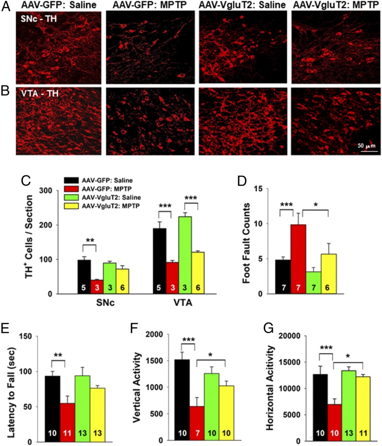
A subset of midbrain dopamine (DA) neurons express vesicular glutamate transporter 2 (VgluT2), which facilitates synaptic vesicle loading of glutamate. Recent studies indicate that such expression can modulate DA-dependent reward behaviors, but little is known about functional consequences of DA neuron VgluT2 expression in neurodegenerative diseases like Parkinson's disease (PD). Here, we report that selective deletion of VgluT2 in DA neurons in conditional VgluT2-KO (VgluT2-cKO) mice abolished glutamate release from DA neurons, reduced their expression of brain-derived neurotrophic factor (BDNF) and tyrosine receptor kinase B (TrkB), and exacerbated the pathological effects of exposure to the neurotoxin 1-methyl-4-phenyl-1,2,3,6-tetrahydropyridine (MPTP). Furthermore, viral rescue of VgluT2 expression in DA neurons of VglutT2-cKO mice restored BDNF/TrkB expression and attenuated MPTP-induced DA neuron loss and locomotor impairment. Together, these findings indicate that VgluT2 expression in DA neurons is neuroprotective. Genetic or environmental factors causing reduced expression or function of VgluT2 in DA neurons may place some individuals at increased risk for DA neuron degeneration. Therefore, maintaining physiological expression and function of VgluT2 in DA neurons may represent a valid molecular target for the development of preventive therapeutic interventions for PD.

Keywords: BDNF; MPTP; Parkinson’s disease; VgluT2; midbrain DA neurons.


Conflict of interest statement

The authors declare no conflict of interest.

Figures

Fig. 1.
Fig. 1.
Identification of VgluT2 expression in midbrain DA neurons in VgluT2-Het (Het) and VgluT2-cKO (cKO) mice. (A and B) Representative VgluT2-mRNA (red) and TH-mRNA (green) staining in a midbrain section under low magnification (10×) illustrates the two major areas of interest in this study, the VTA and SNc. (C and D) Representative confocal images of VgluT2 mRNA and TH mRNA staining in the SNc and VTA in Het and cKO mice. Insets (Lower Right) in images from the Het control mice are the magnified regions in the green boxes illustrating VgluT2 and TH colocalization in the same neurons. No VgluT2 mRNA was detected in TH+ neurons in cKO mice. (E) Mean cell counts per section of total TH+, TH+-only, VgluT2+-only, and TH+-VgluT2+ neurons in Het and cKO mice. There is a significant reduction in TH+-VgluT2+ cell counts between the two phenotypes of mice (Right; SNc, t = 5.18, P < 0.01; VTA, t = 3.89, P < 0.01). (F) cKO mice display a significant reduction in percentage of TH+-VgluT2+ neurons among total TH+ neurons (SNc, t = 4.42, P < 0.001; VTA, t = 3.77, P < 0.001). (G) Electrophysiological recordings of EPSCs in striatal MSNs in brain slices illustrate optogenetic activation (1 ms of 473-nm, 10-mW laser) of DA neuron evoked EPSCs in Het but not in cKO mice. (H) Mean EPSC amplitudes in Het mice (n = 13 cells, three mice) and cKO mice (n = 12 cells, three mice). (I) Coadministration of DNQX, a selective AMPA receptor antagonist, blocked optical stimulation-evoked EPSCs in Het control mice [also see G; n = 6 cells, three mice; **P < 0.01 and ***P < 0.001 vs. Het control mice (E, F, and I) or baseline (H)]. Data indicate mean ± SEM (SI Appendix, Figs. S1 and S2).
Fig. 2.
Fig. 2.
MPTP-induced toxicity in DA neurons in Het and cKO mice. (A and B) Representative TH immunostaining images in Het and cKO mice 14 d after MPTP or saline solution treatment. (C) The mean TH+ DA cell counts per section in the SNc (six brain sections per animal, eight animals per genotype). Two-way ANOVA revealed MPTP treatment main effect (F1,28 = 127.81, P < 0.001), genotype main effect (F1,28 = 14.58, P < 0.001), and treatment × genotype interaction (F1,28 = 4.68, P < 0.05). (D) The mean TH+ DA cell counts per section in the VTA. Two-way ANOVA revealed MPTP treatment main effect (F1,28 = 199.6, P < 0.001), genotype main effect (F1,28 = 7.86, P < 0.01), and treatment × genotype interaction (F1,28 = 6.13, P < 0.05). (E) VgluT2+-TH+ neuron counts illustrating that more DA neurons expressed VgluT2 in the remaining surviving DA neurons in Het mice after MPTP administration than in Het mice after saline solution treatment. (F) Representative TH immunostaining images in the striatum. (G) Mean TH immunostaining density. Two-way ANOVA revealed MPTP treatment main effect (F1,32 = 55.09, P < 0.001), genotype main effect (F1,32 = 17.68, P < 0.001), and treatment × genotype interaction (F1,32 = 4.74, P < 0.05). (H) Original Western blot results. (I) Mean density of DAT band in the striatum. Two-way ANOVA revealed MPTP treatment main effect (F1,28 = 18.49, P < 0.001), genotype main effect (F1,28 = 0.46, P > 0.05), and treatment × genotype interaction (F1,28 = 8.16, P < 0.01) (*P < 0.05, **P < 0.01, and ***P < 0.001 vs. saline solution group or Het control mice; SI Appendix, Figs. S3 and S7).
Fig. 3.
Fig. 3.
MPTP-induced locomotor impairments in Het and cKO mice. (A) Parallel rod floor (foot fault) test results: MPTP produced a significant increase in foot fault counts in the cKO mice compared with the Het control mice (two-way ANOVA, MPTP treatment, F1,31 = 62.57, P < 0.001; genotype, F1,31 = 8.23, P < 0.01; treatment × phenotype interaction, F1,31 = 16.57, P < 0.001). (B) Rotarod test results: MPTP produced significant impairment in rotarod locomotor performance in both phenotypes of mice (F1,24 = 19.41, P < 0.001) as assessed by decreased latency (in seconds) to fall down from elevated rotarod. (C and D) Open-field locomotion data: MPTP produced a significant reduction in vertical and horizontal activity in the cKO mice, but not in the Het mice. Two-way ANOVA revealed significant treatment main effect (C, F1,28 = 8.94, P < 0.01; D, F1,28 = 6.82, P < 0.05), genotype main effect (C, F1,28 = 6.12, P < 0.05; D, F1,28 = 0.08, P > 0.05), and treatment × genotype interaction (C, F1,28 = 4.31, P < 0.05; D, F1,28 = 0.235, P > 0.05). (E) Elevated plus-maze test: MPTP significantly decreased risk-exploring behavior in the open arms in the cKO mice (treatment, F1,20 = 4.76, P < 0.05; genotype, F1,20 = 0.1, P > 0.05; treatment × phenotype interaction, F1,20 = 1.57, P > 0.05) as assessed by increased time spent in the closed arms (*P < 0.05, **P < 0.01, and ***P < 0.001 vs. saline solution group or Het control mice; SI Appendix, Fig. S4).
Fig. 4.
Fig. 4.
Selective deletion of VgluT2 in DA neurons decreases BDNF/TrkB mRNA expression in DA neurons. (A) Representative confocal images in the SNc show a significant reduction in BDNF mRNA expression in cKO mice. (B) Mean TH+ cell counts per section in the SNc and VTA. (C) The mean BDNF+-TH+ cell counts (percentage of total TH+ cells) illustrate a ∼50% reduction in cKO mice compared with Het control mice (SNc, t = 9.05, P < 0.001; VTA, t = 3.9, P < 0.01). (D) BDNF density per DA neuron illustrates a significant reduction in BDNF density in DA neurons in the cKO mice (SNc, t = 11.00, P < 0.001; VTA, t = 3.80, P < 0.01). (E) Representative confocal images of TrkB mRNA staining in the SNc. (F) Mean TrkB+-TH+ cell counts (percentage of total TH+ DA neurons) in the SNc and VTA show a significant decrease in TrkB+-TH+ cells in cKO mice (SNc, t = 5.0, P < 0.01; VTA, t = 3.48, P < 0.05). (G) The mean TrkB+-BDNF+-TH+ cell counts (percentage of total TH+ cells) show a significant reduction in cKO mice compared with Het control mice (SNc, t = 16.93, P < 0.001; VTA, t = 7.84, P < 0.001). (H) TrkB density per DA neuron shows a nonsignificant difference between the two groups of mice (SNc, t = 0.66, P > 0.05; VTA, t = 027, P > 0.05; *P < 0.05, **P < 0.01, and ***P < 0.001 vs. Het control mice; SI Appendix, Figs. S5–S7).
Fig. 5.
Fig. 5.
Viral rescue of VgluT2 expression in VgluT2-cKO mice restored BDNF/TrkB expression in DA neurons. (A) Representative confocal images in the SNc illustrates BDNF expression in VgluT2-overexpressing mice. (B) Quantitative data illustrate that VgluT2 overexpression did not alter total numbers of TH+ DA neurons. (C) VgluT2 overexpression significantly increased the cell counts of BDNF+-TH+ neurons (SNc, t = 7.54, P < 0.001; VTA, t = 3.96, P < 0.01). (D) VgluT2 overexpression significantly increased BDNF density per DA neuron (SNc, t = 3.57, P < 0.05; VTA, t = 4.32, P < 0.01). (E) Representative confocal images of TrkB mRNA staining in the SNc. (F) Quantitative data illustrate that the VgluT2 overexpression significantly increased TrkB expression in TH+ DA neurons (SNc, t = 14.8, P < 0.001; VTA, t = 9.53, P < 0.001). (G) VgluT2 overexpression also significantly increased the cell counts of TrkB+-BDNF+-TH+ neurons (SNc, t = 5.4, P < 0.01; VTA, t = 4.65, P < 0.01). (H) VgluT2 overexpression did not alter TrkB density in DA neurons (SNc, t = 0.33, P > 0.05; VTA, t = 0.44, P > 0.05; *P < 0.05, **P < 0.01, and ***P < 0.001 vs. AAV-GFP control mice).
Fig. 6.
Fig. 6.
Viral rescue of VgluT2 expression in DA neurons in VgluT2-cKO mice attenuated MPTP-induced DA neuron toxicity and locomotor impairment. (A and B) TH immunostaining in the SNc and VTA. (C) Mean TH+ cell counts per section. MPTP produced more DA cell loss in the AAV-GFP control mice than in the VgluT2-rescued mice (SNc, F3,13 = 6.43, P < 0.01; VTA, F3,13 = 17.88, P < 0.001, one-way ANOVA). (D and E) MPTP produced a significant increase in foot fault counts (D, one-way ANOVA, F3,23 = 10.73, P < 0.001) and a significant decrease in rotarod performance (E, F3,43 = 4.74, P < 0.01) in the AAV-GFP control mice but not in VgluT2-overexpressing mice. (F and G) Viral rescue of VgluT2 expression attenuated MPTP-induced reduction in vertical (F, F3,33 = 8.41, P < 0.001) and horizontal (G, F3,40 = 8.87, P < 0.001) locomotor activity (*P < 0.05, **P < 0.01, and ***P < 0.001 between groups labeled by horizontal bars; SI Appendix, Fig. S8)

References

    1. Sgambato-Faure V, Tremblay L. Dopamine and serotonin modulation of motor and non-motor functions of the non-human primate striato-pallidal circuits in normal and pathological states. J Neural Transm (Vienna) 2018;125:485–500. - PubMed
    1. Palmiter RD. Dopamine signaling in the dorsal striatum is essential for motivated behaviors: Lessons from dopamine-deficient mice. Ann N Y Acad Sci. 2008;1129:35–46. - PMC - PubMed
    1. Damier P, Hirsch EC, Agid Y, Graybiel AM. The substantia nigra of the human brain. II. Patterns of loss of dopamine-containing neurons in Parkinson’s disease. Brain. 1999;122:1437–1448. - PubMed
    1. Jabre M, Nohra G, Damier P, Bejjani BP. Does dopamine still have a leading role in advanced Parkinson’s disease after subthalamic stimulation? Stereotact Funct Neurosurg. 2008;86:184–186. - PubMed
    1. Chaudhuri KR, Schapira AH. Non-motor symptoms of Parkinson’s disease: Dopaminergic pathophysiology and treatment. Lancet Neurol. 2009;8:464–474. - PubMed

Publication types

MeSH terms

LinkOut - more resources