. 2018 Nov 27;115(48):E11359-E11368.
doi: 10.1073/pnas.1807123115. Epub 2018 Nov 15.

Permanent neuroglial remodeling of the retina following infiltration of CSF1R inhibition-resistant peripheral monocytes

Affiliations

Permanent neuroglial remodeling of the retina following infiltration of CSF1R inhibition-resistant peripheral monocytes

Eleftherios I Paschalis et al. Proc Natl Acad Sci U S A. .

Abstract

Previous studies have demonstrated that ocular injury can lead to prompt infiltration of bone-marrow-derived peripheral monocytes into the retina. However, the ability of these cells to integrate into the tissue and become microglia has not been investigated. Here we show that such peripheral monocytes that infiltrate into the retina after ocular injury engraft permanently, migrate to the three distinct microglia strata, and adopt a microglia-like morphology. In the absence of ocular injury, peripheral monocytes that repopulate the retina after depletion with colony-stimulating factor 1 receptor (CSF1R) inhibitor remain sensitive to CSF1R inhibition and can be redepleted. Strikingly, consequent to ocular injury, the engrafted peripheral monocytes are resistant to depletion by CSF1R inhibitor and likely express low CSF1R. Moreover, these engrafted monocytes remain proinflammatory, expressing high levels of MHC-II, IL-1β, and TNF-α over the long term. The observed permanent neuroglia remodeling after injury constitutes a major immunological change that may contribute to progressive retinal degeneration. These findings may also be relevant to other degenerative conditions of the retina and the central nervous system.

Keywords: PLX5622; glaucoma; macrophage; neurodegeneration; neuroprotection.


Conflict of interest statement

The authors declare no conflict of interest.

Figures
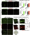
Fig. 1.
Fig. 1.
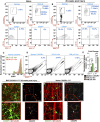
Ocular injury causes permanent engraftment in the retina of peripheral CX3CR1+ cells which adopt microglia morphology. (AC and F–H) Confocal microscopy of busulfan-myelodepleted and bone-marrow–transferred mice with CX3CR1+/EGFP::CCR2+/RFP cells 16 mo after corneal alkali burn injury. Maximum projection of confocal images shows that CX3CR1+ cells cannot enter into the retina of the contralateral noninjured eye 16 mo after injury (A), nor can they populate the retina of naive eyes under physiological conditions (F). Few cells are present around the optic nerve head (red arrow) and along the major retinal vessels (white arrow). Maximum projection of confocal images shows CCR2+ cells are scattered across the retina of the contralateral noninjured (B) and of the naive (G) eye, located mainly in the GCL. C and H show overlay images of CX3CR1 and CCR2 channels. (D, E, I, and J) Quantification of the number of peripheral CX3CR1+ and CCR2+ cells 16 mo after ocular injury shows a marked increase in CX3CR1+ cells (P < 0.003) but normal numbers of CCR2+ cells (P > 0.415) compared with contralateral noninjured and naive eyes. Data in D and E are presented as mean ± SD, paired t test. Data in I and J are presented as mean ± SD, independent t test. **P < 0.01; ****P < 0.0001. (K and L) Ocular injury causes infiltration and permanent engraftment of peripheral CX3CR1+ cells in the retina. (MO) Sixteen months after the injury, infiltrated CX3CR1+ cells migrate into the three distinct microglia strata (GCL, IPL/INL, OPL) and adopt dendritiform microglia morphology. (M) Isometric reconstruction of confocal stacks separated by retinal layers (GCL, IPL/INL, and OPL). (N) A cross-section of the retina shows the presence of CX3CR1+ cells in the GCL, IPL/INL, and OPL. (O) A histogram of CX3CR1+/EGFP expression within the retinal tissue shows intensity peaks of EGFP signal in the GCL, IPL/INL, and OPL. (P and Q) Patrolling CCR2+ cells are present in the retina but do not differentiate into CX3CR1+ cells. (RT) Even though the number of peripheral CCR2+ cells is normal, peripheral CCR2+ cells are present in all three distinct microglia strata (GCL, IPL/INL, and OPL) 16 mo after the injury. In O and T the thickness of the retinal tissue and the relative position of each microglia stratum was derived automatically by the confocal system (Leica SP8) and was calculated by multiplying the number of serial confocal scans by the step of each scan in micrometers. (U and V) Overlay images of CX3CR1 and CCR2 channels 16 mo after ocular injury. (W) A representative schematic of a flat-mount retina as used for confocal imaging. The center white circle represents the optic nerve head, red lines represent the retinal vessels, and green dots represent the peripheral monocytes. (Scale bars: 50 μm in AC, FH, K, L, P, Q, U, and V.) n ≥ 5 mice per group.
Fig. 2.
Fig. 2.
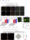
Engrafted peripheral CX3CR1+ cells remain reactive despite their morphometric quiescence. (AD) Flow cytometric analysis of retinal cells from CX3CR1+/EGFP mice. (A) In naive mice, CX3CR1+ cells are predominantly CD45lo (96%), and only a small percentage is CD45hi (4%). CX3CR1+ CD45lo cells do not express MHC-II; however, 2.8% of CX3CR1+ CD45hi cells have baseline MHC-II expression. (B) Twenty weeks after ocular injury, 94% percent of the CX3CR1+ cells are CD45lo, 20% of which express MHC-II. Likewise, 3% of the CX3CR1+ cells are CD45hi, 72% of which express MHC-II. (C) Flow cytometry for the differentiation of yolk-sac–derived retinal microglia (YS-μGlia 16w) from peripherally engrafted bone-marrow CX3CR1+ monocytes (MB μG 16w) 16 wk after ocular injury using RFP-conjugated CX3CR1 antibody against bone-marrow–transferred CX3CR1+/EGFP retinal cells. Both cell populations are strongly expressed. (D) Flow cytometry and cell sorting of splenic CX3CR1+ cells (control), retinal yolk-sac–derived microglia, and retinal peripherally engrafted monocytes 16 wk after ocular injury for subsequent analysis of IL-1β and TNF-α expression with qPCR. (E) Compared with splenic CX3CR1+ cells, both yolk-sac–derived and peripheral microglia had elevated TNF-α and IL-1β gene expression. However, peripherally engrafted monocytes had significantly higher expression than yolk-sac–derived microglia 16 wk after injury. *P < 0.05; ****P < 0.0001; Tukey’s method for multiple comparisons. (F and G) Twenty weeks after the injury, peripheral CX3CR1+ monocytes appear to frequently interact with and engulf β3 tubulin+ neuroretinal tissue (F), despite their otherwise ramified and quiescent morphology (G). (H and I) In contrast, in naive eyes, although ramified CX3CR1+ monocytes come into contact with β3 tubulin+ neuronal tissue, they do not internalize it. (Scale bars: FI, Upper, 50 μm; FI, Lower, 20 μm.) All experiments were performed in triplicate.
Fig. 3.
Fig. 3.
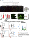
Microglia repopulation after PLX5622 treatment occurs primarily by peripheral CX3CR1+ cells. (A) Schematic of the experiment: Busulfan-myelodepleted C57BL/6 mice received adoptive transfer of CX3CR1+/EGFP bone-marrow cells. Four weeks later they received PLX5622 treatment for 3 wk followed by confocal microscopy 3 wk after the cessation of PLX5622 treatment. (BF) Central color-depth–coded (0 represents innermost retina) confocal images of flat-mount retinas 0, 4, 6 wk, and 3 mo after cessation of PLX5622 treatment (BE) and of naive CX3CR1+/EGFP mice (F). (GK) Midperipheral color-depth–coded confocal images of flat-mount retinas 0, 4, 6 wk, and 3 mo after cessation of PLX5622 treatment (GJ) and of naive CX3CR1+/EGFP mice (K). (Scale bars: 100 μm.) (L) Quantification of CX3CR1+ cell number at the indicated times post PLX5622 treatment. (M) Quantification of EGFP intensity at the indicated times post PLX5622 treatment. **P < 0.01, ****P < 0.0001; Tukey’s multiple-comparisons method. PLX5622 treatment for 3 wk does not cause acute peripheral CX3CR1+ cell infiltration into the retina (B, F, G, L, and M). However, 6 wk after cessation of PLX5622 treatment peripheral CX3CR1+ cells appeared to occupy the majority of the retina area (D, I, L, and M), and at 3 mo, the retina is completely repopulated by peripheral CX3CR1+ cells (E, J, L, and M). The number of repopulated peripheral CX3CR1+ cells at 3 mo after the end of PLX5622 treatment approximates that of naive CX3CR1+/EGFP mice with no PLX5622 treatment (F, K, L, and M). (N) Repopulating peripheral CX3CR1+ cells migrated into all three distinct microglia strata (GCL, IPL/INL, and OPL) 3 mo after the injury. (O) A cross-section of the retina shows the presence of CX3CR1+ cells in the GCL, IPL/INL, and OPL. (P) A histogram of CX3CR1+/EGFP expression within the retinal tissue shows intensity peaks of EGFP signal in the GCL, IPL/INL, and OPL. (Q) Schematic showing reapplication of PLX5622 in repopulated peripheral microglia. (R and S) Peripheral monocytes that repopulate the retina remain sensitive to CSF1R inhibition and can be redepleted by reapplication of PLX5622 treatment. (T) A representative schematic of a flat-mount retina as used for confocal imaging. The center white circle represents the optic nerve head, red lines represent the retinal vessels, and green dots represent the peripheral monocytes. (U) Quantification of peripheral CX3CR1+ cells after reapplication of PLX5622 shows complete depletion of repopulated CX3CR1+ cells. n = 5 mice per group.
Fig. 4.
Fig. 4.
Ocular injury leads to population of the retina by peripheral CX3CR1+ cells that are resistant to the CSF1R inhibitor. (A) Schematic of the experiment: Busulfan-myelodepleted, CX3CR1+/EGFP bone-marrow chimeras received ocular injury and 4 wk later received PLX5622 treatment for 3 wk. (B and G) Immediately after cessation of PLX5622 treatment, peripherally derived, retina-repopulating CX3CR1+ cells remained present in the retina (B) and were resistant to CSF1R inhibition, unlike peripherally derived retina-repopulating CX3CR1+ cells in the absence of ocular injury (G). ****P < 0.0001; independent t test. (CE and H) One week (C), 2 wk (D), and 5 mo (E) after cessation of PLX5622 treatment peripherally populating CX3CR1+ cells remained present in the retina (H) with unchanged cell number. Depth color-coding in BE: 0 represents innermost retina. (Scale bars: 100 μm in BE.) (F) A representative schematic of a flat-mount retina as used for confocal imaging. The center white circle represents the optic nerve head, red lines represent the retinal vessels, and green dots represent the peripheral monocytes. (IK) Inhibition-resistant peripheral CX3CR1+ cells that were localized primarily in the GCL/IPL in the early stages after repopulation (see the depth color-code in BE) migrated into all three distinct microglia strata 5 mo after the injury with unusually increased cell density in the OPL. (LP) Ex vivo functional analysis using flow cytometry of CSF1R+ and CSF1R CX3CR1+ retina cells isolated from CX3CR1+/EGFP mice 3 wk after ocular injury or from naive mice. (L and M) Three weeks after the ocular injury, 86.5% of the CX3CR1+ retinal cells were CSF1R+ (CD115+), and 13.2% were CSF1R (CD115). (O and P) In naive eyes, 97.8% of retinal CX3CR1+ cells were CSF1R+ (CD115+), and 1.97% were CSF1Rlo (CD115lo). (N) CSF1R and CSF1R+ cells were cultured for 5 d with or without recombinant CSF1 and IL-4 stimulation. BrdU was used in the culture to assess cell proliferation. CSF1R cells from both naive and injured eyes were not influenced or rescued by CSF1 and IL-4 stimulation, and cells did not survive the 5-d culture. In contrast, CSF1R+ cells were rescued with CSF1 and IL-4 stimulation and showed increased proliferation and BrdU retention. n = 3 mice per group.

Similar articles

Cited by

References

    1. Paschalis EI, et al. The role of microglia and peripheral monocytes in retinal damage after corneal chemical injury. Am J Pathol. 2018;188:1580–1596. - PMC - PubMed
    1. Cade F, Grosskreutz CL, Tauber A, Dohlman CH. Glaucoma in eyes with severe chemical burn, before and after keratoprosthesis. Cornea. 2011;30:1322–1327. - PubMed
    1. Crnej A, et al. Glaucoma progression and role of glaucoma surgery in patients with Boston keratoprosthesis. Cornea. 2014;33:349–354. - PubMed
    1. Kierdorf K, Katzmarski N, Haas CA, Prinz M. Bone marrow cell recruitment to the brain in the absence of irradiation or parabiosis bias. PLoS One. 2013;8:e58544. - PMC - PubMed
    1. Butovsky O, et al. Identification of a unique TGF-β-dependent molecular and functional signature in microglia. Nat Neurosci. 2014;17:131–143. - PMC - PubMed

Publication types

MeSH terms

Substances