

64Cu-SARTATE PET Imaging of Patients with Neuroendocrine Tumors Demonstrates High Tumor Uptake and Retention, Potentially Allowing Prospective Dosimetry for Peptide Receptor Radionuclide Therapy
- PMID: 30442752
- PMCID: PMC6581229
- DOI: 10.2967/jnumed.118.217745
64Cu-SARTATE PET Imaging of Patients with Neuroendocrine Tumors Demonstrates High Tumor Uptake and Retention, Potentially Allowing Prospective Dosimetry for Peptide Receptor Radionuclide Therapy
Abstract
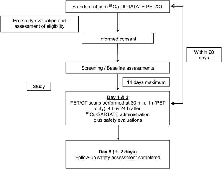
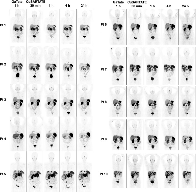
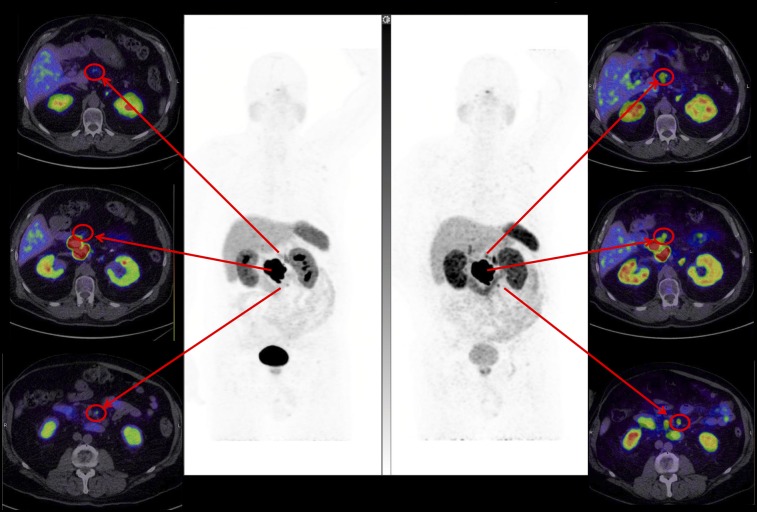
Imaging of somatostatin receptor expression is an established technique for staging of neuroendocrine neoplasia and determining the suitability of patients for peptide receptor radionuclide therapy. PET/CT using 68Ga-labeled somatostatin analogs is superior to earlier agents, but the rapid physical decay of the radionuclide poses logistic and regulatory challenges. 64Cu has attractive physical characteristics for imaging and provides a diagnostic partner for the therapeutic radionuclide 67Cu. Based on promising preclinical studies, we have performed a first-time-in-humans trial of 64Cu-MeCOSar-Tyr3-octreotate (64Cu-SARTATE) to assess its safety and ability to localize disease at early and late imaging time-points. Methods: In a prospective trial, 10 patients with known neuroendocrine neoplasia and positive for uptake on 68Ga-DOTA-octreotate (68Ga-DOTATATE) PET/CT underwent serial PET/CT imaging at 30 min, 1 h, 4 h, and 24 h after injection of 64Cu-SARTATE. Adverse reactions were recorded, and laboratory testing was performed during infusion and at 1 and 7 d after imaging. Images were analyzed for lesion and normal-organ uptake and clearance to assess lesion contrast and perform dosimetry estimates. Results:64Cu-SARTATE was well tolerated during infusion and throughout the study, with 3 patients experiencing mild infusion-related events. High lesion uptake and retention were observed at all imaging time-points. There was progressive hepatic clearance over time, providing the highest lesion-to-liver contrast at 24 h. Image quality remained high at this time. Comparison of 64Cu-SARTATE PET/CT obtained at 4 h to 68Ga-DOTATATE PET/CT obtained at 1 h indicated comparable or superior lesion detection in all patients, especially in the liver. As expected, the highest early physiologic organ uptake was in the kidneys, liver, and spleen. Conclusion:64Cu-SARTATE is safe and has excellent imaging characteristics. High late-retention in tumor and clearance from the liver suggest suitability for diagnostic studies and for prospective dosimetry for 67Cu-SARTATE peptide receptor radionuclide therapy, and the half-life of 64Cu would also facilitate good-manufacturing-practice production and distribution to sites without access to 68Ga.
Keywords: copper-64; neuroendocrine tumor; peptide receptor radionuclide therapy; positron emission tomography; radiopharmaceuticals; theranostics.
© 2019 by the Society of Nuclear Medicine and Molecular Imaging.
Figures
References
-
- Kwekkeboom DJ, de Herder WW, van Eijck CH, et al. Peptide receptor radionuclide therapy in patients with gastroenteropancreatic neuroendocrine tumors. Semin Nucl Med. 2010;40:78–88. - PubMed
-
- Hofman MS, Hicks RJ. Changing paradigms with molecular imaging of neuroendocrine tumors. Discov Med. 2012;14:71–81. - PubMed
-
- Bodei L, Cremonesi M, Ferrari M, et al. Long-term evaluation of renal toxicity after peptide receptor radionuclide therapy with 90Y-DOTATOC and 177Lu-DOTATATE: the role of associated risk factors. Eur J Nucl Med Mol Imaging. 2008;35:1847–1856. - PubMed
Publication types
MeSH terms
Substances
LinkOut - more resources
Full Text Sources
Other Literature Sources
Research Materials