. 2018 Dec 21;362(6421):eaau8872.
doi: 10.1126/science.aau8872. Epub 2018 Nov 15.

Structure of human TFIID and mechanism of TBP loading onto promoter DNA

Affiliations

Structure of human TFIID and mechanism of TBP loading onto promoter DNA

Avinash B Patel et al. Science. .

Abstract

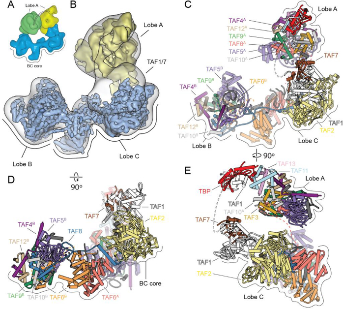
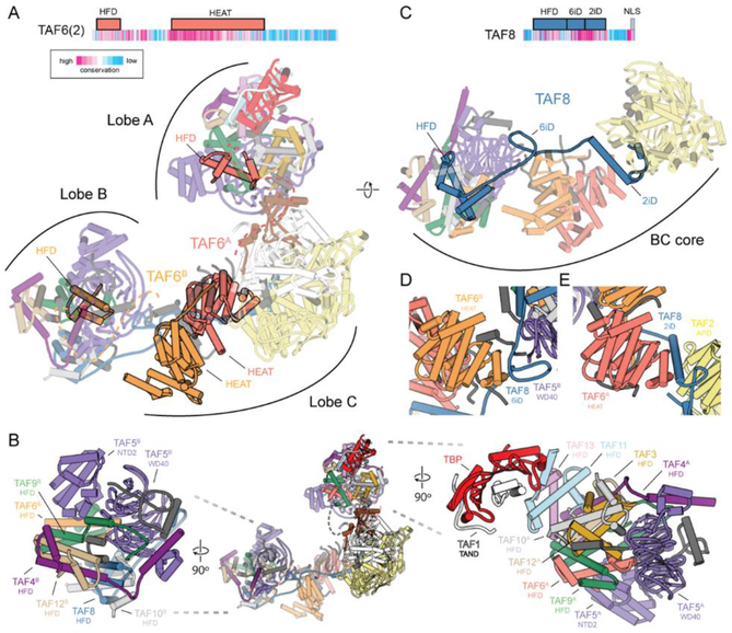
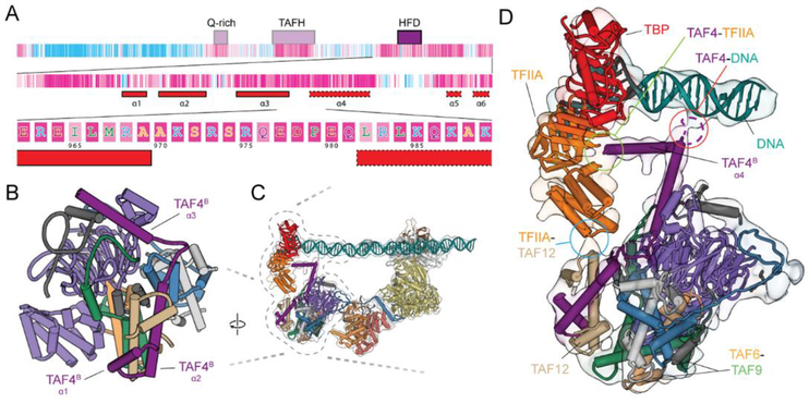
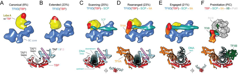
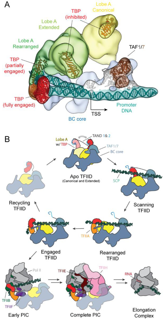
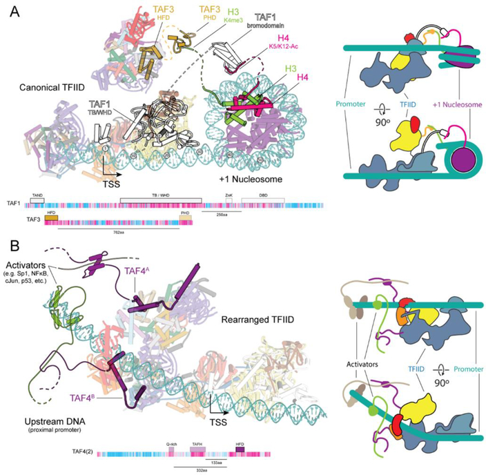
The general transcription factor IID (TFIID) is a critical component of the eukaryotic transcription preinitiation complex (PIC) and is responsible for recognizing the core promoter DNA and initiating PIC assembly. We used cryo-electron microscopy, chemical cross-linking mass spectrometry, and biochemical reconstitution to determine the complete molecular architecture of TFIID and define the conformational landscape of TFIID in the process of TATA box-binding protein (TBP) loading onto promoter DNA. Our structural analysis revealed five structural states of TFIID in the presence of TFIIA and promoter DNA, showing that the initial binding of TFIID to the downstream promoter positions the upstream DNA and facilitates scanning of TBP for a TATA box and the subsequent engagement of the promoter. Our findings provide a mechanistic model for the specific loading of TBP by TFIID onto the promoter.


Conflict of interest statement

Competing interests: The authors declare no competing financial interests.

Figures

Fig. 1.
Fig. 1.. Cryo-EM Structure of TFIID.
(A) Cryo-EM reconstructions of TFIID, with the BC core in blue and Lobe A in yellow (canonical state) and green (extended state). (B) Transparent cryo-EM map of TFIID in the canonical state with fitted cryo-EM maps from focused refinements of the BC core and lobe A in solid blue and yellow, respectively. (C-E) TFIID structural model in front (C), top (D), and side views (E). See also Movie 1.
Fig. 2.
Fig. 2.. Structural organization of human TFIID.
(A) Domain organization of TAF6, with sequence conservation colored based on ConSurf (69) scores (top). Model of TFIID with the TAF6 dimer highlighted (bottom). The dimer of TAF6 FIEAT repeats is centrally located within the complex. Dashed lines are shown connecting the TAF6 FIEAT domains with their corresponding FlFDs in lobes A and B. (B) Model of TFIID (center) and close ups of Lobe B (left) and Lobe A (right). (C) Domain organization of TAF8, with sequence conservation colored based on ConSurf (69) scores (top), and model of the BC core of TFIID with TAF8 highlighted (bottom). (D) The 6iD (TAF6 interacting domain) of TAF8 bridges the WD40 domain of TAF5 in Lobe B and the FIEAT repeat of TAF6 in Lobe C. (E) The 2iD (TAF2 interacting domain) of TAF8 bridges the FIEAT repeat of TAF6 and the APD of TAF2 within Lobe C. See also Movie 1.
Fig. 3.
Fig. 3.. Upstream promoter binding stabilized by Lobe B.
(A) Domain organization and sequence conservation of TAF4 based on ConSurf (69) scores. The first level shows the domain organization of TAF4. The second level zooms in on the C terminus and shows the secondary structure (solid outline corresponds to observed secondary structure and dashed the predicted secondary structure based on PSIPRED (70) results (α4 is not visible in the apo-TFIID structure, but becomes ordered upon interaction with the DNA). The third level shows the amino acid sequence of the loop between helices 3 and 4, which contain several conserved, positively charged residues that could be contacting the DNA. (B) Structure of lobe B. (C) Model of TFIID docked into the 11 DAS reconstruction. (D) Zoom up of part of c, highlighting the loop between helices 3 and 4 as it contacts the DNA (circled in red), helix 4 continuing on toward the TFIIA and TBP (circled in green), and the interaction between the TFIIA and TAF12 (circled in blue).
Fig. 4.
Fig. 4.. Regulation of TBP DNA-binding activity by Lobe A.
Reconstructions of TFIID from the mixed dataset (which includes SCP and TFIIA), showing TFIID in the canonical (A), extended (B), scanning (C), rearranged (D), and engaged (E) states. (F) Human PIC cryo-EM map (EMD-2304) containing Pol II, TFIIA, TFIIB, TBP and promoter DNA (6). Models for TBP (PIC: PDB 5lYA) and its interacting partners are shown below each corresponding reconstructions. See also Movie 2.
Fig. 5.
Fig. 5.. Mechanism of TBP loading by TFIID.
(A) Cryo-EM reconstructions of the canonical, extended, rearranged and engaged states of TFIID superimposed onto the BC core to show the range of motion of Lobe A and TBP. The TAF1/7 module is positioned based on the engaged state reconstruction, and the DNA models for both the engaged and rearranged states are shown. (B) Cartoon schematic for the process of TBP loading onto promoter DNA by TFIID, with subsequent PIC recruitment, assembly and progression to the elongation complex. See also Movie 2.
Fig. 6.
Fig. 6.. Model of TFIID recruitment.
(A) Model of TFIID bound to the promoter including a +1 nucleosome. The model Is compatible with the binding of flexible histone tails of H3 and H4 to the PHD (PDB ID 2K17, ref. (47)) of TAF3 and the bromodomain of BRD2 (PDB ID 2DVR (49)), a homolog of the DBD of TAF1, respectively. Dashed lines Indicate the connections between domains contained In the models of TFIID or the nucleosome, with the flexible domains that bridge the two. Domain architecture maps of TAF1 and TAF3 showing the distance between the structured domains modeled within TFIID and the domains that contact chromatin. A cartoon model of TFIID binding to the +1 nucleosome Is shown to the right. (B) Model of TFIID bound to the core promoter with bound activators at the upstream proximal promoter region. Activators are contacting the N terminus of TAF4 that contains activator Interacting regions, like the Q-rlch and TAFH domains. Domain maps of the highlighted TAFs Illustrate the distance between the domains that were part of the TFIID model (solid) and those domains that were not observed (transparent). Distances between the conserved C terminus and the domains that contact activators (TAFH and Q-rlch) are shown below the domain map. A cartoon model of TFIID binding to activators Is shown on the right.

Comment in

References

    1. Reinberg D, Horikoshi M, Roeder RG, Factors involved in specific transcription in mammalian RNA polymerase II. Functional analysis of initiation factors IIA and IID and identification of a new factor operating at sequences downstream of the initiation site. J. Biol. Chem 262,3322–3330 (1987). - PubMed
    1. Buratowski S, Hahn S, Guarente L, Sharp PA, Five Intermediate complexes in transcription Initiation by RNA polymerase II. Cell 56, 549–561 (1989). doi:10.1016/0092-8674(89)90578-3 - DOI - PubMed
    1. Cortes P, Flores O, Reinberg D, Factors Involved In specific transcription by mammalian RNA polymerase II: Purification and analysis of transcription factor IIA and Identification of transcription factor IIJ. Mol. Cell. Biol 12,413–421(1992). doi:10.1128/MCB.13.1.413 - DOI - PMC - PubMed
    1. Flores O, Lu H, Killeen M, Greenblatt J, Burton ZF, Reinberg D, The small subunit of transcription factor IIF recruits RNA polymerase II into the preinitiation complex. Proc. Natl. Acad. Sci. U.S.A 88, 9999–10003 (1991). doi:10.1073/pnas.88.22.9999 - DOI - PMC - PubMed
    1. Holstege FC, van der Vliet PC, Timmers ΗT, Opening of an RNA polymerase II promoter occurs in two distinct steps and requires the basal transcription factors IIE and IIH. EMB0 J. 15, 1666–1677 (1996). doi:10.1002/j.1460-2075.1996.th00512.x - DOI - PMC - PubMed

Publication types