. 2019 Jan 7;151(1):9-17.
doi: 10.1085/jgp.201812131. Epub 2018 Nov 15.

3-Chlorodiphenylamine activates cardiac troponin by a mechanism distinct from bepridil or TFP

Affiliations

3-Chlorodiphenylamine activates cardiac troponin by a mechanism distinct from bepridil or TFP

Svetlana B Tikunova et al. J Gen Physiol. .

Abstract

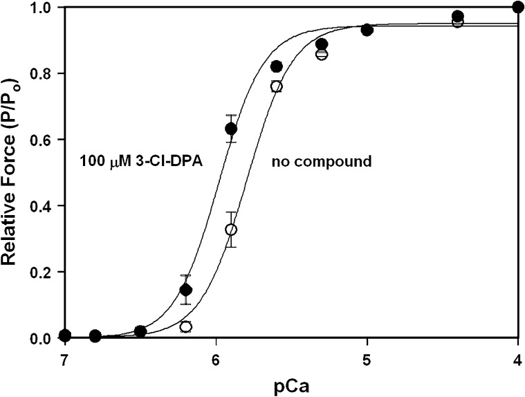
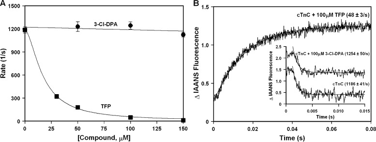
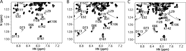
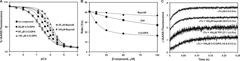
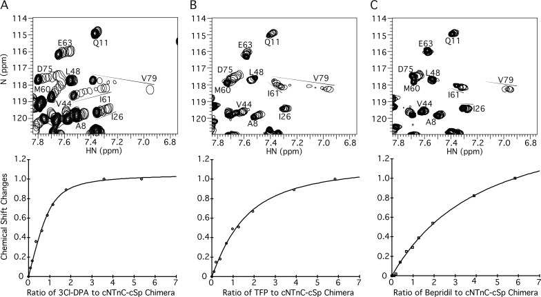
Despite extensive efforts spanning multiple decades, the development of highly effective Ca2+ sensitizers for the heart remains an elusive goal. Existing Ca2+ sensitizers have other targets in addition to cardiac troponin (cTn), which can lead to adverse side effects, such as hypotension or arrhythmias. Thus, there is a need to design Ca2+-sensitizing drugs with higher affinity and selectivity for cTn. Previously, we determined that many compounds based on diphenylamine (DPA) were able to bind to a cTnC-cTnI chimera with moderate affinity (Kd ∼10-120 µM). Of these compounds, 3-chlorodiphenylamine (3-Cl-DPA) bound most tightly (Kd of 10 µM). Here, we investigate 3-Cl-DPA further and find that it increases the Ca2+ sensitivity of force development in skinned cardiac muscle. Using NMR, we show that, like the known Ca2+ sensitizers, trifluoperazine (TFP) and bepridil, 3-Cl-DPA is able to bind to the isolated N-terminal domain (N-domain) of cTnC (Kd of 6 µM). However, while the bulky molecules of TFP and bepridil stabilize the open state of the N-domain of cTnC, the small and flexible 3-Cl-DPA molecule is able to bind without stabilizing this open state. Thus, unlike TFP, which drastically slows the rate of Ca2+ dissociation from the N-domain of isolated cTnC in a dose-dependent manner, 3-Cl-DPA has no effect on the rate of Ca2+ dissociation. On the other hand, the affinity of 3-Cl-DPA for a cTnC-TnI chimera is at least an order of magnitude higher than that of TFP or bepridil, likely because 3-Cl-DPA is less disruptive of cTnI binding to cTnC. Therefore, 3-Cl-DPA has a bigger effect on the rate of Ca2+ dissociation from the entire cTn complex than TFP and bepridil. Our data suggest that 3-Cl-DPA activates the cTn complex via a unique mechanism and could be a suitable scaffold for the development of novel treatments for systolic heart failure.


Figures

Figure 1.
Figure 1.
Effect of 3-Cl-DPA on the Ca2+ sensitivity of force development in skinned ventricular trabeculae. The curves show the Ca2+ dependence of force development in skinned ventricular trabeculae in the absence and presence of 100 µM 3-Cl-DPA. Data represent the mean ± SEM for 10 trabeculae. Data sets were individually normalized and fit with a logistic sigmoid.
Figure 2.
Figure 2.
Effect of 3-Cl-DPA on the rate of Ca2+ dissociation from the regulatory N-domain of intact cTnC. (A) Plot of the apparent rates of Ca2+ dissociation from the N-domain of intact cTnC in the presence of increased concentrations of 3-Cl-DPA or TFP. Each data point represents an average of at least three measurements ± SEM. (B) Representative stopped-flow traces as Ca2+ is removed from the N-domain of intact cTnC in the absence or presence of 100 µM 3-Cl-DPA or TFP. The traces have been normalized and staggered for clarity. Rates of Ca2+ dissociation were measured following fluorescence of IAANS attached to Cys53 of cTnC with C35S, T53C, and C84S substitutions.
Figure 3.
Figure 3.
Titrations of 3-Cl-DPA, TFP, and bepridil into Ca2+-saturated intact cTnC. (A–C) Titrations of 3-Cl-DPA (A), TFP (B), or bepridil (C) into 15N-labeled cTnC, observed by 2D [1H,15N]-HSQC NMR spectra (amide HN 1H chemical shift plotted along x axis, amide 15N chemical shift plotted along y axis). For each titration, all spectra have been overlaid, with peaks plotted as a single contour level so that their progression can be tracked (shown by arrows). The final spectrum at the end of the titration is plotted conventionally with multiple contour levels. For 3-Cl-DPA, note many peaks disappear and do not reappear at the end of the titration.
Figure 4.
Figure 4.
Effect of 3-Cl-DPA on the Ca2+ binding properties of the regulatory N-domain of cTnC reconstituted into the cTn complex. (A) Decreases in IAANS fluorescence occur when Ca2+ binds to labeled cTnC with C35S, T53C, and C84S substitutions, reconstituted into the cTn complex in the absence or presence of increasing concentrations of 3-Cl-DPA or bepridil. Data sets were individually normalized and fit with a logistic sigmoid. (B) Plot of the apparent rates of Ca2+ dissociation from the cTn complex in the presence of increased concentrations of 3-Cl-DPA, TFP, or bepridil. (C) Representative stopped-flow traces as Ca2+ is removed from the cTn complex in the absence or presence of 100 µM 3-Cl-DPA, TFP, or bepridil. The traces have been normalized and staggered for clarity. Rates of Ca2+ dissociation were measured following the fluorescence of IAANS attached to Cys53 of cTnC with C35S, T53C, and C84S substitutions after reconstitution into the cTn complex.
Figure 5.
Figure 5.
Titrations of 3-Cl-DPA, TFP, and bepridil into Ca2+-saturated cNTnC–cSp chimera. (A–C) Titrations of 3-Cl-DPA (A), TFP (B), or bepridil (C) into 15N-labeled cNTnC–cSp chimera, as observed by 2D [1H,15N]-HSQC NMR spectra (amide HN 1H chemical shift plotted along x axis, amide 15N chemical shift plotted along y axis). For each titration, all spectra have been overlaid, with peaks plotted as a single contour level so that their progression can be tracked (shown by arrows). The final spectrum at the end of the titration is plotted conventionally with multiple contour levels. Below each spectrum is a representative plot of chemical shift changes as a function of compound added, from which Kd is calculated. Note that the ratio of compound to cNTnC–cSp chimera, rather than the absolute concentration of compound, is on the x axis. This is because plots of saturation versus compound concentration are typically performed when the protein concentration is much smaller than the Kd. However, because of the sensitivity limitation of multidimensional solution NMR, the protein concentration (0.11 mM) is greater than the Kd (10 µM in the case of 3-Cl-DPA). Thus, to avoid confusion, the protein/compound ratio is used instead.

Similar articles

Cited by

References

    1. Bai Y., Milne J.S., Mayne L., and Englander S.W.. 1993. Primary structure effects on peptide group hydrogen exchange. Proteins. 17:75–86. 10.1002/prot.340170110 - DOI - PMC - PubMed
    1. Benjamin E.J., Virani S.S., Callaway C.W., Chamberlain A.M., Chang A.R., Cheng S., Chiuve S.E., Cushman M., Delling F.N., Deo R., et al. American Heart Association Council on Epidemiology and Prevention Statistics Committee and Stroke Statistics Subcommittee . 2018. Heart Disease and Stroke Statistics-2018 Update: A Report From the American Heart Association. Circulation. 137:e67–e492. 10.1161/CIR.0000000000000558 - DOI - PubMed
    1. Black D.J., Tikunova S.B., Johnson J.D., and Davis J.P.. 2000. Acid pairs increase the N-terminal Ca2+ affinity of CaM by increasing the rate of Ca2+ association. Biochemistry. 39:13831–13837. 10.1021/bi001106+ - DOI - PubMed
    1. Boyle K.L., and Leech E.. 2012. A review of the pharmacology and clinical uses of pimobendan. J. Vet. Emerg. Crit. Care (San Antonio). 22:398–408. 10.1111/j.1476-4431.2012.00768.x - DOI - PubMed
    1. Cai F., Li M.X., Pineda-Sanabria S.E., Gelozia S., Lindert S., West F., Sykes B.D., and Hwang P.M.. 2016. Structures reveal details of small molecule binding to cardiac troponin. J. Mol. Cell. Cardiol. 101:134–144. 10.1016/j.yjmcc.2016.10.016 - DOI - PMC - PubMed

Publication types