. 2018 Nov 16;362(6416):eaat9528.
doi: 10.1126/science.aat9528.

SFXN1 is a mitochondrial serine transporter required for one-carbon metabolism

Affiliations

SFXN1 is a mitochondrial serine transporter required for one-carbon metabolism

Nora Kory et al. Science. .

Abstract

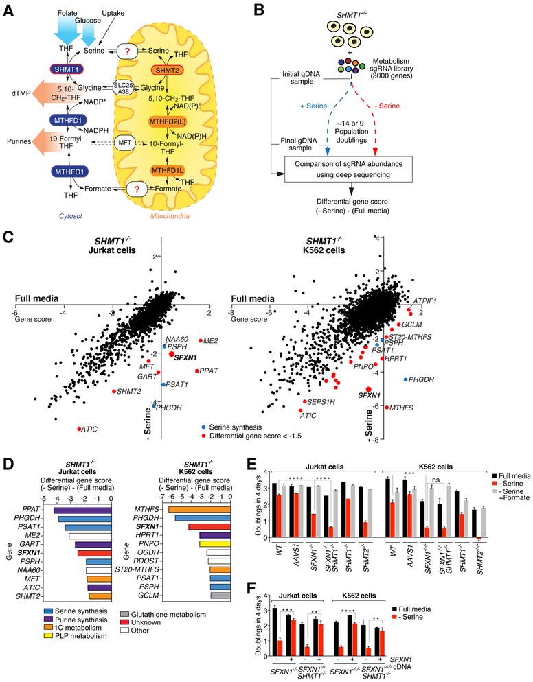
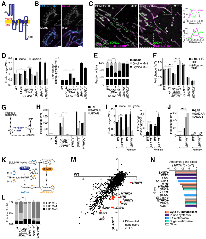
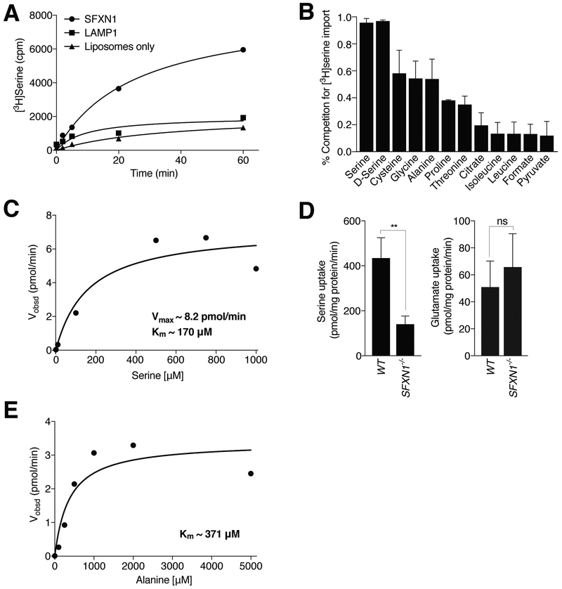
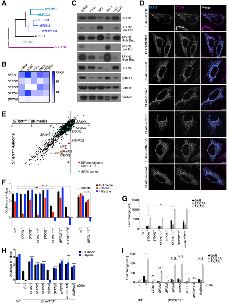
One-carbon metabolism generates the one-carbon units required to synthesize many critical metabolites, including nucleotides. The pathway has cytosolic and mitochondrial branches, and a key step is the entry, through an unknown mechanism, of serine into mitochondria, where it is converted into glycine and formate. In a CRISPR-based genetic screen in human cells for genes of the mitochondrial pathway, we found sideroflexin 1 (SFXN1), a multipass inner mitochondrial membrane protein of unclear function. Like cells missing mitochondrial components of one-carbon metabolism, those null for SFXN1 are defective in glycine and purine synthesis. Cells lacking SFXN1 and one of its four homologs, SFXN3, have more severe defects, including being auxotrophic for glycine. Purified SFXN1 transports serine in vitro. Thus, SFXN1 functions as a mitochondrial serine transporter in one-carbon metabolism.


Conflict of interest statement

Competing interests: N.K. and D.M.S. are inventors on a patent application filed by the Whitehead Institute relating to work described in this paper. None of the authors have a competing interest. Data and materials availability: All data are available in the manuscript or the supplementary materials. All expression plasmids were deposited at Addgene.

Figures

Fig. 1.
Fig. 1.. A genetic screen for components of the one-carbon metabolism pathway yields SFXN1.
(A) Schematic of the one-carbon metabolism pathway. dTMP, deoxythymidine monophosphate; THF, tetrahydrofolate; CH2THF, methyleneTHF. NAD(P)H, nicotinamide adenine dinucleotide (phosphate); SHMT, serine hydroxymethyltransferase; MFT, mitochondrial folate transporter/carrier; MTHFD, methylenetetrahydrofolate dehydrogenase. The dashed arrows indicate that the exact nature of the substrate for MFT is unknown. (B) CRISPRCas9–based screening strategy designed to identify new components of the mitochondrial one-carbon pathway. The cells in the serine-free media were collected after ~9 population doublings because they proliferated more slowly than cells in full media, which were collected after ~14 doublings. For each gene, we calculated its gene score as the mean log2 foldchange in the abundance of the 10 sgRNAs targeting the gene. The differential gene score is the difference in scores in the absence versus presence of serine. sgRNA, single guide RNA; gDNA, genomic DNA. (C) SFXN1 emerges as a hit in both the Jurkat and K562 screens. Gene scores in full media were plotted against those in serine-deficient media. Genes with a differential score of < 0.001, ****P < 0.0001; ns, not significant). AAVS1 indicates control cells that were treated with an sgRNA targeting the AAVS1 locus as described previously (14). SFXN1- and SHMT2-null K562 cells are designated as −/−/− as they are triploid for these genes. (F) Expression of an sgRNA-resistant cDNA for SFXN1 in the SFXN1-null cells restores their proliferation rate in serine-deficient media (mean ± SD; n = 3; **P < 0.01, ****P < 0.0001). Two-tailed t tests were used for comparisons between groups.
Fig. 2.
Fig. 2.. Loss of SFXN1 phenocopies mutants in mitochondrial one-carbon metabolism.
(A) Model of the predicted topology of SFXN1 in the mitochondrial inner membrane. Transmembrane helices are indicated by numbers. IMS, intermembrane space. (B) FLAG-tagged SFXN1 localizes to mitochondria. Wild-type HeLa cells transiently expressing FLAG-SFXN1 were processed for immunofluorescence detection of the FLAG epitope (cyan) and the mitochondrial inner membrane marker cytochrome c oxidase subunit 4 (COX4) (magenta). The merged image shows the overlap of both channels in white. Scale bar is 10 mm in the full image and 2 mm in the inset. (C) Super-resolution microscopy confirms SFXN1 localization to the inner membrane of mitochondria. Wild-type HeLa cells transiently expressing FLAG-SFXN1 were processed for immunofluorescence detection of the FLAG epitope (magenta) and the outer mitochondrial membrane marker Tom20 (left panel, green) or the mitochondrial inner membrane marker cytochrome c oxidase subunit 4 (COX4) (right panel, green) and imaged by STED microscopy. Overlap of magenta and green channels is shown in white, and line profiles show fluorescent signals of each channel across mitochondria where marked by the dotted rectangles. Scale bars are 2 mm in the full images and 1 mm in the insets. (D) As in cells lacking known components of the mitochondrial one-carbon pathway, glycine levels are reduced and the cellular serine/glycine ratio is increased in SFXN1-null cells. Serine and glycine levels were measured by gas chromatography–mass spectrometry (GC-MS) in extracts from wild-type Jurkat cells or single-cell–derived control and knockout clones (mean ± SD; n = 3; **P < 0.01, ***P < 0.001). (E) Loss of SFXN1 causes a glycine synthesis defect. GC-MS was used to measure glycine in the culture media of wild-type Jurkat cells or single-cell–derived knockout clones incubated for 12 hours with 2,3,3–2 H3-serine as the only serine source. The glycine M+0 species is the unlabeled species. The glycine M+1 species is derived from 2,3,3–2 H3-serine (mean ± SD; n = 3; **P < 0.01, ***P < 0.001). (F) Levels of charged folate species are decreased in SFXN1-null cells. Metabolites were measured by LC-MS in extracts from wild-type Jurkat cells or single-cell–derived control and knockout clones (mean ± SD; n = 3; **P < 0.01, ***P < 0.001, ****P < 0.0001). 5,10-CH+ -THF, 5,10-methenyl-THF. (G) Schematic of the purine synthesis pathway, indicating steps using one-carbon units in the form of 10-formyl-THF. GAR, 5′-phosphoribosyl-glycinamide. SAICAR, phosphoribosylaminoimidazolesuccinocarboxamide. AICAR, 5-aminoimidazole-4- carboxamide ribonucleotide. IMP, inosine monophosphate. (H) The purine synthesis intermediates GAR, SAICAR, and AICAR accumulate in SFXN1-null cells. Purine synthesis intermediates were measured by LC-MS in extracts from wild-type Jurkat cells or single-cell–derived control and knockout clones (mean ± SD; n = 3; ****P < 0.0001). (I) Addition of 1 mM formate does not rescue glycine levels and serine/glycine ratio of SFXN1- null cells. Serine and glycine levels were measured by LC-MS in extracts from wild-type Jurkat cells or single-cell–derived SFXN1-null cells incubated for 24 hours in the indicated media (mean ± SD; n = 3; *P < 0.05, **P < 0.01). (J) Addition of 1 mM formate reverses the accumulation of purine synthesis intermediates in SFXN1-null cells. Intermediates were measured by LC-MS in extracts from wild-type Jurkat cells or single-cell– derived SFXN1-null cells incubated for 24 hours in the indicated media (mean ± SD; n = 3; ****P < 0.0001; N.D., not detected). (K) Tracing strategy to differentiate contribution of cytosolic and mitochondrial pathways to cytosolic TTP synthesis. Oxidation of 2,3,3–2 H3-serine by SHMT2 and subsequent enzymes in mitochondria gives rise to a singly labeled formate species, and thus singly labeled (one mass unit heavier, M+1) TTP. Oxidation by SHMT1 in the cytosol gives rise to doubly labeled (two mass units heavier, M+2) TTP. The difference between unlabeled (M+0), M+1, and M+2 TTP can be resolved on a high-resolution mass spectrometer. The ratio of M+1 to M+2 is indicative of the contribution of mitochondria- versus cytosol-derived one-carbon units to nucleotide synthesis. Adapted from (8). TTP, thymidine triphosphate. (L) The relative contribution of the cytosolic and mitochondrial one-carbon pathways to TTP synthesis is inverted in SFXN1-null compared to wild-type cells. Wild-type Jurkat or single-cell-derived knockout cells were cultured for 12 hours in media containing 2,3,3–2 H3-serine as the only serine source before harvesting and LC-MS analysis (mean ± SD; n = 3, ****P < 0.0001). (M) Genes of the cytosolic one-carbon pathway are selectively required for the optimal proliferation of SFXN1-null cells. Gene scores in wild-type cells were plotted against those in SFXN1-null cells. Genes with a differential gene score of <−1.5 are shown in red. (N) Serine Hydroxymethyltransferase 1 (SHMT1) was the top hit from the SFXN1 synthetic lethality screen. Genes were ranked according to differential gene score between wild-type and SFXN1-null cells. Cyto 1C metabolism, cytosolic one-carbon metabolism; FA, fatty acid. Two-tailed t tests were used for comparisons between metabolites.
Fig. 3.
Fig. 3.. SFXN1 transports serine in vitro.
(A) Time course of radioactive serine uptake into proteoliposomes containing SFXN1. LAMP1-containing or empty liposomes were used as controls. Values are the averages of two replicates. Cpm, counts per minute. (B) Competition of serine uptake after 60 min by different metabolites at 500 mM (mean ± SD; n = 3). (C) Steady-state kinetic analysis of SFXN1-mediated serine transport reveals a Vmax of ~ 8.2 pmol/min and a Km of ~170 mM. Velocity, as shown, was calculated as a function of the serine concentration. Each data point was calculated from three replicate data points. (D) Radioactive serine uptake into mitochondria purified from SFXN1-null cells is reduced compared to that into mitochondria purified from wild-type cells, whereas glutamate uptake is unchanged (mean ± SD; n = 3, **P < 0.01; ns, not significant). (E) Steady-state kinetic analysis of SFXN1-mediated alanine transport reveals a Km of ~371 mM for alanine. Each data point was calculated from three replicate data points
Fig. 4.
Fig. 4.. SFXN3 and fly and yeast sideroflexin homologs can substitute for SFXN1 loss.
(A) Phylogenetic tree of human, Drosophila melanogaster, and Saccharomyces cerevisae sideroflexins. (B) mRNA levels of the five human sideroflexins in commonly used cell lines. RPKM (reads per kilobase million) levels were extracted from the Cancer Cell Line Encyclopedia. (C) Sideroflexin protein levels in commonly used cell lines. Cell lysates were equalized for total protein amounts and analyzed by immunoblotting for the levels of the indicated proteins. (D) FLAG-tagged sideroflexin homologs localize to mitochondria. Wild-type HeLa cells transiently expressing FLAG-sideroflexin homologs were processed for immunofluorescence detection of the FLAG epitope (cyan) and the mitochondrial inner membrane marker COX4 (magenta). The merged image shows the overlap of both channels in white. (E) CRISPR-Cas9–based genetic screen reveals that SFXN3 is required for proliferation in the absence of SFXN1 and glycine. Gene scores in SFXN1-null cells cultured in the presence or absence of glycine were plotted against each other. Except for SFXN3, genes with a differential gene score of < 0.001, ****P < 0.0001). (G) The accumulation of purine synthesis intermediates is exacerbated in cells lacking both SFXN1 and SFXN3 compared to their single-knockout counterparts. The asterisk denotes a cell clone lacking SFXN1 and SFXN2 and with incomplete deletion of SFXN3. Purine intermediates were measured by LC-MS in extracts from the indicated cells (mean ± SD; n = 3; **P < 0.01). Abbreviations as in Fig. 2C. (H) Human, yeast, and Drosophila sideroflexin homologs, with the exception of SFXN4, rescue the glycine auxotrophy of cells lacking both SFXN1 and SFXN3. Single cell–derived doubleknockout Jurkat cells were transduced with an empty vector (EV) or cDNAs of human, yeast, and Drosophila sideroflexin homologs. Asterisks denote statistically significant differences in proliferation in media lacking glycine between the cells expressing the empty vector and the sideroflexin homologs. Mean ± SD; n = 3; ***P < 0.001, ****P < 0.0001). (I) Sideroflexin homologs rescue to varying degrees the purine synthesis defects of cells lacking SFXN1 and SFXN3. Purine intermediates were measured by LC-MS in extracts from wild-type Jurkat cells or the double-knockout Jurkat cells expressing an empty vector (EV) or cDNAs of human, yeast, and Drosophila sideroflexin homologs. Asterisks denote statistically significant differences between the cells expressing the empty vector and the sideroflexin homologs. Values were normalized to the average value of the wild-type samples in (G) because purine synthesis intermediates were not detected in the wild-type samples in this experiment (mean ± SD; n = 3, **P < 0.01; N.D., not detected; N.S., not significant). Two-tailed t tests were used for comparisons between metabolites.

References

    1. Yang M, Vousden KH, Serine and one-carbon metabolism in cancer. Nat. Rev. Cancer 16, 650–662 (2016). doi: 10.1038/nrc.2016.81; pmid: 27634448 - DOI - PubMed
    1. Locasale JW, Serine, glycine and one-carbon units: Cancer metabolism in full circle. Nat. Rev. Cancer 13, 572–583 (2013). doi: 10.1038/nrc3557; pmid: 23822983 - DOI - PMC - PubMed
    1. Ducker GS, Rabinowitz JD, One-Carbon Metabolism in Health and Disease. Cell Metab. 25, 27–42 (2017). doi: 10.1016/j.cmet.2016.08.009; pmid: 27641100 - DOI - PMC - PubMed
    1. Tibbetts AS, Appling DR, Compartmentalization of Mammalian folate-mediated one-carbon metabolism. Annu. Rev. Nutr 30, 57–81 (2010). doi: 10.1146/annurev.nutr.012809.104810; pmid: 20645850 - DOI - PubMed
    1. Barlowe CK, Appling DR, In vitro evidence for the involvement of mitochondrial folate metabolism in the supply of cytoplasmic one-carbon units. Biofactors 1, 171–176 (1988). pmid: 2475123 - PubMed

Publication types