. 2018 Nov 16;362(6416):829-834.
doi: 10.1126/science.aau0976.

Protein assemblies ejected directly from native membranes yield complexes for mass spectrometry

Affiliations

Protein assemblies ejected directly from native membranes yield complexes for mass spectrometry

Dror S Chorev et al. Science. .

Abstract

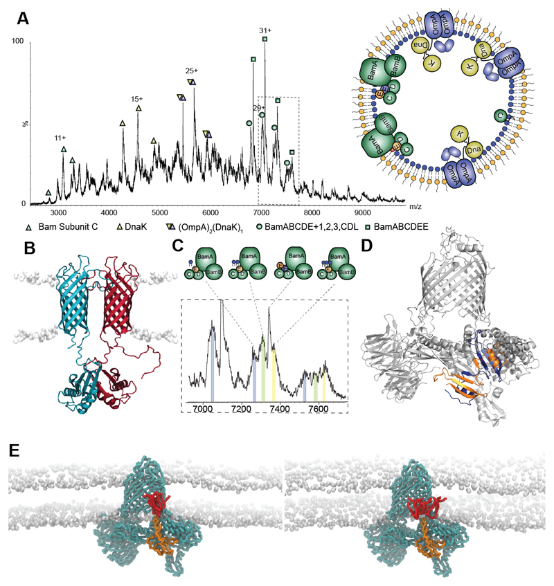
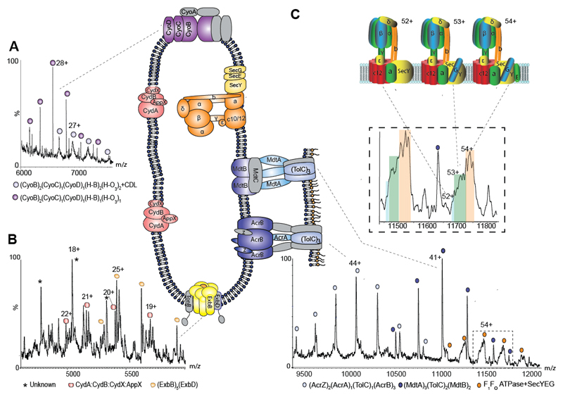
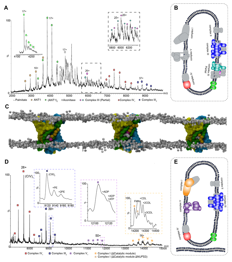
Membrane proteins reside in lipid bilayers and are typically extracted from this environment for study, which often compromises their integrity. In this work, we ejected intact assemblies from membranes, without chemical disruption, and used mass spectrometry to define their composition. From Escherichia coli outer membranes, we identified a chaperone-porin association and lipid interactions in the β-barrel assembly machinery. We observed efflux pumps bridging inner and outer membranes, and from inner membranes we identified a pentameric pore of TonB, as well as the protein-conducting channel SecYEG in association with F1FO adenosine triphosphate (ATP) synthase. Intact mitochondrial membranes from Bos taurus yielded respiratory complexes and fatty acid-bound dimers of the ADP (adenosine diphosphate)/ATP translocase (ANT-1). These results highlight the importance of native membrane environments for retaining small-molecule binding, subunit interactions, and associated chaperones of the membrane proteome.


Conflict of interest statement

Competing interests:

Authors declare no competing interests.

Figures

Fig. 1
Fig. 1. Protein complexes ejected directly from E. coli outer membranes
A Mass spectrum recorded at 400 V assigned to BamC, DnaK, DnaK:OmpA:pro-OmpA, and two states of the Bam complex with inset an outer membrane vesicle with complexes observed. B Model of the OmpA dimer (15) with the hydrophobic pro-sequence (red) a potential binding site for DnaK. C Expansion of the mass spectrum assigned to the Bam complex with monomeric BamE (Bam ABCDE) binding to one, two and three cardiolipins (grey, green, yellow respectively). D Atomic structure of BamE dimer (PDB: 2YH9) (orange and blue) docked into the Bam complex (PDB: 5D0O) with BamE monomer removed. E MD simulations of the BamABCDE complex (cyan) with monomeric BamE (orange) and two (lhs) and three (rhs) CDL molecules (red).
Fig. 2
Fig. 2. Regions of the mass spectrum recorded for inner membranes from E. coli yield cytochromes, the Ton complex multi-drug transporters and the intact ATP synthase in complex with the SecYEG translocon.
A and B Expanded regions of the spectrum assigned to the cytochromes bo3 and cytochrome bd oxidase showing peak splitting due to binding of quinol and heme groups (fig. S8). The pentameric ExbB complex, with one copy of ExbD located in the center of the pore, forming part of the TonB complex is also observed (yellow). C High m/z region of the mass spectrum assigned to multidrug efflux pumps AcrAB and MdtAB and the intact ATP synthase. Expansion of the peaks assigned to the ATPase reveals binding of SecY (blue) SecYG (green) and SecYEG (orange) charge states 52+, 53+ and 54+ shown schematically.
Fig. 3
Fig. 3. Intact Mitochondria and inner membranes yield complexes I, III, IV and V as well as the adenine nucleotide translocator 1 (Ant1) with palmitate transport through the dimer interface.
A Mass spectrum of bovine mitochondrial inner membranes recorded at 600 V reveals the ANT-1 dimer, complexes III and IV, as well as aconitase. The left inset, recorded at 400 V, shows an expanded view of the spectrum at 4100 to 4300 m/z revealing multiple palmitate anions bounds to the dimer of ANT-1. The right inset shows an expansion of the boxed area in the main panel. B Depiction of the protein assemblies ejected from sonicated mitochondrial inner membranes. Subunits shown in gray have dissociated. C MD simulation of ANT-1 after 2.1, 2.5, and 2.6 μs (left to right) in an asymmetric membrane containing phosphatidylcholine (PC), phosphatidylethanolamine (PE), and CDL (only in the matrix leaflet). The protein surface is colored according to the three pseudo-repeats (R1, yellow; R2, green; R3, cyan), and the charged palmitate headgroup (magenta) is buried between helices three and four. D Complexes I, III, IV, and V are expelled from intact mitochondria. Intact complex V is observed with associated nucleotides, together with a dimer of complex IV (fig. S15) and partial assemblies of complex I (the charge state of the subassembly lacking NUFS3 is z = 56+), in the absence of the catalytic core, bound to FMN. Inset schematics represent assigned membrane complexes, color coded according to labels on the peaks. E Depiction of the protein assemblies ejected from sonicated intact mitochondrial membranes color coded according to the labels on the peaks. Gray subunits were not observed.

Comment in

References

    1. Vinothkumar KR, Henderson R. Single particle electron cryomicroscopy: trends, issues and future perspective. Q Rev Biophys. 2016 Jan;49:e13. - PubMed
    1. Heck AJ. Native mass spectrometry: a bridge between interactomics and structural biology. Nat Methods. 2008 Nov;5:927. - PubMed
    1. Gupta K, et al. The role of interfacial lipids in stabilizing membrane protein oligomers. Nature. 2017 Jan 19;541:421. - PMC - PubMed
    1. Laganowsky A, et al. Membrane proteins bind lipids selectively to modulate their structure and function. Nature. 2014 Jun 5;510:172. - PMC - PubMed
    1. Hopper JT, et al. Detergent-free mass spectrometry of membrane protein complexes. Nat Methods. 2013 Dec;10:1206. - PMC - PubMed

Publication types

MeSH terms

LinkOut - more resources