. 2018 Nov 15;8(1):16855.
doi: 10.1038/s41598-018-35303-9.

Androgen induced cellular proliferation, neurogenesis, and generation of GnRH3 neurons in the brain of mature female Mozambique tilapia

Affiliations

Androgen induced cellular proliferation, neurogenesis, and generation of GnRH3 neurons in the brain of mature female Mozambique tilapia

Yasuto Narita et al. Sci Rep. .

Abstract

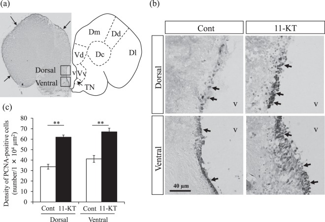
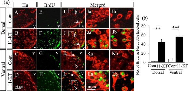
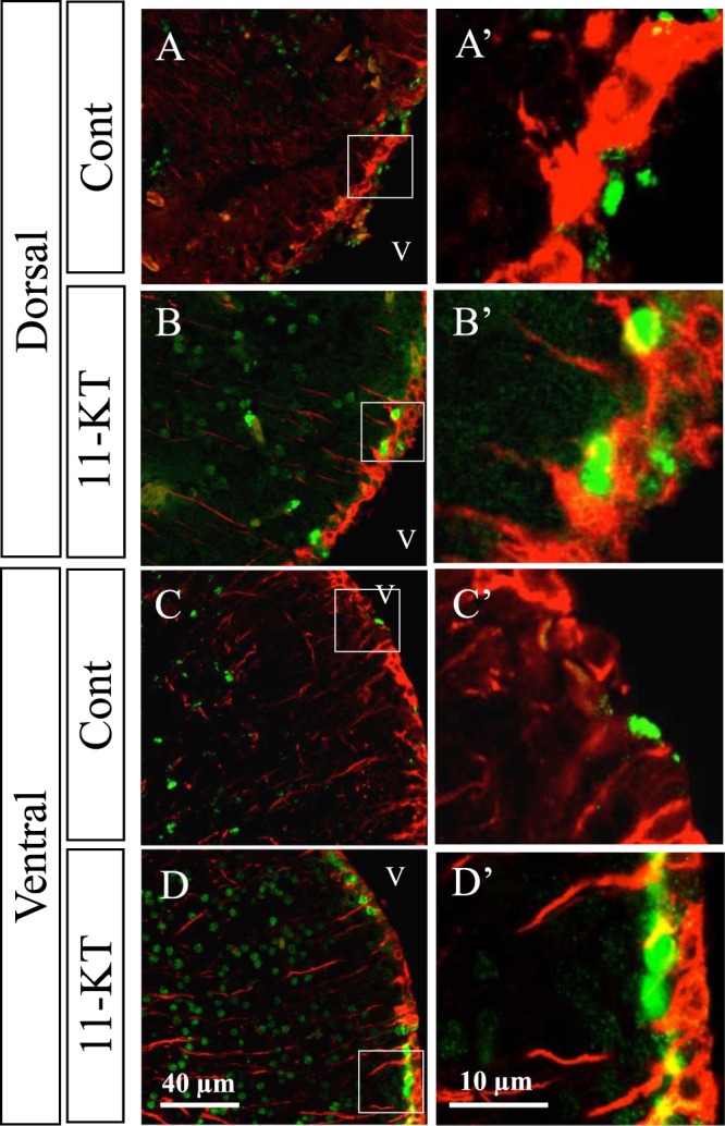
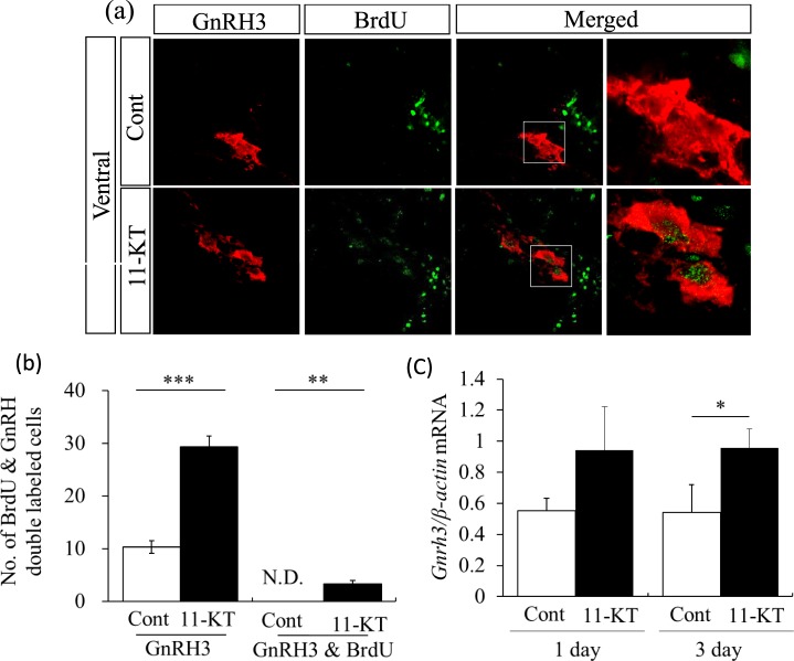
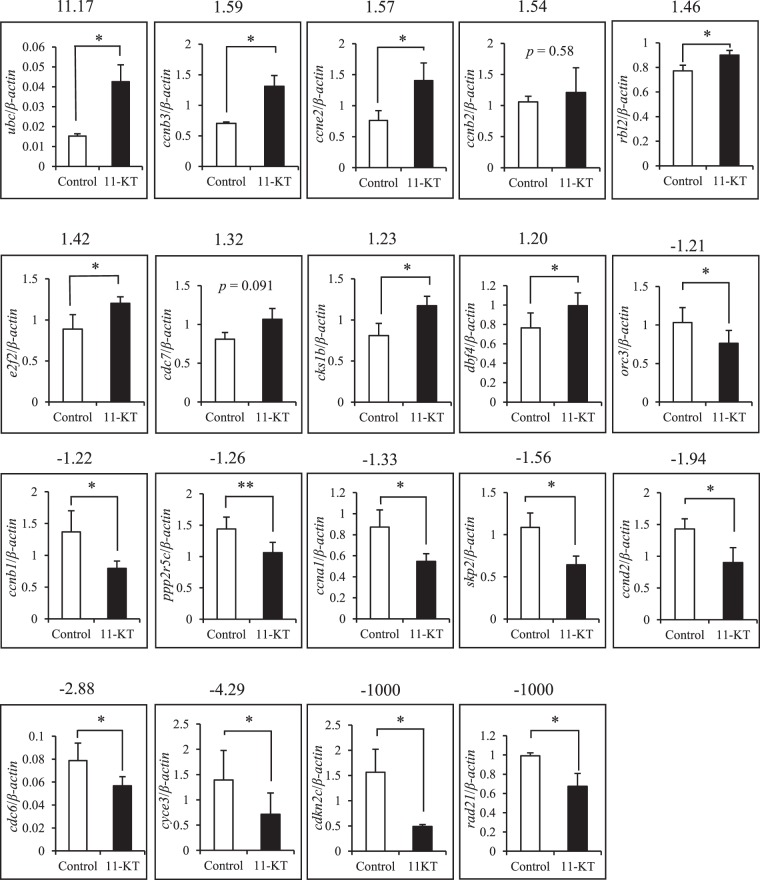
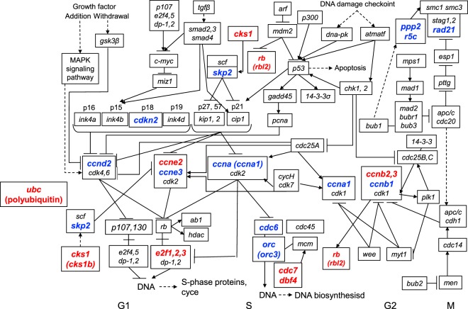
The neuroplastic mechanisms in the fish brain that underlie sex reversal remain unknown. Gonadotropin-releasing hormone 3 (GnRH3) neurons control male reproductive behaviours in Mozambique tilapia and show sexual dimorphism, with males having a greater number of GnRH3 neurons. Treatment with androgens such as 11-ketotestosterone (KT), but not 17β-estradiol, increases the number of GnRH3 neurons in mature females to a level similar to that observed in mature males. Compared with oestrogen, the effect of androgen on neurogenesis remains less clear. The present study examined the effects of 11-KT, a non-aromatizable androgen, on cellular proliferation, neurogenesis, generation of GnRH3 neurons and expression of cell cycle-related genes in mature females. The number of proliferating cell nuclear antigen-positive cells was increased by 11-KT. Simultaneous injection of bromodeoxyuridine and 11-KT significantly increased the number of newly-generated (newly-proliferated) neurons, but did not affect radial glial cells, and also resulted in newly-generated GnRH3 neurons. Transcriptome analysis showed that 11-KT modulates the expression of genes related to the cell cycle process. These findings suggest that tilapia could serve as a good animal model to elucidate the effects of androgen on adult neurogenesis and the mechanisms for sex reversal in the fish brain.


Conflict of interest statement

The authors declare no competing interests.

Figures

Figure 1
Figure 1
PCNA immunostaining in the female tilapia brain. (a) Left: Representative frontal section of the female tilapia brain crossing the terminal nerve (TN) immunostained with PCNA antibody. The two inserted squares correspond to regions in the dorsal and ventral areas used for counting cells displaying a PCNA-positive nucleus (each area, 140 µm × 140 µm) around the ventricle. Arrows indicate PCNA-positive cells. Right: Atlas of the brain corresponding to the left image. Dm = medial zone of dorsal telencephalic area; Dd = dorsal zone of dorsal telencephalic area; Dl = lateral zone of dorsal telencephalic area; Vd = dorsal nucleus of ventral telencephalic area; Vv, = ventral nucleus of ventral telencephalic area; v = ventricle. (b) Representative image of PCNA-positive cells in the dorsal and ventral areas around the ventricle at the level of the TN in 11-KT-treated and control (Cont) female tilapia. Arrows indicate PCNA-positive nuclei. (c) Density of PCNA-positive cells in control and 11-KT-treated females in the dorsal and ventral areas around the ventricle. **Indicates p < 0. 01 (unpaired Student’s t-test). Data are expressed as mean ± SEM.
Figure 2
Figure 2
Representative images obtained from double immunohistochemistry with BrdU and Hu antibodies. (a) Hu-positive cells (red) observed in dorsal and ventral areas around the ventricle in control (cont) and 11-KT-treated (11-KT) females (A,B,C,D). Although few BrdU-labelled nuclei (green) were detected in control animals (E,G), BrdU-labelled nuclei were frequently observed in 11-KT-treated females (F,H). I,J,K and L show merged images. Ia,b,Ja,b,Ka,b and La,b show enlarged images of the areas inset in I,J,K, and L, respectively; v = ventricle. (b) The number of Hu-positive cells with a BrdU-labelled nucleus. White and black bars indicate the numbers of double-labelled cells in control (Cont) and 11-KT-treated (11-KT) females. **Indicates p < 0. 01; ***Indicates p < 0. 001 (unpaired Student’s t-test). Data are expressed as mean ± SEM.
Figure 3
Figure 3
Representative merged images of sections double-stained with BrdU and GFAP antibodies. GFAP-positive cells (red) visualized on the inner surface of the ventricle, with long processes in the dorsal and ventral areas around the ventricle in control (Cont) and 11-KT-treated (11-KT) females. (A,B) dorsal and ventral areas, respectively, of control animals. (C,D) dorsal and ventral areas, respectively, of 11-KT-treated females. A’,B’,C’ and D’ show enlarged views of inset squares in A,B,C, and D, respectively; v = ventricle.
Figure 4
Figure 4
Representative images of sections double-stained with BrdU and GnRH3 antibodies. (a) GnRH3 neurons (red), BrdU-labelling nuclei (green) and merged images in control (Cont) and 11-KT treated (11-KT) females. GnRH3 neurons with BrdU-labelling in the nucleus were observed in 11-KT treated females, but not in controls. The rightmost column shows enlarged views of inserted squares in the second from the right column. (b) Number of GnRH3 neurons in control (Cont, white column) and 11-KT-treated (11-KT, black column) females (left) three days after 11-KT injection. Number of double labelled cells (GnRH & BrdU) in control (Cont, white column) and 11-KT treated (11-KT, black column) animals (right); N. D. = not detected. (c) Relative expression levels of GnRH3 mRNA in control (Cont, white column) and 11-KT treated (11-KT, black column) females one day (left) and three days (right) after 11-KT injection. **Indicates p < 0.01; ***Indicates p < 0.001 (unpaired Student’s t-test). Data are expressed as mean ± SEM.
Figure 5
Figure 5
Changes in expressions of genes related to cell cycle after 11-KT treatment. Expressions of 19 genes listed in Table 1 were examined by real-time RT-qPCR analysis and compared between control (white column) and 11-KT treated (11-KT, black column) females. The numeric values on the top of each graph indicates the fold change as revealed by the transcriptome analysis. *Indicates p < 0. 05, **Indicates p < 0.01 (unpaired Student’s t-test). Data are expressed as mean ± SEM.
Figure 6
Figure 6
Cell cycle regulation pathway and the genes whose expressions were altered by 11-KT. The figure was slightly modified from ‘Cell cycle’ pathway (map04110, KEGG). Genes whose expressions were increased (red, fold change >1. 2) or decreased (blue, fold change < -1. 2) by 11-KT treatment were marked in the figure. Ubc coding polyubiquitin-C precursor regulates each phase of the cell cycle through the degradation of cyclins.

Similar articles

Cited by

References

    1. Munakata A, Kobayashi M. Endocrine control of sexual behavior in teleost fish. Gen Comp Endocrinol. 2010;165:456–468. doi: 10.1016/j.ygcen.2009.04.011. - DOI - PubMed
    1. Kobayashi M, Nakanishi T. 11-ketotestosterone induces male-type sexual behavior and gonadotropin secretion in gynogenetic crucian carp, Carassius auratus langsdorfii. Gen Comp Endocrinol. 1999;115:178–187. doi: 10.1006/gcen.1999.7314. - DOI - PubMed
    1. Saoshiro S, Kawaguchi Y, Hayakawa Y, Kobayashi M. Sexual bipotentiality of behavior in male and female goldfish. Gen Comp Endocrinol. 2013;181:265–270. doi: 10.1016/j.ygcen.2012.08.022. - DOI - PubMed
    1. Pandian TJ, Sheela SG. Hormonal induction of sex reversal in fish. Aquaculture. 1995;138:1–22. doi: 10.1016/0044-8486(95)01075-0. - DOI
    1. Phelps, R. P. & Popma, T. J. Sex reversal of tilapia. Tilapia aquaculture in the Americas, Vol. 2. The World Aquaculture Society, 34–59 (2000).

Publication types

MeSH terms