. 2018 Nov 15;9(12):1140.
doi: 10.1038/s41419-018-1189-2.

c-Jun N-terminal kinases differentially regulate TNF- and TLRs-mediated necroptosis through their kinase-dependent and -independent activities

Affiliations

c-Jun N-terminal kinases differentially regulate TNF- and TLRs-mediated necroptosis through their kinase-dependent and -independent activities

Mengtao Cao et al. Cell Death Dis. .

Abstract

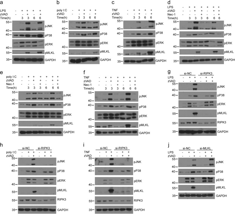
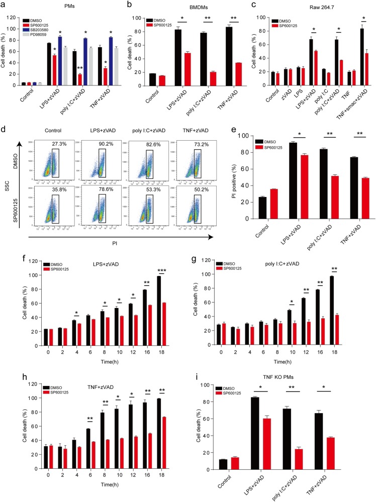
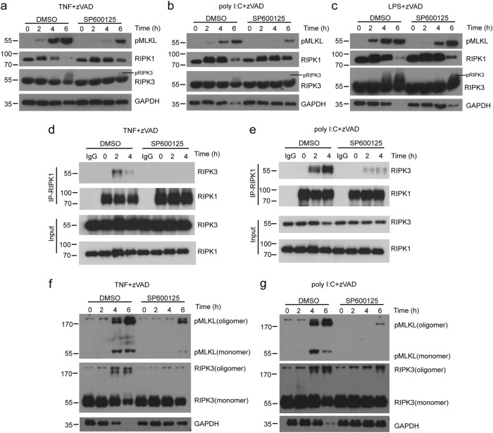
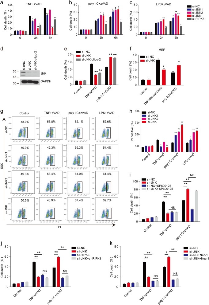
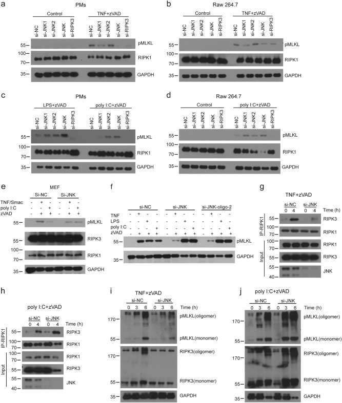
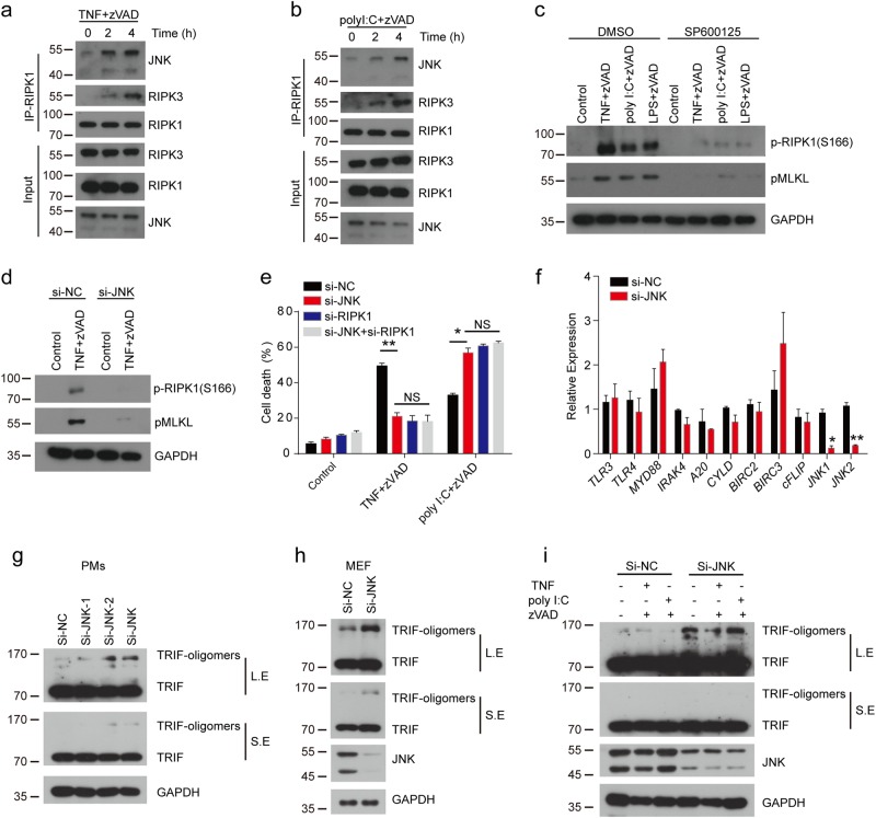
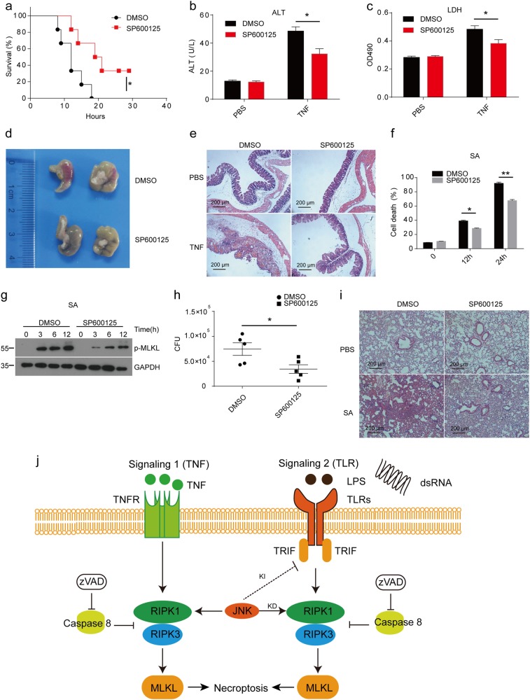
Tumor necrosis factor (TNF) and Toll-like receptor (TLR)3/TLR4 activation trigger necroptotic cell death through downstream signaling complex containing receptor-interacting protein kinase 1 (RIPK1), RIPK3, and pseudokinase mixed lineage kinase-domain-like (MLKL). However, the regulation of necroptotic signaling pathway is far less investigated. Here we showed that c-Jun N-terminal kinases (JNK1 and JNK2) displayed kinase-dependent and -independent functions in regulating TNF- and TLRs-mediated necroptosis. We found that RIPK1 and RIPK3 promoted cell-death-independent JNK activation in macrophages, which contributed to pro-inflammatory cytokines production. Meanwhile, blocking the kinase activity of JNK dramatically reduced TNF and TLRs-induced necroptotic cell death. Consistently, inhibition of JNK activity protected mice from TNF-induced death and Staphylococcus aureus-mediated lung damage. However, depletion of JNK protein using siRNA sensitized macrophages to necroptosis that was triggered by LPS or poly I:C but still inhibited TNF-induced necroptosis. Mechanistic studies revealed that RIPK1 recruited JNK to the necrosome complex and their kinase activity was required for necrosome formation and the phosphorylation of MLKL in TNF- and TLRs-induced necroptosis. Loss of JNK protein consistently suppressed the phosphorylation of MLKL and necrosome formation in TNF-triggered necroptosis, but differentially promoted the phosphorylation of MLKL and necrosome formation in poly I:C-triggered necroptosis by promoting the oligomeration of TRIF. In conclusion, our findings define a differential role for JNK in regulating TNF- and TLRs-mediated necroptosis by their kinase or scaffolding activities.


Conflict of interest statement

The authors declare that they have no competing interests.

Figures

Fig. 1
Fig. 1. Necroptosis-activated RIPK1 and RIPK3 promote cell-death-independent P38 and JNK activation in macrophages.
ac Peritoneal macrophages from wild-type mice were left untreated or pretreated with zVAD (20μM) for 30min and then treated with LPS (25ng/ml) (a), poly I:C (30μg/ml) (b), or recombinant mouse TNF (50ng/ml) (c) for the indicated times. MAPKs activation and MLKL phosphorylation were assessed by immunoblotting with the indicated antibodies. df Peritoneal macrophages were left untreated or pretreated with zVAD or zVAD+Nec-1 (30μM) for 30min and then treated with LPS (d), poly I:C (e), or TNF (f) for the indicated times. MAPKs activation and MLKL phosphorylation were assessed by immunoblotting with the indicated antibodies. gi Peritoneal macrophages were transfected with scramble siRNA (si-NC) or RIPK3 siRNA (si-RIPK3) for 3 days. Then the cells were left untreated or pretreated with zVAD for 30 min followed by LPS (g), poly I:C (h), or TNF (i) treatment for an additional 3h. Lysates were analyzed by immunoblotting with the indicated antibodies. j Peritoneal macrophages were transfected with si-NC or si-MLKL for 3 days, and then untreated or treated with zVAD for 30 min followed by LPS treatment for 3h. Lysates were analyzed by immunoblotting with the indicated antibodies. Data are representative of at least three independent experiments
Fig. 2
Fig. 2. Inhibition of JNK with SP600125 restricts both TNF-induced and TLRs-induced necroptosis in macrophages.
a Peritoneal macrophages were pretreated with zVAD, DMSO, SP600125 (20μM), SB203580 (10μM), or PD98059 (10μM) for 30 min, followed by LPS, poly I:C, or TNF treatment for an additional 12h. Cell death was determined by released lactate dehydrogenase (LDH). b, c Bone marrow-derived macrophages (BMDMs) (b) and Raw 264.7 cells (c) were pretreated with zVAD, DMSO, smac, or SP600125 for 30 min, followed by LPS, poly I:C, or TNF treatment for an additional 12h. Cell death was determined by released LDH. d, e Peritoneal macrophages were stimulated as in b for 12h and then were stained with propidium iodide (PI). The cells were analyzed with fluorescence-activated cell sorting (FACS). Representative plots of data were shown in d. The percentage of PI-positive cells was presented in e. fh Peritoneal macrophages were treated as in b for the indicated time points. The cell death of peritoneal macrophages triggered by LPS (f), poly I:C (g), or TNF (h) was measured by released LDH. i Peritoneal macrophages from wild-type and TNF-deficient mice were treated as in b for 12h. Cell death was determined by released LDH. Data are representative of at least three independent experiments and shown as mean±SEM in graphs ac and e–i. * p< 0.05, **p < 0.01, and *** p< 0.001 by Student’s t test
Fig. 3
Fig. 3. Inhibition of JNK using SP600125 reduces necrosome formation in macrophages.
(a–c) Peritoneal macrophages were pretreated with zVAD, DMSO, or SP600125 for 30 min, followed by TNF (a), poly I:C (b), or LPS (c) treatment for the indicated times. Lysates were analyzed by immunoblotting with the indicated antibodies. d, e Immunoblot analysis with indicated antibodies of RIPK1 or mouse IgG immunoprecipitates and total lysates from peritoneal macrophages treated with TNF+zVAD (d) and poly I:C+zVAD (e) for indicated periods of time. f, g Peritoneal macrophages were treated by TNF (f) or poly I:C (g) as in d or e. Lysates were analyzed by immunoblotting with antibodies against pMLKL, RIPK3, or GAPDH. Data are representative of at least three independent experiments
Fig. 4
Fig. 4. JNK knockdown attenuates TNF-induced necroptosis but augments TLRs-induced necroptosis.
ac Peritoneal macrophages were transfected with indicated siRNA for 3 days. Cells were then treated with TNF+zVAD (a), poly I:C+zVAD (b), or LPS+zVAD (c) for the indicated time. Cell death was determined by released LDH. d Peritoneal macrophages were transfected with indicated siRNA oligos for 3 days. Lysate were analyzed by immunoblotting with antibodies against JNK or GAPDH. e Cells as in d were treated with TNF+zVAD, poly I:C+zVAD for 6 h, and cell death was determined by released LDH. f MEF cells were transfected with control siRNA or JNK-specific siRNA for 3 days, and then treated with TNF+Smac+zVAD or poly I:C+zVAD for 12 h. Cell death was measured by released LDH. g, h Peritoneal macrophages were transfected with indicated siRNA for 3 days. Cells were then treated with TNF+zVAD, LPS+zVAD, or poly I:C+zVAD for 4 h. The cells were stained with PI, and followed by FACS analysis. Representative plots of data are shown in g. Percentage of PI-positive cells is presented in h. i Peritoneal macrophages were transfected with si-NC or si-JNK for 3 days and then pretreated with DMSO, SP600125, or zVAD for 30 min, followed by poly I:C or TNF treatment for 6 h. Cell death was measured by released LDH. j Peritoneal macrophages were transfected with indicated siRNA for 3 days and then pretreated with zVAD for 30 min, followed by poly I:C and TNF treatment for 6 h. Cell death was measured by released LDH. k Peritoneal macrophages were transfected with si-NC or si-JNK for 3 days and then pretreated with Nec-1 or zVAD for 30 min, followed by poly I:C and TNF treatment for 6 hours. Cell death was measured by released LDH. Data are representative of at least three independent experiments and shown as mean±SEM in graphs a–c, e, f, and h–k. *p < 0.05, **p < 0.01 by Student’s t test. JNK: JNK1+JNK2, NS: no significance
Fig. 5
Fig. 5. JNK knockdown attenuates TNF-induced necrosome formation but augments TLRs-induced necrosome formation.
ad Peritoneal macrophages (a, c) or Raw 264.7 cells (b, d) were transfected with indicated siRNA for 3 days and then treated with TNF+zVAD, LPS+zVAD, or poly I:C+zVAD for 3 h. MLKL phosphorylation was assessed by immunoblotting with the indicated antibodies. (e) MEF cells were transfected with indicated siRNA for 3 days and then treated with TNF+Smac+zVAD, or poly I:C+zVAD for 3 h. MLKL phosphorylation was assessed by immunoblotting with the indicated antibodies. f Peritoneal macrophages were transfected with control siRNA, si-JNK, or si-JNK-oligo-2 for 3 days, and then treated with TNF+zVAD, LPS+zVAD, or poly I:C+zVAD for 3 h. MLKL phosphorylation was assessed by immunoblotting with the indicated antibodies. g, h Peritoneal macrophages were transfected with si-NC or si-JNK for 3 days and then treated with TNF+zVAD (g) or poly I:C+zVAD (h) for 4 h. The cell lysates were immunoprecipitated using anti-RIPK1 antibody and then analyzed by immunoblotting with indicated antibodies. i, j Peritoneal macrophages were transfected with each siRNA for 3 days and then treated with TNF+zVAD (i) or poly I:C+zVAD (j) for the indicated times. Lysates were analyzed by immunoblotting with indicated antibodies. Data are representative of at least three independent experiments
Fig. 6
Fig. 6. JNK is recruited to necrosome and then regulates necroptosis through RIPK1 and TRIF.
a, b Peritoneal macrophages were treated with TNF+zVAD (a) or poly I:C+zVAD (b) for the indicated time. Cell lysates were immunoprecipitated with anti-RIPK1 antibody and then analyzed by immunoblotting using the indicated antibodies. c Peritoneal macrophages were pretreated with zVAD, DMSO, or SP600125 for 30 min, followed by TNF, poly I:C, or LPS treatment for 3 h. Lysates were analyzed by immunoblotting with indicated antibodies. d Immunoblot analysis of peritoneal macrophages transfected with indicated siRNA for 3 days and then treated with TNF+zVAD for 3h. e Peritoneal macrophages were transfected with indicated siRNA for 3 days and then treated with TNF+zVAD and poly I:C+zVAD for 6h. Cell death was measured by released LDH. f Peritoneal macrophages transfected with control siRNA and JNK-specific siRNA for 3 days and then the expression of TLR3, TLR4, Myd88, IRAK4, A20, cyld, BIRC2, BIRC3, cFlip, JNK1, and JNK2 determined via real-time PCR. g, h Immunoblot analysis of TRIF in peritoneal macrophages (g) or MEF cells (h) transfected with indicated siRNA for 3 days. i Immunoblot analysis of TRIF in peritoneal macrophages transfected with indicated siRNA for 3 days, and then treated with TNF+zVAD or poly I:C+zVAD for 3 h. Data are representative of at least three independent experiments and shown as mean±SEM in graph e, f. *p < 0.05 by Student’s t test. NS no significance, LE long exposure, SE short exposure
Fig. 7
Fig. 7. JNK inhibition with SP600125 protects mice from TNF-induced SIRS and Staphylococcus aureus-mediated lung damage.
a Wild-type mice were injected i.p. with DMSO (n = 6) or SP600125 (n = 6) for 30 min prior to a challenge with 15μg of TNF per mice through the tail vein. Mouse survival was monitored. b, c Plasma samples of mice treated with DMSO or SP600125 were collected 12h after challenge with TNF or PBS and analyzed for activities of ALT (b) and LDH (c). d Macroscopic view of the representative cecums from mice treated as in a for 12h. e H&E histology of the representative cecums from mice treated as in b and c. Photomicrographs of histology are shown at 100× magnification. f Peritoneal macrophages were pretreated with DMSO or SP600125 for 30 min and then treated with MOI 1 Staphylococcus aureus (SA) for the indicated time. Cell death was measured by released LDH. g Immunoblot analysis of peritoneal macrophages pretreated with DMSO or SP600125 for 30 min and then treated with MOI 1 SA for the indicated time. h Wild-type mice were treated with DMSO or SP600125 and then infected with SA. CFU in the lung was assayed 24h after infection. i H&E histology of the representative lungs from mice treated with DMSO+PBS, SP600125+PBS, DMSO+SA, and SP600125+SA for 24h. Photomicrographs of histology are shown at ×100 magnification. j Model of the kinase-dependent (KD) and -independent (KI) functions of JNK in necroptosis. Data are representative of at least three independent experiments and shown as mean±SEM in graphs b, c, f, and h. *p < 0.05, **p < 0.01 by Student’s t test

Similar articles

Cited by

References

    1. Weinlich R, Oberst A, Beere HM, Green DR. Necroptosis in development, inflammation and disease. Nat. Rev. Mol. Cell Biol. 2017;18:127–136. doi: 10.1038/nrm.2016.149. - DOI - PubMed
    1. Newton K, Manning G. Necroptosis and Inflammation. Annu. Rev. Biochem. 2016;85:743–763. doi: 10.1146/annurev-biochem-060815-014830. - DOI - PubMed
    1. He S, Liang Y, Shao F, Wang X. Toll-like receptors activate programmed necrosis in macrophages through a receptor-interacting kinase-3-mediated pathway. Proc. Natl Acad. Sci. USA. 2011;108:20054–20059. doi: 10.1073/pnas.1116302108. - DOI - PMC - PubMed
    1. Thapa RJ, et al. Interferon-induced RIP1/RIP3-mediated necrosis requires PKR and is licensed by FADD and caspases. Proc. Natl Acad. Sci. USA. 2013;110:E3109–E3118. doi: 10.1073/pnas.1301218110. - DOI - PMC - PubMed
    1. Upton JW, Kaiser WJ, Mocarski ES. DAI/ZBP1/DLM-1 complexes with RIP3 to mediate virus-induced programmed necrosis that is targeted by murine cytomegalovirus vIRA. Cell Host Microbe. 2012;11:290–297. doi: 10.1016/j.chom.2012.01.016. - DOI - PMC - PubMed

Publication types

MeSH terms