. 2019 Aug;26(8):1411-1427.
doi: 10.1038/s41418-018-0217-1. Epub 2018 Nov 15.

Dysregulation of autophagy and stress granule-related proteins in stress-driven Tau pathology

Affiliations

Dysregulation of autophagy and stress granule-related proteins in stress-driven Tau pathology

Joana Margarida Silva et al. Cell Death Differ. 2019 Aug.

Abstract

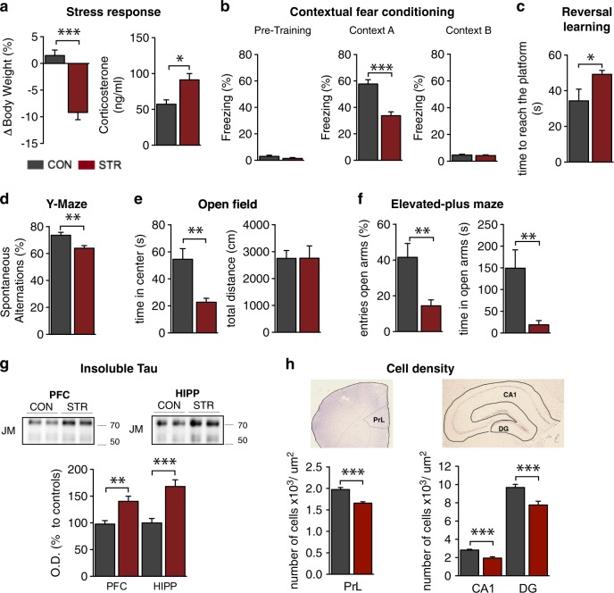
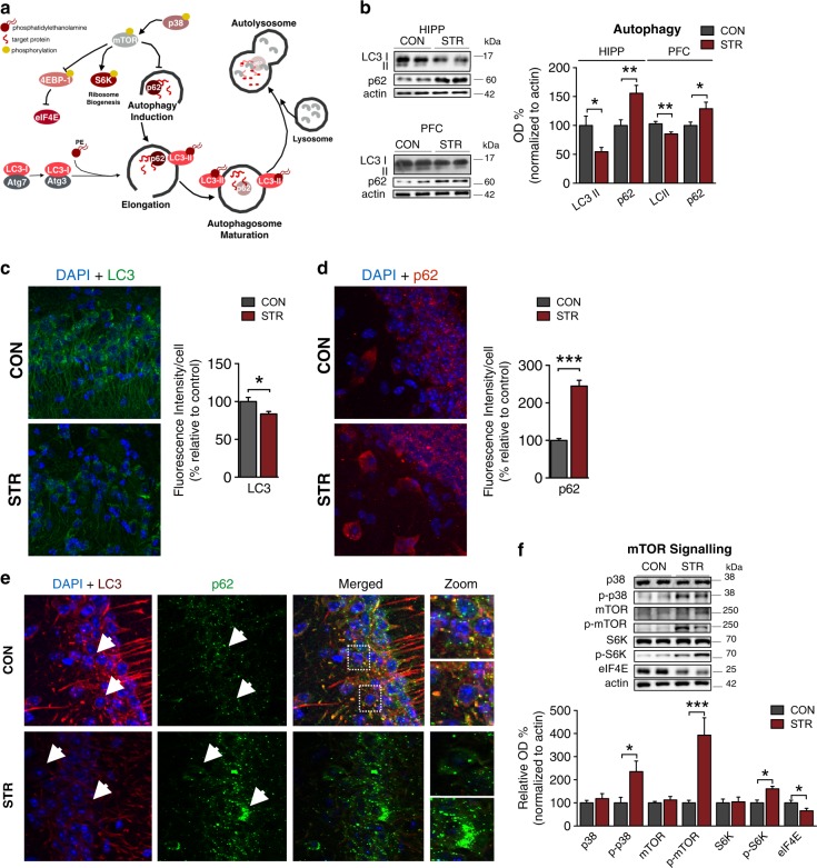
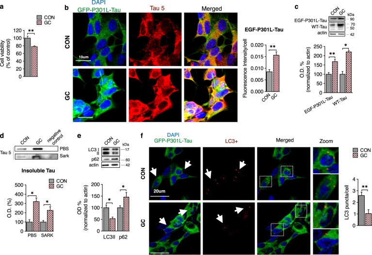
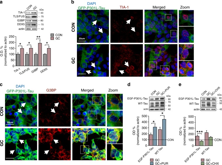
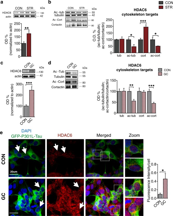
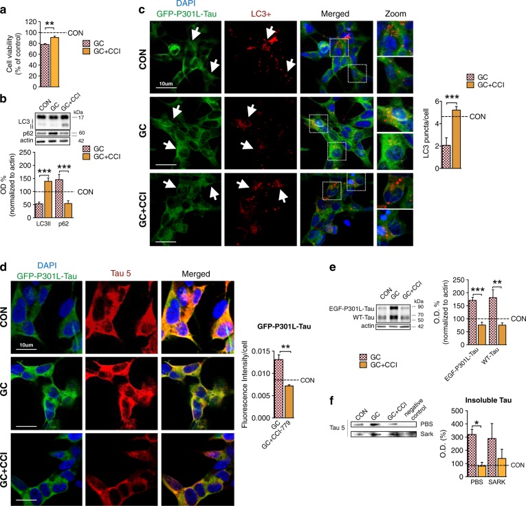
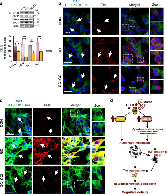
Imbalance of neuronal proteostasis associated with misfolding and aggregation of Tau protein is a common neurodegenerative feature in Alzheimer's disease (AD) and other Tauopathies. Consistent with suggestions that lifetime stress may be an important AD precipitating factor, we previously reported that environmental stress and high glucocorticoid (GC) levels induce accumulation of aggregated Tau; however, the molecular mechanisms for such process remain unclear. Herein, we monitor a novel interplay between RNA-binding proteins (RBPs) and autophagic machinery in the underlying mechanisms through which chronic stress and high GC levels impact on Tau proteostasis precipitating Tau aggregation. Using molecular, pharmacological and behavioral analysis, we demonstrate that chronic stress and high GC trigger mTOR-dependent inhibition of autophagy, leading to accumulation of Tau aggregates and cell death in P301L-Tau expressing mice and cells. In parallel, we found that environmental stress and GC disturb cellular homeostasis and trigger the insoluble accumulation of different RBPs, such as PABP, G3BP1, TIA-1, and FUS, shown to form stress granules (SGs) and Tau aggregation. Interestingly, an mTOR-driven pharmacological stimulation of autophagy attenuates the GC-driven accumulation of Tau and SG-related proteins as well as the related cell death, suggesting a critical interface between autophagy and the response of the SG-related protein in the neurodegenerative potential of chronic stress and GC. These studies provide novel insights into the RNA-protein intracellular signaling regulating the precipitating role of environmental stress and GC on Tau-driven brain pathology.


Conflict of interest statement

B.W. is co-founder and chief scientific officer of Aquinnah Pharmaceutics Inc. The other authors declare that they have no conflict of interest.

Figures

Fig. 1
Fig. 1
Chronic stress evokes accumulation of neurotoxic Tau aggregates causing cognitive and emotional deficits in P301L-Tau Tg mice. a P301L-Tg mice exhibited reduced body weight (p = 0.005) and increased corticosterone (p < 0.001) levels after chronic environmental stress. b In contrast to pre-training session (p = 0.171), stressed animals exhibited a significant decrease in percentage of freezing time in test section (context A) of CFC in comparison to control animals indicating hippocampus-dependent associative memory impairment (p < 0.001); note that both animal groups exhibit similar freezing levels in context B which is not associated with the adverse stimuli animals received in context A (p = 0.640). c Chronic stress increased the time that animals swum to reach the new (opposite) place of the escaping platform indicating PFC-dependent deficits of behavioral flexibility (p = 0.046). d Stress also reduced percentage of spontaneous alternations in the arms of a Y-maze as compared with control animals pointing to deficits of working memory (p = 0.007). ef Whereas no different in total distance traveled by animals in OF apparatus (p = 0.988), stressed animals exhibited a decrease in time spent in the center of the OF arena (p = 0.003) (e) followed by reduced time (p = 0.004) and entries (p = 0.001) spend in the open arms of EPM apparatus (f); these behavioral parameters suggest increased levels of anxiety in stressed animals compared with controls. gh Chronic stress elevated the levels of sarkosyl-insoluble Tau in both hippocampus and PFC of P301LTau mice (Hipp: p = 0.028; PFC: p < 0.001) (g); an effect that was accompanied by decreased cell density in PFC (prelimbic cortex; PrL: p < 0.001) and hippocampus (DG: p < 0.001; CA1: p < 0.001) (h). All numeric data represent mean ± SEM, *p < 0.05; **p < 0.01; ***p < 0.001
Fig. 2
Fig. 2
Prolonged exposure to environmental stress inhibits autophagy in a mTOR-dependent manner. a Schematic representation of autophagy highlighting the role of mTOR, LC3, and p62 in this cellular process. be Stressed animals exhibited reduced LC3 (PFC: p = 0.003; HIPP: p = 0.026) and increased p62 (PFC: p = 0.029; HIPP: p = 0.004) protein levels as assessed by WB analysis (b); these findings were confirmed by corresponding changes in LC3 (p = 0.044) and p62 (p = 0.044) fluorescence intensity/cell number (cd) an decrease in their co-localization (e), indicating a stress-driven inhibition of autophagic process. f In line with the above findings, the levels of phosphorylated S6K (p = 0.014), p38 (p = 0.030), and mTOR (p < 0.001) proteins were increased by stress, with decreased levels of eIF4E (p = 0.024), which are indicative of mTOR activation. All numeric data represent mean ± SEM, *p < 0.05; **p < 00.01; ***p < 00.001
Fig. 3
Fig. 3
Exposure to glucocorticoids causes Tau accumulation and autophagy inhibition in vitro. a GC treatment (10−6 M; 48 h) of EGF-P301L-hTau SH-SY5Y cells decreased cell viability (p = 0.004). bd Glucocorticoid (GC) treatment triggered cytoplasmic accumulation of exogenously expressed mutated human Tau (EGF-P301L-Tau) and endogenous human Tau (WT-Tau) as assessed by IF (f) (p = 0.044) (b) and WB analysis (c) (WT-Tau: p < 0.001; EGF-P301L-Tau: p < 0.001), and lead to increased levels of insoluble Tau aggregates (p = 0.042) (d). e-f GC decreased LC3II (p = 0.013) protein levels with parallel increase of p62 (p = 0.039) (e); immunofluorescence analysis confirmed the GC-induced reduction in LC3 (p = 0.004) puncta (f). All numeric data represent mean ± SEM, *p < 0.05; **p < 0.01; ***p < 0.001
Fig. 4
Fig. 4
Chronic stress evokes dysregulation of RBPs and their insoluble accumulation in P301L-Tau Tg mice. a, b Chronic stress triggered an increase in the protein levels of several RBPs and SG markers in soluble fraction [TIA-1 (p = 0.013), TLS/FUS (p = 0.017), DDX5 (p < 0.001) and EWRS1 (p = 0.005)] (a), as well in insoluble fraction of P301L-Tau mice [DDX5 (p = 0.02) and PABP (p = 0.011)] (b). c, d Chronic stress causes the cytoplasmic accumulation of the SG marker TIA-1 in hippocampus (c) and the accumulation of PABP, a SG marker (d), increasing their co-localization with p-Tau (PHF1). e Moreover, chronic stress leads to an increase co-localization of TIA-1 and PABP in perinuclear region of hippocampal neurons, indicating the presence of TIA-1 positive stress granules. f Immunofluroresecent analysis of PABP and DDX with p-Tau in human AD brain. All numeric data are represented as mean ± SEM, *p < 0.05; **p < 0.01; ***p < 0.001
Fig. 5
Fig. 5
Dysruption and insoluble accumulation of RBPs in GC-driven Tau accumulation. Treatment of EGF-P301L-hTau SH-SY5Y cells with GC (10−6 M; 48 h) elevated the cytoplasmic levels of the RBPs, TIA-1 (p = 0.043), TLS/FUS (p = 0.034), G3BP (p = 0.004), and DDX5 (p = 0.038) as assessed by WB analysis. b, c IF staining of TIA-1 (b) and G3BP (c) demonstrate that GC triggered their accumulation and cytoplasmic appearance in EGF-P301L-hTau SH-SY5Y cells. d Co-treatment of GC and puromycin (PUR), a well-known SG inducer, aggravate levels of EGF-P301L-Tau (p = 0.013) and WT-Tau (p = 0.023) when compared to GC treatment. e While co-treament of GC and CHX (the later inhibits SG formation) blocked the GC-driven Tau increase (p < 0.001). All numeric data are represented as mean ± SEM, *p < 0.05; **p < 0.01; ***p < 0.001
Fig. 6
Fig. 6
Stress and glucocorticoids induce HDAC6 reducing the acetylation levels of its cytoplasmic targets. a, b Chronic stress elevated HDAC6 levels (p = 0.004) (a) and reduced acetylation of tubulin (p = 0.016) and cortactin (p = 0.048), two cytoskeletal targets of HDAC6, in P301L-Tau mice (b). ce GC treatment increased HDAC6 (p < 0.001) as analyzed by WB (c) and elevated HDAC6 staining in P301LTau cells (p = 0.041) (e), in parallel with decreased levels of acetylated forms of tubulin (p = 0.004) and cortactin (p < 0.001), two cytoskeletal targets of HDAC6 (d). All numeric data are represented as mean ± SEM, *p < 0.05; **p < 0.01; ***p < 0.001
Fig. 7
Fig. 7
mTOR-driven pharmacological stimulation of autophagy blocked GC-triggered Tau accumulation. a, b Combined treatment of GC with CCI-779 (Temsirolimus), a rapamycin analog, attenuated the GC-driven decrease of cell viability (p = 0.002) in EGF-P301LTau SH-SY5Y cells (a) and reversed the GC effect on autophagy markers, increasing protein levels of LC3II (p < 0.001) with parallel decrease of p62 levels (p = 0.001) (b). c IF staining of LC3 confirmed the CCI-779-evoked blockage of GC-driven reduction of LC3 puncta (p = 0.0012). df CCI-779 treatment reduced the GC-driven elevated IF levels of GFP-P301LTau (p = 0.002) (d); WB analysis revealed that CCI-779 attenuated the GC-driven accumulation of both EGF-P301L-Tau (p < 0.005) and WT-Tau (p < 0.001) levels (e), also leading to decrease in insoluble Tau levels (f). All numeric data are represented mean ± SEM, *p < 0.05; **p < 0.01; ***p < 0.0001
Fig. 8
Fig. 8
The CCI-779 inhibitor of mTOR attenuated GC-induced dysregulation of RBPs. ac GC-driven increase of different RBPs [TLS/FUS G3BPand DDX5] was attenuated by co-treatment with CCI-779 (p = 0.022; p = 0.007; p = 0.045, respectively) (a); IF staining of TIA-1 (b) and G3BP (c) confirmed the blockage of GC-driven induction of RBPs by CCI-779. c Working/hypothetical model integrating autophagy inhibition and dysregulation of RBPs in the cellular mechanisms through which chronic stress and/or high levels of stress hormones, glucocorticoids (GC), precipitate Tau pathology. Chronic exposure to environmental stress and/or prolonged signaling of GC receptors (GR) evoke the activation of mTOR signaling and the induction of Histone deacetylase 6 (HADC6) and subsequently, reduce acetylation of proteins related to cytoskeletal instability. These cellular events lead to inhibition of autophagic process that, together with the dysregulation and insolubilization of RBPs and the potential formation of SGs, may contribute to the accumulation of Tau and its neurotoxic aggregates causing cell death and cognitive deficits

Similar articles

Cited by

References

    1. Sotiropoulos I, Catania C, Pinto LG, Silva R, Pollerberg GE, Takashima A, et al. Stress acts cumulatively to precipitate Alzheimer’s disease-like Tau pathology and cognitive deficits. J Neurosci. 2011;31:7840–7. doi: 10.1523/JNEUROSCI.0730-11.2011. - DOI - PMC - PubMed
    1. Sotiropoulos I, Cerqueira J, Catania C, Takashima A, Sousa N, Almeida O. Stress and glucocorticoid footprints in the brain—the path from depression to Alzheimer’s disease. Neurosci Biobehav Rev. 2008;32:1161–73. doi: 10.1016/j.neubiorev.2008.05.007. - DOI - PubMed
    1. Launer LJ, Andersen K, Dewey ME, Letenneur L, Ott A, Amaducci LA, et al. Rates and risk factors for dementia and Alzheimer’s disease: results from EURODEM pooled analyses. Neurology. 1999;52:78–84. doi: 10.1212/WNL.52.1.78. - DOI - PubMed
    1. Hatzinger M, Z’Brun A, Hemmeter U, Seifritz E, Baumann F, Holsboer-Trachsler E, et al. Hypothalamic–pituitary–adrenal system function in patients with Alzheimer’s disease. Neurobiol Aging. 1995;16:205–9. doi: 10.1016/0197-4580(94)00159-6. - DOI - PubMed
    1. Csernansky JG, Dong H, Fagan AM, Wang L, Xiong C, Holtzman DM, et al. Plasma cortisol and progression of dementia in subjects with Alzheimer-type dementia. Am J Psychiatry. 2006;163:2164–9. doi: 10.1176/ajp.2006.163.12.2164. - DOI - PMC - PubMed

Publication types