Review
. 2019 Feb;16(2):106-120.
doi: 10.1038/s41575-018-0079-5.

Control and dysregulation of redox signalling in the gastrointestinal tract

Affiliations
Review

Control and dysregulation of redox signalling in the gastrointestinal tract

Eric L Campbell et al. Nat Rev Gastroenterol Hepatol. 2019 Feb.

Abstract

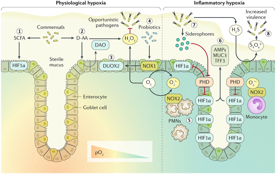
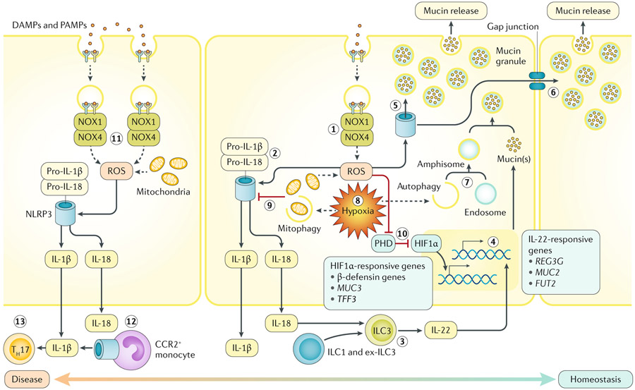
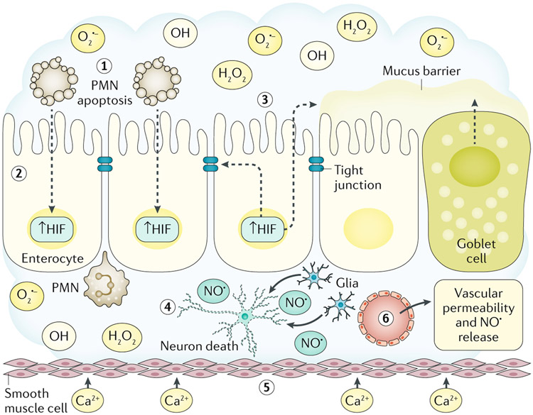
Redox signalling in the gastrointestinal mucosa is held in an intricate balance. Potent microbicidal mechanisms can be used by infiltrating immune cells, such as neutrophils, to protect compromised mucosae from microbial infection through the generation of reactive oxygen species. Unchecked, collateral damage to the surrounding tissue from neutrophil-derived reactive oxygen species can be detrimental; thus, maintenance and restitution of a breached intestinal mucosal barrier are paramount to host survival. Redox reactions and redox signalling have been studied for decades with a primary focus on contributions to disease processes. Within the past decade, an upsurge of exciting findings have implicated subtoxic levels of oxidative stress in processes such as maintenance of mucosal homeostasis, the control of protective inflammation and even regulation of tissue wound healing. Resident gut microbial communities have been shown to trigger redox signalling within the mucosa, which expresses similar but distinct enzymes to phagocytes. At the fulcrum of this delicate balance is the colonic mucosal epithelium, and emerging evidence suggests that precise control of redox signalling by these barrier-forming cells may dictate the outcome of an inflammatory event. This Review will address both the spectrum and intensity of redox activity pertaining to host-immune and host-microbiota crosstalk during homeostasis and disease processes in the gastrointestinal tract.


Figures

Fig. 1 ∣
Fig. 1 ∣. Host-microbial redox signalling during hypoxia.
Enzymatic utilization of molecular oxygen (O2) within the intestinal mucosa facilitates redox signalling and results in both spatial and dynamic patterns of O2 availability. In the healthy intestinal mucosa, a steep O2 gradient exists between the highly vascularized mucosa and the anoxic lumen. Thus, cells within the crypt stem cell niche normally experience higher partial pressures of oxygen (pO2; ~100 mmHg) than the luminal-facing epithelia (<10 mmHg), which are known to normally experience hypoxia at homeostasis. This physiological hypoxia is experienced by epithelia adjacent to the lumen and results in stabilization of hypoxia-inducible factor (HIF). Gut microbiota secreting short chain fatty acids (SCFAs), particularly butyrate, contribute to this physiological hypoxia and associated stabilization of HIF1α through increased oxidative phosphorylation (step 1). Luminal redox signalling initiated by resident microorganisms releasing d-amino acids (D-AA) stimulates the epithelium to secrete D-AA oxidase (DAO) into the lumen, which subsequently yields hydrogen peroxide (H2O2) (step 2). Apical expression of epithelial dual oxidase 2 (DUOX2) probably results in luminal secretion of H2O2, which contributes to limiting opportunistic pathogen niche expansion (step 3). Probiotic lactobacilli upregulate epithelial NADPH oxidase 1 (NOX1) expression, which in turn induces DUOX2 (step4). Epithelial-expressed NOX1 and DUOX2, utilizing microenvironmental O2, generate oxygen radicals to further contribute to luminal release of H2O2. During inflammatory hypoxia, infiltrating polymorphonuclear leukocytes (PMNs) and monocytes expressing NOX2 generate superoxide (O2·−), resulting in inhibition of prolyl hydroxylase enzymes (PHD) and stabilization of HIF deep into the crypt (step 5). HIF transcriptional activity induces expression of barrier protective factors such as antimicrobial peptides (AMPs), mucin 3 (MUC3) and trefoil factor 3 (TFF3) (step 6). Certain opportunistic pathogens release siderophores, sequestering iron and inhibiting PHD (step 7). Sulfur metabolism of the mucosa can be hijacked by opportunistic pathogens. Hydrogen sulfide (H2S) is routinely detoxified to thiosulfate; however, high levels of reactive oxygen species within the mucosa can result in tetrathionate (S4O62−) generation, which can be utilized by Salmonella serotypes to provide a competitive advantage (step 8).
Fig. 2 ∣
Fig. 2 ∣. Host redox–hypoxia crosstalk in the gastrointestinal mucosa.
The two major sources of endogenous reactive oxygen species (ROS) within the intestinal epithelium originate from mitochondria and NADPH oxidase 1 (NOX1) or NOX4 (step 1). In response to pathogen-associated molecular patterns (PAMPs) and danger-associated molecular patterns (DAMPs), epithelia recruit and activate the NOX1–NOX4 complex, stimulating superoxide and hydrogen peroxide generation (sources of ROS). Both enzymatic and mitochondria-derived ROS can trigger the activity of epithelial inflammasomes. In colonic epithelia, ROS-stimulated NOD-, LRR- and pyrin domain-containing 3 (NLRP3) inflammasome assembly leads to IL-18 (and IL-1β) production (step 2). Although excessive mature secreted IL-18 is detrimental to epithelial integrity, the presence of IL-18 is necessary for IL-22 release by type 3 innate lymphoid cells (ILC3s) (step 3). ILC3-derived IL-22 promotes mucosal barrier protection by inducing mucin synthesis and goblet cell function (step 4). In goblet cells, ROS triggers the NLRP6 inflammasome to elicit mucin granule secretion (step 5). Sentinel goblet cells responding to microbial triggers can signal to adjacent goblet cells to degranulate via gap junctions (step 6). A combination of autophagic proteins, endosomes and NOX-derived ROS are necessary for mucin granule formation in goblet cells (step 7). Both autophagy and mitophagy are induced by hypoxia (step 8). Mitophagy might decrease NLRP3 inflammasome activity, reducing processing of IL-1β and IL-18 (step 9). Inhibition of prolyl hydroxylase enzymes (PHD) by ROS or hypoxia stabilizes hypoxia-inducible factor-1α (HIF1α), regulating barrier protective genes (step 10). Unimpeded or excessive ROS generation during active inflammation can lead to abundant maturation of IL-1β or IL-18 or even inflammasome-mediated cell death (necroptosis and pyroptosis) (step 11). Inflammasome-activation of infiltrating CC-chemokine receptor 2 (CCR2)+ monocytes contributes to active IL-1β (step 12). Mucosal IL-1β may lead to accumulation of IL-17A-secreting immune cells, such as T helper 17 cells (TH17) (step 13).
Fig. 3 ∣
Fig. 3 ∣. ROS collateral damage and gastrointestinal disease.
During active inflammation, reactive oxygen species (ROS; O2·−, OH· and H2O2) and reactive nitrogen species generated in the local microenvironment cause collateral tissue damage. Activated, transmigrating polymorphonuclear leukocytes (PMNs) consume large amounts of O2 in the generation of ROS in the local milieu (step 1). Under these conditions, PMN ROS generation is limited by rapid induction of PMN apoptosis. Such O2 consumption results in localized hypoxia and the stabilization of epithelial hypoxia-inducible factor (HIF) (step 2). Epithelial HIF stabilization activates a cascade of gene transcription that promotes expression of barrier protective function genes (for example, TFF3, ABCB1 and CLDN1) and mucins in goblet cells (step 3). Within the lamina propria, activation of glial cell inducible nitric oxide synthase and the generation of nitric oxide (NO·) leads to enteric nerve cell death, resulting in intestinal dysmotility (step 4). Smooth muscle responses to oxidative stress include increased Ca2+ permeability that perpetuates intestinal dysmotility (step 5). An early event in acute mucosal inflammation within the gastrointestinal tract is increased vascular permeability through the generation of NO· by multiple cell types, such as smooth muscle cells, endothelial cells and enteric glia (step 6).

References

    1. Luissint AC, Parkos CA & Nusrat A Inflammation and the intestinal barrier: leukocyte-epithelial cell interactions, cell junction remodeling, and mucosal repair. Gastroenterology 151, 616–632 (2016). - PMC - PubMed
    1. Crosnier C, Stamataki D & Lewis J Organizing cell renewal in the intestine: stem cells, signals and combinatorial control. Nat. Rev. Genet 7, 349–359 (2006). - PubMed
    1. Clevers HC & Bevins CL Paneth cells: maestros of the small intestinal crypts. Annu. Rev. Physiol 75, 289–311 (2013). - PubMed
    1. Johansson ME & Hansson GC Immunological aspects of intestinal mucus and mucins. Nat. Rev. Immunol 16, 639–649 (2016). - PMC - PubMed
    1. Tailford LE, Crost EH, Kavanaugh D & Juge N Mucin glycan foraging in the human gut microbiome. Front. Genet 6, 81 (2015). - PMC - PubMed

Publication types

MeSH terms

Substances