Review
. 2018 Dec;15(1):33-51.
doi: 10.1038/s41574-018-0115-0.

The calcium-sensing receptor in physiology and in calcitropic and noncalcitropic diseases

Affiliations
Review

The calcium-sensing receptor in physiology and in calcitropic and noncalcitropic diseases

Fadil M Hannan et al. Nat Rev Endocrinol. 2018 Dec.

Abstract

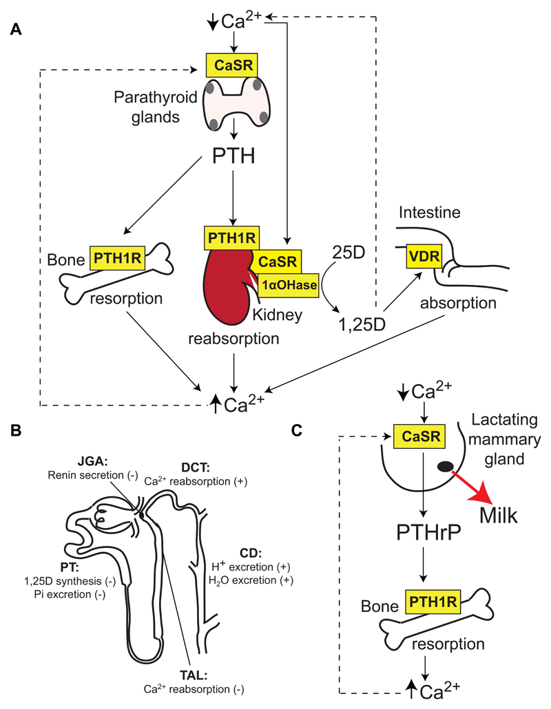
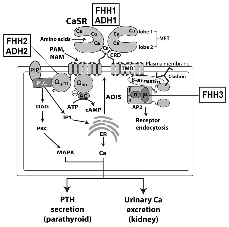
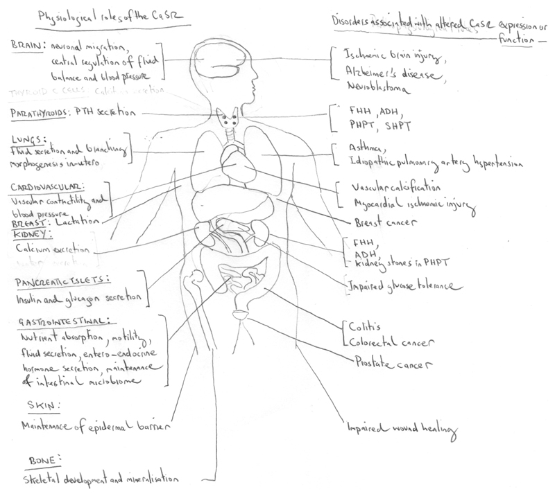
The Ca2+-sensing receptor (CaSR) is a dimeric family C G protein-coupled receptor that is expressed in calcitropic tissues such as the parathyroid glands and the kidneys and signals via G proteins and β-arrestin. The CaSR has a pivotal role in bone and mineral metabolism, as it regulates parathyroid hormone secretion, urinary Ca2+ excretion, skeletal development and lactation. The importance of the CaSR for these calcitropic processes is highlighted by loss-of-function and gain-of-function CaSR mutations that cause familial hypocalciuric hypercalcaemia and autosomal dominant hypocalcaemia, respectively, and also by the fact that alterations in parathyroid CaSR expression contribute to the pathogenesis of primary and secondary hyperparathyroidism. Moreover, the CaSR is an established therapeutic target for hyperparathyroid disorders. The CaSR is also expressed in organs not involved in Ca2+ homeostasis: it has noncalcitropic roles in lung and neuronal development, vascular tone, gastrointestinal nutrient sensing, wound healing and secretion of insulin and enteroendocrine hormones. Furthermore, the abnormal expression or function of the CaSR is implicated in cardiovascular and neurological diseases, as well as in asthma, and the CaSR is reported to protect against colorectal cancer and neuroblastoma but increase the malignant potential of prostate and breast cancers.


Conflict of interest statement

Competing interests:

F.M.H and R.V.T. have received grant funding from NPS/Shire Pharmaceuticals and GlaxoSmithKline for studies involving the use of calcium-sensing receptor allosteric inhibitors.

Figures

Figure 1
Figure 1. Role of the CaSR in Ca2+o homeostasis.
A. The CaSR is highly expressed in the parathyroid glands (grey), which are located adjacent and posterior to the thyroid gland (pink). The parathyroid CaSR detects reductions in Ca2+o, which leads to the release of PTH. PTH acts on the PTH1 receptor (PTH1R) to increase resorption of Ca2+ from bone, promote urinary Ca2+ reabsorption, and enhance expression of the renal 1-α-hydroxylase (1αOHase) enzyme, which converts the 25-hydroxyvitamin D (25D) precursor metabolite to biologically active 1,25-dihydroxyvitamin D (1,25D). The elevated 1,25D increases absorption of dietary calcium by acting on the intestinal vitamin D receptor (VDR). The kidney CaSR acts independently of PTH to regulate urinary Ca2+ reabsorption,. Increases in Ca2+o and 1,25D concentrations lead to negative feedback on the parathyroid glands, thereby inhibiting further PTH release. B. Nephron segment-specific roles of the CaSR. The CaSR is expressed in the: apical membrane of the proximal tubule (PT), where it regulates 1,25D synthesis and phosphate (Pi) excretion; basolateral membrane of the cortical thick ascending limb (TAL) of the Loop of Henle, and apical and basolateral membranes of the distal convoluted tubule (DCT), where it regulates Ca2+ reabsorption; apical and basolateral membranes of the collecting duct (CD), where it regulates H+ and water excretion; and juxtaglomerular apparatus (JGA), where it regulates renin secretion,. (+), stimulatory action of CaSR; (-), inhibitory action of CaSR. C. During lactation, the mammary gland CaSR detects reductions in Ca2+o, which leads to increased PTHrP secretion from mammary epithelial cells into the circulation. PTHrP acts on the PTH1R to increase bone resorption, which in turn releases Ca2+o for milk production. Stimulatory and inhibitory actions are indicated by solid lines and dashed lines, respectively.
Figure 2
Figure 2. CaSR signalling and trafficking.
The CaSR is functionally active as a constitutive homodimer, and may also form heterodimers with other family C GPCRs such as the metabotropic glutamate receptors and gamma-amino butryric acid (GABA) type B receptors in growth plate chondrocytes and the central nervous system–. The CaSR ECD binds calcium (Ca2+) at multiple sites within the lobes of the venus flytrap (VFT) and cysteine-rich domain (CRD). The CaSR also binds amino acids within the VFT cleft. Synthetic positive allosteric modulators (PAMs) and negative allosteric modulators (NAMs) bind to the CaSR transmembrane domain (TMD). The binding of Ca2+ leads to Gq/11-dependent activation of phospholipase C (PLC) and the production of diacylglycerol (DAG) and inositol 1,4,5-trisphosphate (IP3) from membrane bound phosphatidylinositol 4,5-bisphosphate (PIP2). The increase in intracellular IP3 levels facilitates the release of Ca2+ from intracellular stores such as the endoplasmic reticulum (ER). DAG activates protein kinase C (PKC) and the mitogen-activated protein kinase (MAPK) pathway. The CaSR also activates the Gi/o protein, which leads to inhibition of adenylate cyclase (AC)-mediated production of cAMP. These signalling events cause a decrease in parathyroid hormone (PTH) secretion and reduction in renal tubular Ca2+ reabsorption. CaSR cell-surface expression is regulated by agonist-driven insertional signalling (ADIS), which mediates anterograde receptor trafficking; and also by an endocytic complex comprising clathrin, β-arrestin and the heterotetrameric adaptor-related protein complex 2 (AP2) complex, which mediates retrograde receptor trafficking. Loss- and gain-of function mutations of the CaSR lead to FHH1 and ADH1, respectively; loss- and gain-of function mutations of the Gα11 subunit are associated with FHH2 and ADH2, respectively; and loss-of-function mutations of the AP2σ subunit are associated with FHH3.
Figure 3
Figure 3. Disruption of a salt-bridge within the CaSR TMD causes biased signalling.
The salt-bridge is formed by the Arg680 residue located in the proximal portion of transmembrane helix 3 (TM3, shaded blue) and the Glu767 residue located in extracellular loop 2 (ECL2). The Arg680-Glu767 salt-bridge is situated at the entrance of the allosteric modulator binding pocket, which is formed by residues from TM3 and TM5-TM7. The Arg680 residue mediates the binding of the NPS 2143 calcilytic compound. The Arg680-Glu767 salt-bridge is associated with G-protein-mediated signalling, whereas disruption of the salt-bridge by the ADH-causing CaSR mutation, Arg680Gly, selectively increases β-arrestin-mediated signalling, as well as abrogating the inhibitory effect of the NPS 2143 compound.
Figure 4
Figure 4. Physiological roles and disease associations of the CaSR.
The CaSR has key physiological roles in calcitropic tissues (e.g. parathyroid glands, kidneys and bone) and in non-calcitropic tissues (e.g. brain, cardiovascular system, lungs, breast, intestine and pancreas, and skin). Altered CaSR activity in calcitropic tissues causes inherited and sporadic diseases of Ca2+ homeostasis, whereas alterations in CaSR function or expression in non-calcitropic tissues is associated with cardiovascular disease, asthma and malignancy. ADH, autosomal dominant hypocalcaemia; FHH, familial hypocalciuric hypercalcaemia; PHPT, primary hyperparathyroidism; SHPT, secondary hyperparathyroidism.

References

    1. Brown EM, MacLeod RJ. Extracellular calcium sensing and extracellular calcium signaling. Physiol Rev. 2001;81:239–297. - PubMed
    1. Regard JB, Sato IT, Coughlin SR. Anatomical profiling of G protein-coupled receptor expression. Cell. 2008;135:561–571. doi: 10.1016/j.cell.2008.08.040. - DOI - PMC - PubMed
    1. Hannan FM, Babinsky VN, Thakker RV. Disorders of the calcium-sensing receptor and partner proteins: insights into the molecular basis of calcium homeostasis. J Mol Endocrinol. 2016;57:R127–142. doi: 10.1530/JME-16-0124. - DOI - PMC - PubMed
    1. Hannan FM, Thakker RV. Calcium-sensing receptor (CaSR) mutations and disorders of calcium, electrolyte and water metabolism. Best Pract Res Clin Endocrinol Metab. 2013;27:359–371. doi: 10.1016/j.beem.2013.04.007. - DOI - PubMed
    1. Ward DT, Brown EM, Harris HW. Disulfide bonds in the extracellular calcium-polyvalent cation-sensing receptor correlate with dimer formation and its response to divalent cations in vitro. J Biol Chem. 1998;273:14476–14483. - PubMed

Publication types

MeSH terms

Substances

Supplementary concepts