Review
. 2018 Oct 6;8(5):20180023.
doi: 10.1098/rsfs.2018.0023. Epub 2018 Aug 17.

The hallmarks of living systems: towards creating artificial cells

Affiliations
Review

The hallmarks of living systems: towards creating artificial cells

N Amy Yewdall et al. Interface Focus. .

Abstract

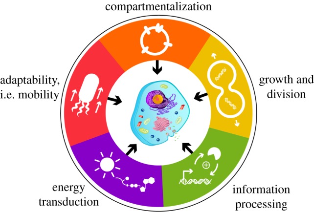
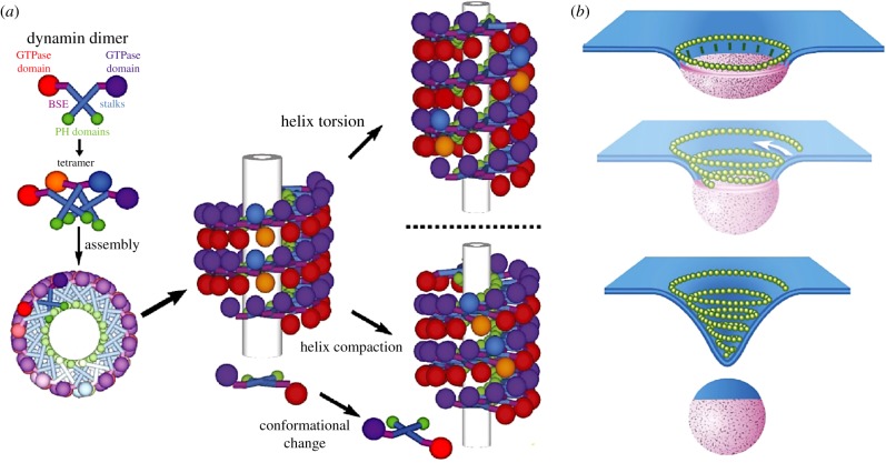
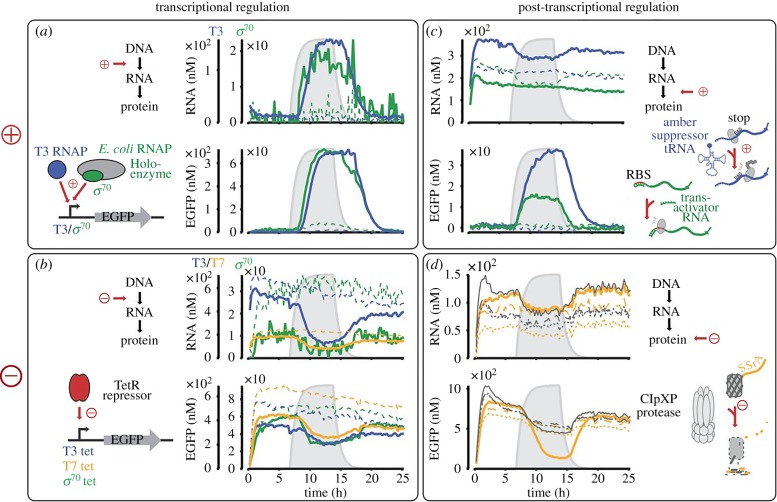
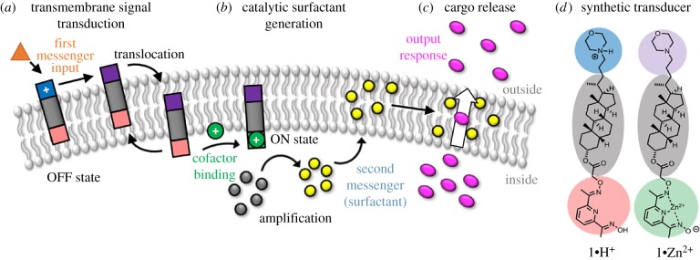
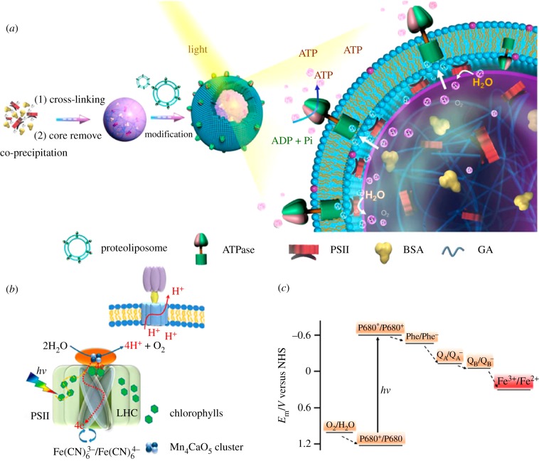
Despite the astonishing diversity and complexity of living systems, they all share five common hallmarks: compartmentalization, growth and division, information processing, energy transduction and adaptability. In this review, we give not only examples of how cells satisfy these requirements for life and the ways in which it is possible to emulate these characteristics in engineered platforms, but also the gaps that remain to be bridged. The bottom-up synthesis of life-like systems continues to be driven forward by the advent of new technologies, by the discovery of biological phenomena through their transplantation to experimentally simpler constructs and by providing insights into one of the oldest questions posed by mankind, the origin of life on Earth.

Keywords: adaptability; artificial cell; compartmentalization; division; energy transduction; information processing.


Conflict of interest statement

We have no competing interests.

Figures

Figure 1.
Figure 1.
The hallmarks of life. A summary of the five characteristics required for systems to live and thrive. In the last decade, developments in bio-analogous and bio-mimetic bottom-up technologies have emulated aspects of each hallmark to inform us about the functional mechanisms behind each process, and about the opportunities to construct integrated artificial cell platforms.
Figure 2.
Figure 2.
Schematic of G-protein-coupled receptor proteopolymersomes formed using IVTT. (a) Spontaneous insertion of G-protein-coupled receptors into polymersomes during cell-free expression. (b) Antibody and ligand-binding studies indicate that the G-protein-coupled receptor is reconstituted in an active conformation. Reproduced with permission from Wiley [15].
Figure 3.
Figure 3.
The osmotically induced formation of nanotubes from spherical polymersomes. DLL, D,L-lactide; DBU, 1,8-diazabicyclo[5.4.0]undec-7-ene; DCM, dichloromethane; RT, room temperature; PEG-PDDLA, poly(ethylene glycol)-poly(D,L-actide). Reproduced with permission from the American Chemical Society [30].
Figure 4.
Figure 4.
Hierarchical self-assembly of a terpolymer-stabilized coacervate protocell. Oppositely charged amylose biopolymers undergo complex coacervation and droplet formation, followed by interfacial self-assembly of terpolymer 1. (a) Confocal micrograph of terpolymer/coacervate protocells with internalized bovine serum albumin–fluorescein isothiocyante conjugate (purple) and terpolymer membrane (green, Nile Red). (b) Three-dimensional representation of the interfacial assembly of terpolymers. Reproduced with permission from the American Chemical Society [44].
Figure 5.
Figure 5.
The mechanoenzymes involved in membrane scission. (a) The helical arrangement of the dynamin dimers along the surface of a protein tube changes with protein conformation change to further constrict the membrane. (b) The proposed model for membrane constriction mediated by spiralling ESCRT proteins (in yellow). BSE, bundle signalling element; PH, pleckstrin homology. Reproduced with permission from the National Academy of Sciences [76] and eLife Science Publications [77].
Figure 6.
Figure 6.
Cell-free transcription and translation systems enable the expansion of engineered genetic circuits, to produce regulatory feedback networks for artificial cells. Regulation can be at the transcriptional or post-transcriptional level using proteins or RNA. Reproduced with permission from the National Academy of Sciences [110].
Figure 7.
Figure 7.
Bio-analogous membrane receptors that cause triggered release of cargo in response to an external signal. This process is mediated by hydrophobic small molecules rather than membrane proteins and provides an excellent alternative route to engineering artificial cell mimics. Reprinted with permission from the American Chemical Society [131].
Figure 8.
Figure 8.
(a) Co-assembly of ATPase and PSII in lipid-coated microspheres results in the formation of artificial chloroplasts. (b) The proton gradient generated by PSII drives the rotation of ATPase. (c) Redox potential scheme of active elements in the system. ADP, adeosine diphosphate; Pi, inorganic phosphate. Reproduced with permission from the American Chemical Society [142].
Figure 9.
Figure 9.
Schematic depiction of stomatocyte-based enzyme-driven nanomotors. (a) Solvent addition method for stomatocyte formation. (b) Assembly of the nanomotor with multiple enzymes entrapped inside the structure. The enzymes are responsible for generating the propelling jet of oxygen gas during the catalytic reaction. THF, tetrahydrofuran. Reproduced with permission from the American Chemical Society [149].

Similar articles

Cited by

References

    1. Mason AF, Thordarson P. 2017. Polymersomes as protocellular constructs. J. Polym. Sci. Part A Polym. Chem. 55, 3817–3825. (10.1002/pola.28780) - DOI
    1. Elani Y. 2016. Construction of membrane-bound artificial cells using microfluidics: a new frontier in bottom-up synthetic biology. Biochem. Soc. Trans. 44, 723–730. (10.1042/BST20160052) - DOI - PMC - PubMed
    1. Brea RJ, Hardy MD, Devaraj NK. 2015. Towards self-assembled hybrid artificial cells: novel bottom-up approaches to functional synthetic membranes. Chem. Eur. J. 21, 12 564–12 570. (10.1002/chem.201501229) - DOI - PMC - PubMed
    1. Buddingh’ BC, van Hest JCM. 2017. Artificial cells: synthetic compartments with life-like functionality and adaptivity. Acc. Chem. Res. 50, 769–777. (10.1021/acs.accounts.6b00512) - DOI - PMC - PubMed
    1. Spoelstra WK, Deshpande S, Dekker C. 2018. Tailoring the appearance: what will synthetic cells look like? Curr. Opin. Biotechnol. 51, 47–56. (10.1016/j.copbio.2017.11.005) - DOI - PubMed

LinkOut - more resources