

Alterations of brain local functional connectivity in amnestic mild cognitive impairment
- PMID: 30443345
- PMCID: PMC6220503
- DOI: 10.1186/s40035-018-0134-8
Alterations of brain local functional connectivity in amnestic mild cognitive impairment
Erratum in
-
Correction to: Alterations of brain local functional connectivity in amnestic mild cognitive impairment.Transl Neurodegener. 2018 Dec 24;7:33. doi: 10.1186/s40035-018-0140-x. eCollection 2018. Transl Neurodegener. 2018. PMID: 30603084 Free PMC article.
Abstract
Background: Resting-state functional magnetic resonance imaging studies using a regional homogeneity (ReHo) method have reported that amnestic mild cognitive impairment (aMCI) was associated with abnormalities in local functional connectivity. However, their results were not conclusive.
Methods: Seed-based d Mapping was used to conduct a coordinate-based meta-analysis to identify consistent ReHo alterations in aMCI.
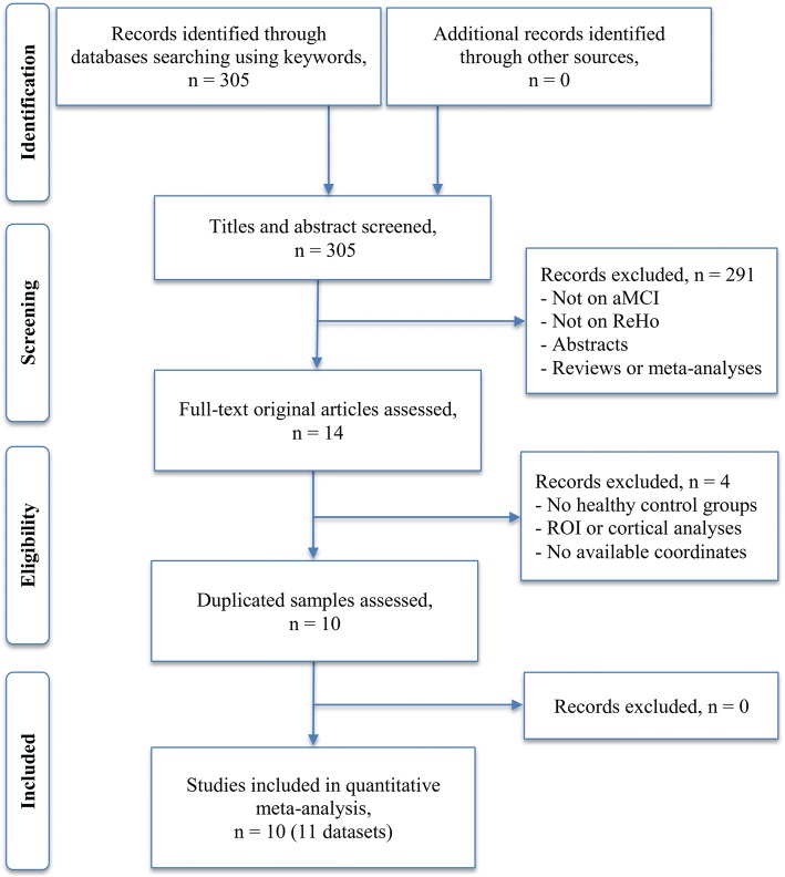
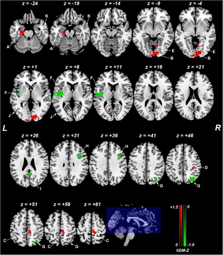
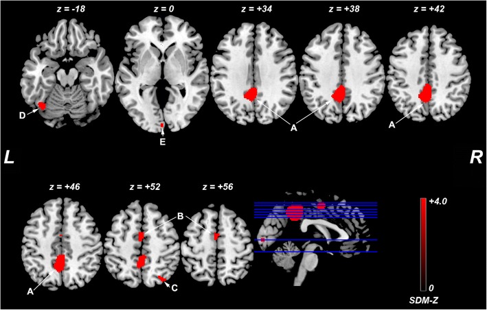
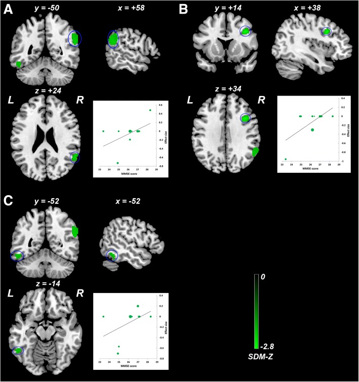
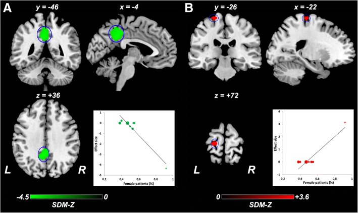
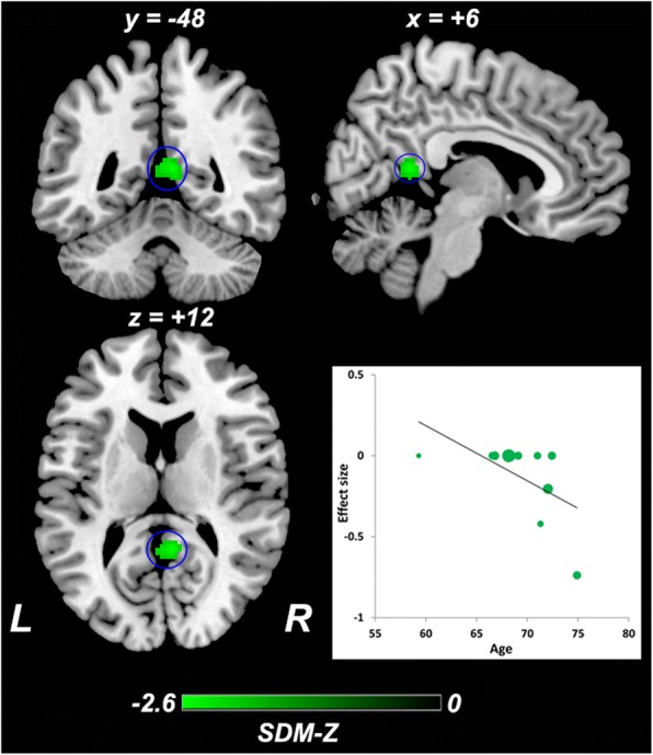
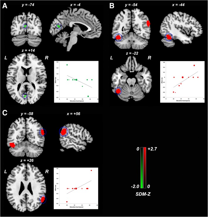
Results: We identified 10 studies with 11 datasets suitable for inclusion, including 378 patients with aMCI and 435 healthy controls. This meta-analysis identified significant ReHo alterations in patients with aMCI relative to healthy controls, mainly within the default mode network (DMN) (bilateral posterior cingulate cortex [PCC], right angular gyrus, bilateral middle temporal gyri, and left parahippocampal gyrus/hippocampus), executive control network (right superior parietal lobule and dorsolateral prefrontal cortex), visual network (right lingual gyrus and left middle occipital gyrus), and sensorimotor network (right paracentral lobule/supplementary motor area, right postcentral gyrus and left posterior insula). Significant heterogeneity of ReHo alterations in the bilateral PCC, left parahippocampal gyrus/hippocampus, and right superior parietal lobule/angular gyrus was observed. Exploratory meta-regression analyses indicated that general cognitive function, gender distribution, age, and education level partially contributed to this heterogeneity.
Conclusions: This study provides provisional evidence that aMCI is associated with abnormal ReHo within the DMN, executive control network, visual network, and sensorimotor network. These local functional connectivity alterations suggest coexistence of functional deficits and compensation in these networks. These findings contribute to the modeling of brain functional connectomes and to a better understanding of the neural substrates of aMCI. Confounding factors merit much attention and warrant future investigations.
Keywords: Amnestic mild cognitive impairment; Default mode network; Meta-analysis; Regional homogeneity; Resting-state functional magnetic resonance imaging; Seed-based d mapping.
Conflict of interest statement
This article does not contain any studies with human participants performed by any of the authors.Not applicable.The authors declare that they have no competing interests.
Figures
Similar articles
-
Regional homogeneity changes in amnestic mild cognitive impairment patients.Neurosci Lett. 2016 Aug 26;629:1-8. doi: 10.1016/j.neulet.2016.06.047. Epub 2016 Jun 23. Neurosci Lett. 2016. PMID: 27345927
-
Aberrant default mode network in amnestic mild cognitive impairment: a meta-analysis of independent component analysis studies.Neurol Sci. 2018 May;39(5):919-931. doi: 10.1007/s10072-018-3306-5. Epub 2018 Mar 6. Neurol Sci. 2018. PMID: 29511960
-
Mediation of episodic memory performance by the executive function network in patients with amnestic mild cognitive impairment: a resting-state functional MRI study.Oncotarget. 2016 Oct 4;7(40):64711-64725. doi: 10.18632/oncotarget.11775. Oncotarget. 2016. PMID: 27589839 Free PMC article.
-
Functional MRI-Specific Alterations in Salience Network in Mild Cognitive Impairment: An ALE Meta-Analysis.Front Aging Neurosci. 2021 Jul 26;13:695210. doi: 10.3389/fnagi.2021.695210. eCollection 2021. Front Aging Neurosci. 2021. PMID: 34381352 Free PMC article.
-
Aberrant spontaneous low-frequency brain activity in amnestic mild cognitive impairment: A meta-analysis of resting-state fMRI studies.Ageing Res Rev. 2017 May;35:12-21. doi: 10.1016/j.arr.2016.12.001. Epub 2016 Dec 23. Ageing Res Rev. 2017. PMID: 28017880 Review.
Cited by
-
Structural and functional brain abnormal alteration in patients with type 2 diabetes mellitus: A coordinate-based meta-analysis.Transl Psychiatry. 2025 Aug 6;15(1):269. doi: 10.1038/s41398-025-03488-z. Transl Psychiatry. 2025. PMID: 40770174 Free PMC article.
-
Diagnosis of Amnesic Mild Cognitive Impairment Using MGS-WBC and VGBN-LM Algorithms.Front Aging Neurosci. 2022 May 30;14:893250. doi: 10.3389/fnagi.2022.893250. eCollection 2022. Front Aging Neurosci. 2022. PMID: 35707699 Free PMC article.
-
Convergent Functional Changes of Default Mode Network in Mild Cognitive Impairment Using Activation Likelihood Estimation.Front Aging Neurosci. 2021 Oct 5;13:708687. doi: 10.3389/fnagi.2021.708687. eCollection 2021. Front Aging Neurosci. 2021. PMID: 34675797 Free PMC article.
-
Interactions between cigarette smoking and cognitive status on functional connectivity of the cortico-striatal circuits in individuals without dementia: A resting-state functional MRI study.CNS Neurosci Ther. 2022 Aug;28(8):1195-1204. doi: 10.1111/cns.13852. Epub 2022 May 4. CNS Neurosci Ther. 2022. PMID: 35506354 Free PMC article.
-
Alterations of Regional Homogeneity in Children With Congenital Sensorineural Hearing Loss: A Resting-State fMRI Study.Front Neurosci. 2021 Oct 6;15:678910. doi: 10.3389/fnins.2021.678910. eCollection 2021. Front Neurosci. 2021. PMID: 34690668 Free PMC article.
References
LinkOut - more resources
Full Text Sources
