. 2018 Nov 1:8:497.
doi: 10.3389/fonc.2018.00497. eCollection 2018.

Membrane Hsp70-A Novel Target for the Isolation of Circulating Tumor Cells After Epithelial-to-Mesenchymal Transition

Affiliations

Membrane Hsp70-A Novel Target for the Isolation of Circulating Tumor Cells After Epithelial-to-Mesenchymal Transition

Stephanie Breuninger et al. Front Oncol. .

Abstract

The presence of circulating tumor cells (CTCs) in the peripheral blood is a pre-requisite for progression, invasion, and metastatic spread of cancer. Consequently, the enumeration and molecular characterization of CTCs from the peripheral blood of patients with solid tumors before, during and after treatment serves as a valuable tool for categorizing disease, evaluating prognosis and for predicting and monitoring therapeutic responsiveness. Many of the techniques for isolating CTCs are based on the expression of epithelial cell surface adhesion molecule (EpCAM, CD326) on tumor cells. However, the transition of adherent epithelial cells to migratory mesenchymal cells (epithelial-to-mesenchymal transition, EMT)-an essential element of the metastatic process-is frequently associated with a loss of expression of epithelial cell markers, including EpCAM. A highly relevant proportion of mesenchymal CTCs cannot therefore be isolated using techniques that are based on the "capture" of cells expressing EpCAM. Herein, we provide evidence that a monoclonal antibody (mAb) directed against a membrane-bound form of Hsp70 (mHsp70)-cmHsp70.1-can be used for the isolation of viable CTCs from peripheral blood of tumor patients of different entities in a more quantitative manner. In contrast to EpCAM, the expression of mHsp70 remains stably upregulated on migratory, mesenchymal CTCs, metastases and cells that have been triggered to undergo EMT. Therefore, we propose that approaches for isolating CTCs based on the capture of cells that express mHsp70 using the cmHsp70.1 mAb are superior to those based on EpCAM expression.

Keywords: EpCAM (CD326); circulating tumor cells (CTCs); cmHsp70.1 antibody; epithelial-to-mesenchymal transition (EMT); membrane Hsp70.


Figures
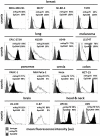
Figure 1
Figure 1
Membrane Hsp70 and EpCAM expression by different cancer cell lines. Cell lines from different cancer entities (breast, lung, melanoma, pancreas, cervix, colon, brain, head, and neck) were screened for their expression of mHsp70 and EpCAM using flow cytometry. Gray histograms represent isotype control, white histograms mHsp70 staining and black histograms EpCAM staining. The numbers in the histograms indicate the proportion of cells stained positively for mHsp70 and EpCAM. The data show one representative result of at least three independent experiments.
Figure 2
Figure 2
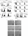
Influence of TGFβ- and L-lactic-acid (Lac-Ac)-induced EMT on mHsp70 and EpCAM expression by DU145 (prostate), UP154 (head and neck), UD5 (head and neck), A549 (lung) cancer cells, and LS174T melanoma cells. (A) DU145 cells were cultured with TGFβ for 10 days, UP154 and UD5 cells were cultured for 2 days with L-lactic-acid (Lac-Ac), after which the expression of mHsp70 and EpCAM was determined. (B) A549 and LS174T cells were cultured either with TGFβ for 10 days or with L-lactic-acid (Lac-Ac) for 2 days, after which the expression of mHsp70 and EpCAM was determined on viable cells using flow cytometry. Moreover, non-adherent and adherent cells were analyzed separately in A549 cells for their mHsp70 and EpCAM expression. Gray histograms represent isotype control, white histograms mHsp70 staining and black histograms EpCAM staining. The proportion of cells stained positively for mHsp70 and EpCAM are indicated in each histogram. The data show one representative result of at least three independent experiments. (C) Representative photomicrographs of untreated and TGFβ treated A549 cells on day 10, day 10 + 4 days recovery without TGFβ and day 10 + 7 days recovery without TGFβ.
Figure 3
Figure 3
Recovery of tumor cells from buffer and a mixture with EDTA blood. Two breast cancer cell lines were chosen for the recovery experiments with cmHsp70.1 mAb-functionalized magnetic bead system: SK-BR-3 with a high surface expression of Hsp70 and T47D with a low surface expression of Hsp70. (A) Tumor cells were spiked into buffer and incubated with the beads for 1 h at 37°C. The percentage of recovered cells aligned with the mHsp70 status of the cells (see Figure 1), as determined by flow cytometry, with 80 ± 11% of SK-BR-3 and 28 ± 13% of T47D cells being recovered. (B) Tumor cells were spiked into blood from healthy donors after depletion of erythrocytes and incubated with cmHsp70.1 mAb-functionalized magnetic beads for 1 h at 37°C. Recovery from blood was very similar to that in buffer, with an 83 ± 5% recovery of SK-BR-3 cells and a 34 ± 7% recovery of T47D cells. Data are from at least 3 independent experiments and are expressed as means ± SD.
Figure 4
Figure 4
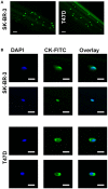
Capture of SK-BR-3 and T47D cells with the cmHsp70.1 mAb-functionalized CellCollector® system. (A) SK-BR-3 cells were stained with CFSE (green) and incubated with the cmHsp70.1 mAb-functionalized CellCollector® system. Captured cells were analyzed with a fluorescence microscope. Scale bar 100 μm. (B) SK-BR-3 or T47D cells were incubated with the cmHsp70.1 mAb-functionalized CellCollector® system for 30 min, fixed and stained with cytokeratin antibodies (CK-FITC, green) and Hoechst 33342 (DAPI, blue). Scale bar 25 μm.
Figure 5
Figure 5
Capture of SK-BR-3 tumor cells with the cmHsp70.1 mAb-functionalized CellCollector® system and subsequent cultivation and analysis. SK-BR-3 cells were captured from a mixture of the tumor cell line with EDTA blood. The functional part of the wire was cut from the detector and transferred to a cell culture flask. Cells that became adherent in the flask were expanded and finally analyzed with flow cytometry. Cells captured with the cmHsp70.1 mAb-functionalized wire (right panel) showed characteristics comparable to those of SK-BR-3 cells from cell culture (left panel). Gray histograms represent isotype control, white histograms mHsp70 staining and black histograms EpCAM staining. The numbers in the histograms indicate the proportion of cells stained positive for mHsp70 and EpCAM. Data are a representative result of at least three independent experiments.
Figure 6
Figure 6
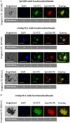
Capture of CTCs from the EDTA blood of patients using EpCAM mAb- or cmHsp70.1 mAb-functionalized magnetic beads. Representative views of singular (A) and clustered (B) CTCs derived from EDTA blood (7.5 ml, each) of patients with squamous cell carcinoma of the head and neck (SCCHN) and non-small cell lung carcinoma (NSCLC) bound to the EpCAM mAb- and cmHsp70.1 mAb-functionalized beads: brightfield, DAPI (blue), FITC-labeled cytokeratin antibody (CK-FITC, green), PE-labeled EpCAM (EpCAM-PE, red), overlay derived thereof. Scale bar 20 μm.

References

    1. Kan Z, Jaiswal BS, Stinson J, Janakiraman V, Bhatt D, Stern HM, et al. . Diverse somatic mutation patterns and pathway alterations in human cancers. Nature (2010) 466:869–73. 10.1038/nature09208 - DOI - PubMed
    1. Hanahan D, Weinberg RA. Hallmarks of cancer: the next generation. Cell (2011) 144:646–74. 10.1016/j.cell.2011.02.013 - DOI - PubMed
    1. Vignot S, Besse B, Andre F, Spano JP, Soria JC. Discrepancies between primary tumor and metastasis: a literature review on clinically established biomarkers. Crit Rev Oncol Hematol. (2012) 84:301–13. 10.1016/j.critrevonc.2012.05.002 - DOI - PubMed
    1. Viswanath B, Kim S. Influence of nanotoxicity on human health and environment: the alternative strategies. Rev Environ Contamin Toxicol. (2017) 242:61–104. 10.1007/398_2016_12 - DOI - PubMed
    1. Kuhlmann J, Wensing G. The applications of biomarkers in early clinical drug development to improve decision-making processes. Curr Clin Pharmacol. (2006) 1:185–91. 10.2174/157488406776872578 - DOI - PubMed