Review
. 2018 Nov 1:6:309.
doi: 10.3389/fped.2018.00309. eCollection 2018.

Pediatric Central Nervous System Tumors: State-of-the-Art and Debated Aspects

Affiliations
Review

Pediatric Central Nervous System Tumors: State-of-the-Art and Debated Aspects

Mitchell T Foster et al. Front Pediatr. .

Abstract

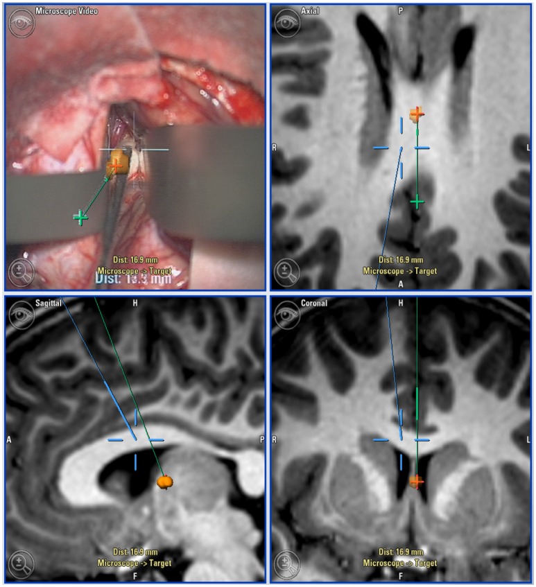
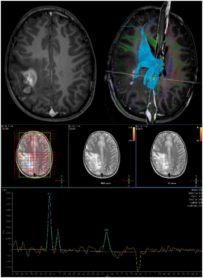
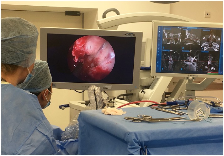
Pediatric neuro-oncology surgery continues to progress in sophistication, largely driven by advances in technology used to aid the following aspects of surgery: operative planning (advanced MRI techniques including fMRI and DTI), intraoperative navigation [preoperative MRI, intra-operative MRI (ioMRI) and intra-operative ultrasound (ioUS)], tumor visualization (microscopy, endoscopy, fluorescence), tumor resection techniques (ultrasonic aspirator, micro-instruments, micro-endoscopic instruments), delineation of the resection extent (ioMRI, ioUS, and fluorescence), and intraoperative safety (neurophysiological monitoring, ioMRI). This article discusses the aforementioned technological advances, and their multimodal use to optimize safe pediatric neuro-oncology surgery.

Keywords: intraoperative magnetic resonance imaging; neurooncology; pediatric brain tumors; pediatric neuroimaging; technology in surgery.


Figures

Figure 1
Figure 1
Neuro-navigation guided surgical approach: Top left: microscope view with navigation overlaid. Top right (axial), Bottom left (sagittal), and bottom right (coronal): Views of target lesion with planned trajectory (green) and microscope line of sight (blue).
Figure 2
Figure 2
Preoperative imaging for planning surgical approach and resection extent: Top Left: Axial post gadolinium T1 MRI. Top Right: DTI derived tractography to map white matter bundles in vicinity of tumor. Bottom: Multi-voxel MR Spectroscopy to define resection target.
Figure 3
Figure 3
Multimodal use of technology: The use of intraoperative endoscopy for an extended endo-nasal approach, in tandem with neuro-navigation and intraoperative MRI was vital in maximizing resection of this complex recurrent atypical meningioma.

References

    1. Colafati GS, Voicu IP, Carducci C, Miele E, Carai A, Di Loreto S, et al. . MRI features as a helpful tool to predict the molecular subgroups of medulloblastoma: state of the art. Ther Adv Neurol Disord. (2018) 11:1–14. 10.1177/1756286418775375 - DOI - PMC - PubMed
    1. Perreault S, Ramaswamy V, Achrol AS, Chao K, Liu TT, Shih D, et al. . MRI surrogates for molecular subgroups of medulloblastoma. Am J Neuroradiol. (2014) 35:1263–69. 10.3174/ajnr.A3990 - DOI - PMC - PubMed
    1. Marcus HJ, Vakharia VN, Ourselin S, Duncan J, Tisdall M, Aquilina K. Robot-assisted stereotactic brain biopsy: systematic review and bibliometric analysis. Childs Nerv Syst. (2018) 34:1299–1309. 10.1007/s00381-018-3821-y - DOI - PMC - PubMed
    1. Verploegh IS, Volovici V, Haitsma IK, Schouten JW, Dirven CM, Kros JM, et al. . Contemporary frameless intracranial biopsy techniques: Might variation in safety and efficacy be expected? Acta Neurochir. (2015) 157:2011–16; discussion 2016. 10.1007/s00701-015-2543-0 - DOI - PMC - PubMed
    1. Gempt J, Buchmann N, Ryang YM, Krieg S, Kreutzer J, Meyer B, et al. . Frameless image-guided stereotaxy with real-time visual feedback for brain biopsy. Acta Neurochir. (2012) 154:1663–7. 10.1007/s00701-012-1425-y - DOI - PubMed

LinkOut - more resources