Review
. 2018 Nov 15;114(1):2.
doi: 10.1007/s00395-018-0711-0.

Of mice and men: models and mechanisms of diabetic cardiomyopathy

Affiliations
Review

Of mice and men: models and mechanisms of diabetic cardiomyopathy

Christian Riehle et al. Basic Res Cardiol. .

Abstract

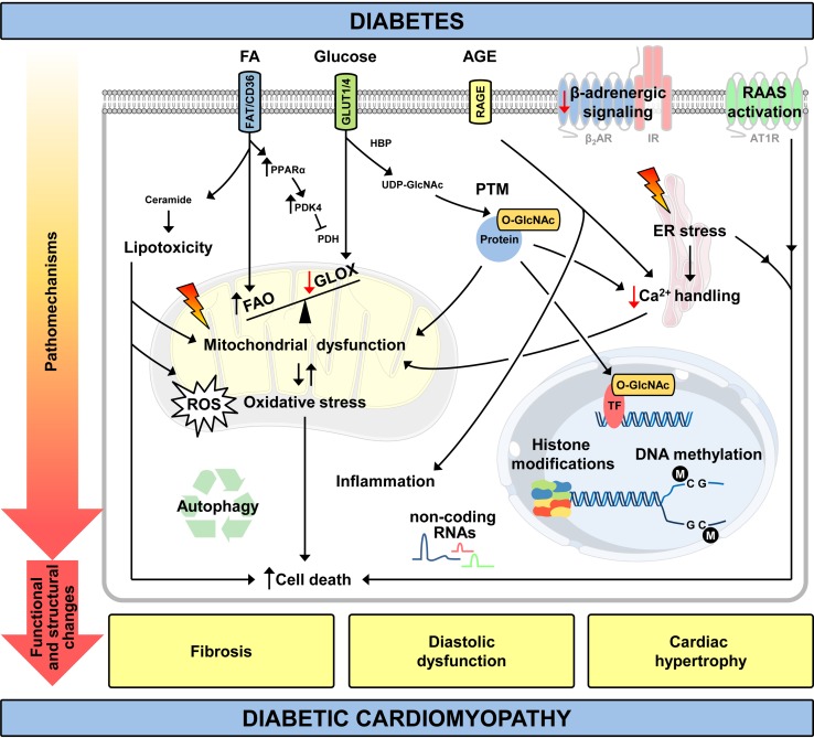
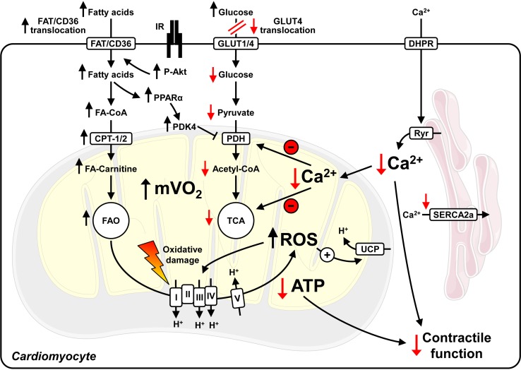
Diabetes mellitus increases the risk of heart failure independent of co-existing hypertension and coronary artery disease. Although several molecular mechanisms for the development of diabetic cardiomyopathy have been identified, they are incompletely understood. The pathomechanisms are multifactorial and as a consequence, no causative treatment exists at this time to modulate or reverse the molecular changes contributing to accelerated cardiac dysfunction in diabetic patients. Numerous animal models have been generated, which serve as powerful tools to study the impact of type 1 and type 2 diabetes on the heart. Despite specific limitations of the models generated, they mimic various perturbations observed in the diabetic myocardium and continue to provide important mechanistic insight into the pathogenesis underlying diabetic cardiomyopathy. This article reviews recent studies in both diabetic patients and in these animal models, and discusses novel hypotheses to delineate the increased incidence of heart failure in diabetic patients.

Keywords: Animal models; Cardiac energetics; Diabetes mellitus; Diabetic cardiomyopathy; Heart failure; Mitochondria.


Conflict of interest statement

The authors declare that they have no conflict of interest.

Figures

Fig. 1
Fig. 1
Pathomechanisms and clinical features of diabetic cardiomyopathy. ↑ increased/↓ decreased relative to normal conditions; AGE, advanced glycation end products; AT1R, angiotensin II receptor type 1; ER, endoplasmic reticulum; FAO, fatty acid (FA) oxidation; FAT/CD36, fatty acid translocase; GLOX, glucose oxidation; HBP, hexosamine biosynthetic pathway; IR, insulin receptor; PDH, pyruvate dehydrogenase; PDK4, pyruvate dehydrogenase kinase 4; PPARα, peroxisome proliferator activated receptor α; PTM, posttranslational modification; RAAS, renin–angiotensin–aldosterone system; RAGE, receptor for advanced glycation end products; ROS, reactive oxygen species; TF, transcription factor; UDP-GlcNAc, uridine diphosphate-N-acetylglucosamine; β2AR, β2-adrenergic receptor
Fig. 2
Fig. 2
Mitochondrial uncoupling and perturbed Ca2+ dynamics in cardiomyocytes of type 2 diabetic hearts. Hyperinsulinemia activates insulin receptors (IR) and Akt, contributing to increased fatty acid translocase (FAT/CD36) transport to the plasma membrane, increased fatty acid (FA) uptake and fatty acid oxidation (FAO). Impaired GLUT4 expression and translocation attenuate glucose uptake and utilization, which further increases FAO and myocardial oxygen consumption (mVO2). Increased FAO stimulates the generation of reactive oxygen species (ROS), which may induce damage to proteins involved in oxidative phosphorylation and may activate uncoupling proteins (UCPs). Increased mitochondrial uncoupling enhances mVO2 and FAO, and decreases mitochondrial ATP production. As the increase in mVO2 is not paralleled by increased ATP production and contractility, cardiac efficiency (cardiac work/mVO2) decreases. Perturbed intracellular Ca2+ handling (reduced sarcoplasmic reticulum Ca2+ release by ryanodine receptors (Ryr) and impaired re-uptake by SERCA2a) reduce peak cytosolic Ca2+ levels, which may further decrease contractility and intramitochondrial Ca2+ levels. This limits the activity of mitochondrial enzymes and further compromises contractile function. Note that mitochondrial uncoupling, increased ROS and decreased cardiac efficiency are not observed in rodent models of type 1 diabetes. I–V, mitochondrial electron transport chain complexes I–V; CoA, Coenzyme A; CPT, carnitine palmitoyltransferase; DHPR, dihydropyridine receptor; PDK4, pyruvate dehydrogenase kinase 4; PPARα, peroxisome proliferator activated receptor α; TCA, tricarboxylic acid cycle. ↑ increased/↓ decreased relative to normal conditions
Fig. 3
Fig. 3
Diastolic dysfunction in the absence of coronary artery disease in a patient with type 2 diabetes. a Preserved diastolic function in a normal subject as indicated by the E/A wave ratio (E: peak velocity blood flow in early diastole, A: peak velocity blood flow in late diastole caused by atrial contraction), b E′/A′ wave ratio (E′: peak mitral annular velocity during early diastolic filling, A′: peak mitral annular velocity during late diastolic filling caused by atrial contraction). Diastolic dysfunction in a patient with type 2 diabetes as indicated by c an abnormal high (“pseudonormal”) E/A wave ratio and E/E’ wave ratio as calculated from the values presented in panels (c) and (d). Images were adjusted to the same scales. Coronary angiogram of the e right coronary artery system and f left main coronary artery system from the same patient presented in panels (c/d) indicating no concomitant coronary artery disease

Similar articles

Cited by

References

    1. Aasum E, Belke DD, Severson DL, Riemersma RA, Cooper M, Andreassen M, Larsen TS. Cardiac function and metabolism in Type 2 diabetic mice after treatment with BM 17.0744, a novel PPAR-alpha activator. Am J Physiol Heart Circ Physiol. 2002;283:H949–H957. doi: 10.1152/ajpheart.00226.2001. - DOI - PubMed
    1. Aasum E, Hafstad AD, Severson DL, Larsen TS. Age-dependent changes in metabolism, contractile function, and ischemic sensitivity in hearts from db/db mice. Diabetes. 2003;52:434–441. doi: 10.2337/diabetes.52.2.434. - DOI - PubMed
    1. Action to Control Cardiovascular Risk in Diabetes Study G. Gerstein HC, Miller ME, Byington RP, Goff DC, Jr, Bigger JT, Buse JB, Cushman WC, Genuth S, Ismail-Beigi F, Grimm RH, Jr, Probstfield JL, Simons-Morton DG, Friedewald WT. Effects of intensive glucose lowering in type 2 diabetes. N Engl J Med. 2008;358:2545–2559. doi: 10.1056/NEJMoa0802743. - DOI - PMC - PubMed
    1. Alrob OA, Sankaralingam S, Ma C, Wagg CS, Fillmore N, Jaswal JS, Sack MN, Lehner R, Gupta MP, Michelakis ED, Padwal RS, Johnstone DE, Sharma AM, Lopaschuk GD. Obesity-induced lysine acetylation increases cardiac fatty acid oxidation and impairs insulin signalling. Cardiovasc Res. 2014;103:485–497. doi: 10.1093/cvr/cvu156. - DOI - PMC - PubMed
    1. Anderson EJ, Kypson AP, Rodriguez E, Anderson CA, Lehr EJ, Neufer PD. Substrate-specific derangements in mitochondrial metabolism and redox balance in the atrium of the type 2 diabetic human heart. J Am Coll Cardiol. 2009;54:1891–1898. doi: 10.1016/j.jacc.2009.07.031. - DOI - PMC - PubMed

Publication types