. 2019 Feb;13(2):338-357.
doi: 10.1002/1878-0261.12406. Epub 2019 Jan 10.

O-GlcNAc-induced nuclear translocation of hnRNP-K is associated with progression and metastasis of cholangiocarcinoma

Affiliations

O-GlcNAc-induced nuclear translocation of hnRNP-K is associated with progression and metastasis of cholangiocarcinoma

Chatchai Phoomak et al. Mol Oncol. 2019 Feb.

Abstract

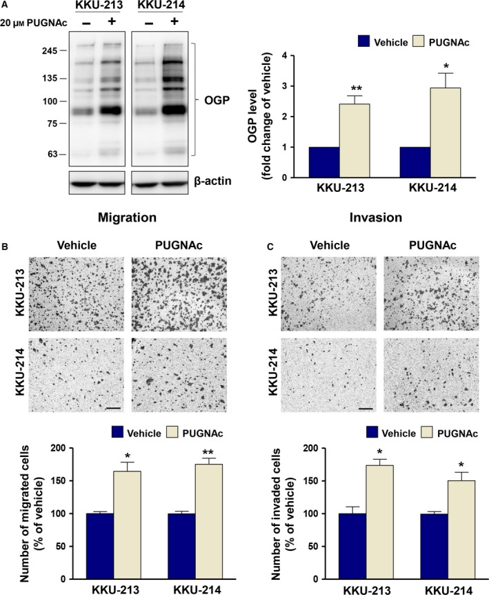
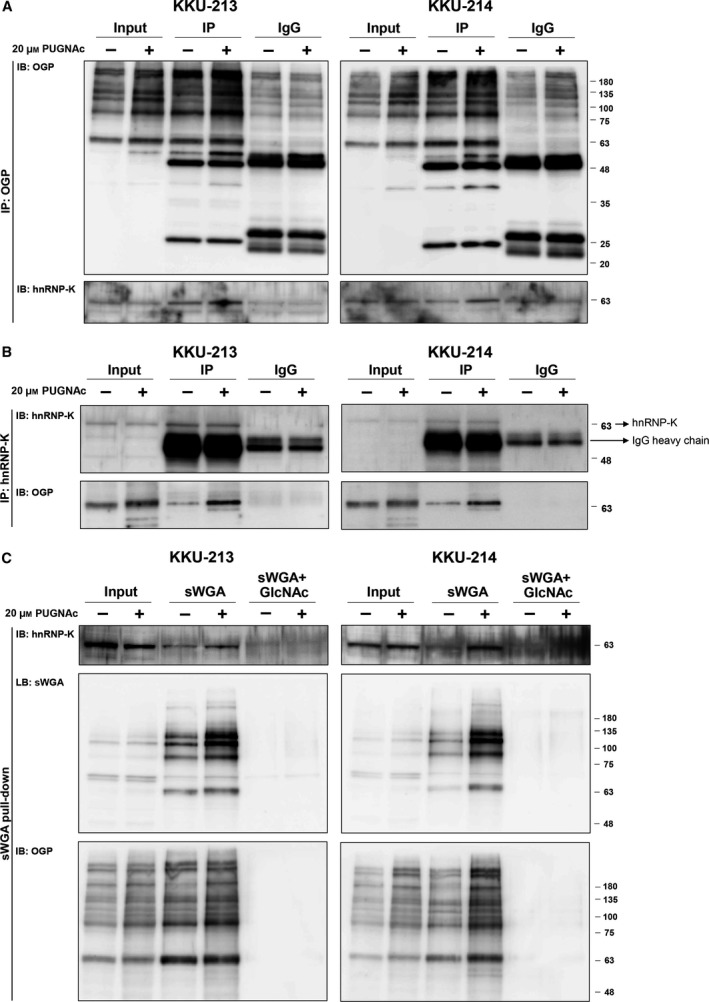
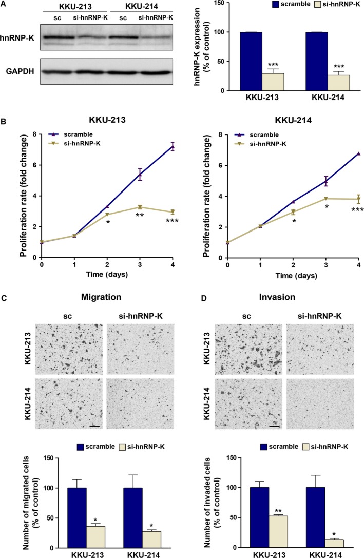
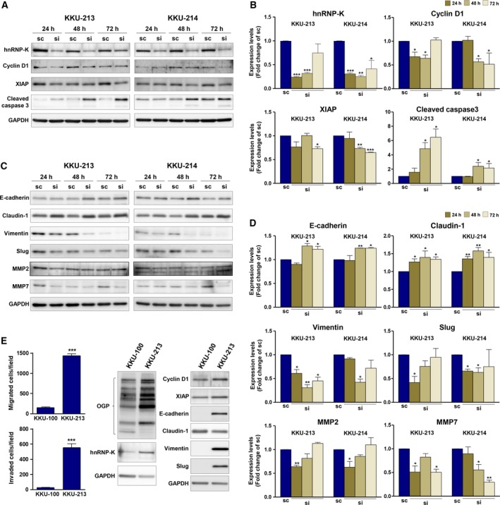
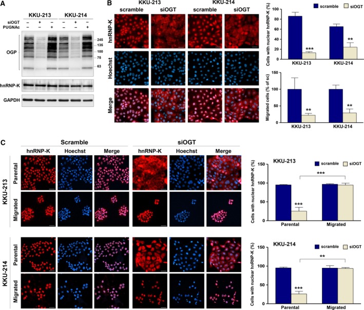
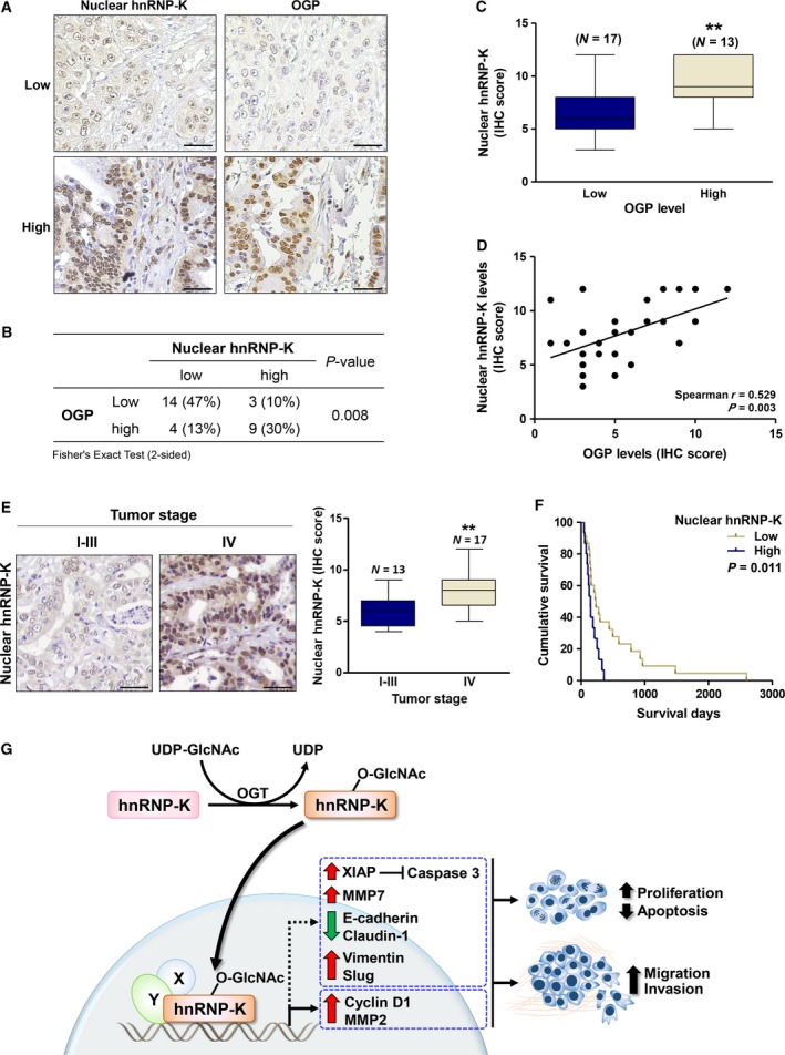
O-GlcNAcylation is a key post-translational modification that modifies the functions of proteins. Associations between O-GlcNAcylation, shorter survival of cholangiocarcinoma (CCA) patients, and increased migration/invasion of CCA cell lines have been reported. However, the specific O-GlcNAcylated proteins (OGPs) that participate in promotion of CCA progression are poorly understood. OGPs were isolated from human CCA cell lines, KKU-213 and KKU-214, using a click chemistry-based enzymatic labeling system, identified using LC-MS/MS, and searched against an OGP database. From the proteomic analysis, a total of 21 OGPs related to cancer progression were identified, of which 12 have not been previously reported. Among these, hnRNP-K, a multifaceted RNA- and DNA-binding protein known as a pre-mRNA-binding protein, was one of the most abundantly expressed, suggesting its involvement in CCA progression. O-GlcNAcylation of hnRNP-K was further verified by anti-OGP/anti-hnRNP-K immunoprecipitations and sWGA pull-down assays. The perpetuation of CCA by hnRNP-K was evaluated using siRNA, which revealed modulation of cyclin D1, XIAP, EMT markers, and MMP2 and MMP7 expression. In native CCA cells, hnRNP-K was primarily localized in the nucleus; however, when O-GlcNAcylation was suppressed, hnRNP-K was retained in the cytoplasm. These data signify an association between nuclear accumulation of hnRNP-K and the migratory capabilities of CCA cells. In human CCA tissues, expression of nuclear hnRNP-K was positively correlated with high O-GlcNAcylation levels, metastatic stage, and shorter survival of CCA patients. This study demonstrates the significance of O-GlcNAcylation on the nuclear translocation of hnRNP-K and its impact on the progression of CCA.

Keywords: O-GlcNAcylated proteins; bile duct cancer; heterogeneous nuclear ribonucleoprotein-K; metastasis.


Figures

Figure 1
Figure 1
O‐GlcNAcylation promotes CCA migration and invasion. CCA cells, KKU‐213 and KKU‐214, were treated with 20 μm PUGNAc for 24 h. (A) OGP levels were determined using western blot. (B) Migration and (C) invasion abilities of PUGNAc‐treated CCA cells were compared with those of the vehicle control cells. The results represent one of two independent experiments (mean ± SEM, *P < 0.05; **P < 0.01, Students’ t‐test). The images shown are 100 ×  magnification with 50 μm of scale bar.
Figure 2
Figure 2
Validation of hnRNP‐K O‐GlcNAcylation. CCA cells (KKU‐213 and KKU‐214) were treated with PUGNAc or vehicle for 24 h. The cell lysates were immunoprecipitated with either (A) anti‐OGP or (B) anti‐hnRNP‐K and probed with anti‐hnRNP‐K and anti‐OGP. Human immunoglobulin G (IgG) isotype was used as the controls of the specificity of the antibodies that were used in the immunoprecipitation assay. (C) The sWGA pull‐down assay was performed using sWGA‐conjugated agarose and probed with anti‐hnRNP‐K, sWGA, and anti‐OGP. GlcNAc neutralization was used to examine the specific binding of sWGA to the OGPs.
Figure 3
Figure 3
Suppression of hnRNP‐K reduces proliferation, migration, and invasion of CCA cells. The expression of hnRNP‐K was transiently suppressed by siRNA for 48 h prior to the migration and invasion assays. (A) The expression of hnRNP‐K was determined using western blot. (B) Cell proliferation, (C) migration, and (D) invasion abilities of si‐hnRNP‐K‐treated CCA cells were compared with those of the scramble siRNA (sc)‐treated cells. The migration and invasion assays were conducted for 9 h in KKU‐213 and 24 h in KKU‐214. The images shown are 100 ×  magnification with 50 μm scale bar. Data are mean ± SEM (*P < 0.05; **P < 0.01; ***P < 0.001, Students’ t‐test).
Figure 4
Figure 4
hnRNP‐K mediates the expression of growth‐ and metastasis‐related proteins in CCA cells. CCA cell lines, KKU‐213 and KKU‐214, were treated with si‐hnRNP‐K for 24, 48, and 72 h. The expression levels of growth‐ and metastasis‐related proteins were determined using western blot. Expression of GAPDH was used as an internal control. The expression of (A) hnRNP‐K, cyclin D1, XIAP, and cleaved caspase 3 as well as (C) EMT markers (E‐cadherin, claudin‐1, vimentin, and slug), and MMP2 and MMP7 was determined in si‐hnRNP‐K‐treated cells in comparison with those of scramble control cells. B and D are the quantitative analysis of (A) and (C) presented as mean ± SD from two independent experiments. (E) The endogenous expression of hnRNP‐K and its downstream targets was compared in 2 CCA cell lines (KKU‐100 and KKU‐213) with different migration/invasion abilities and O‐GlcNAcylation levels. (*P < 0.05; **P < 0.01, Students’ t‐test)
Figure 5
Figure 5
hnRNP‐K nuclear localization is regulated by O‐GlcNAcylation and correlated with migratory activity of CCA cells. (A) The expression of OGT was suppressed by siRNA for 24 h. Expression levels of OGP and hnRNP‐K in siOGT‐ and PUGNAc‐treated cells were compared with those of control cells using western blot analysis. (B) Localization of hnRNP‐K and nuclei was observed using immunocytofluorescent staining. hnRNP‐K was stained using PE (red), and cell nuclei were visualized using Hoechst 33342 (blue). Nuclear localization of hnRNP‐K is demonstrated by the purple nuclei in the merged images. The quantification of scramble siRNA‐ and siOGT‐treated cells with nuclear hnRNP‐K is shown in the upper panel graph. The migratory ability of cells treated with scramble siRNA and siOGT is shown in the lower panel graph. (C) The scramble siRNA‐ and siOGT‐treated cells were allowed to migrate to the lower chamber in a Transwell culture system for 48 h. Localization of hnRNP‐K was determined in the parental and migrated cells using immunocytofluorescent staining. Cells with nuclear hnRNP‐K were counted as shown in the graphs. The images are 200 ×  magnification and scale bars = 20 μm. Data are mean ± SD with **P < 0.01; ***P < 0.001 (Students’ t‐test).
Figure 6
Figure 6
The expression and localization of hnRNP‐K in CCA tissues correlated with OGP levels. (A) IHC staining of hnRNP‐K and OGP in two representative pairs of CCA tissues. (B) The correlations between hnRNP‐K expression and OGP were analyzed by Fisher's exact test (N = 30). (C) Mean expression of hnRNP‐K in nucleus of the high OGP group was significantly higher than those of the low OGP group (**P < 0.01; Mann–Whitney test). (D) The positive correlation between nuclear hnRNP‐K and OGP levels was shown by Spearman's rank correlation test. (E) Nuclear hnRNP‐K in CCA patient tissues with stage I‐III and IV were compared. (F) High level of nuclear hnRNP‐K was correlated with shorter CCA patients with high nuclear hnRNP‐K level exhibited the shorter survival (median survival = 147 days, 95% CI = 106–187 days), than those with low nuclear hnRNP‐K level (median survival = 233 days, 95% CI = 128‐338 days) (P = 0.011, Kaplan–Meier plot and log‐rank test). The IHC images are showed in 200 ×  magnification with 20 μm of scale bar. (G) Schematic diagram presents molecular mechanism by which O‐GlcNAcylation regulated nuclear translocation of hnRNP‐K in association with progression of CCA.

References

    1. Ali A, Kim SH, Kim MJ, Choi MY, Kang SS, Cho GJ, Kim YS, Choi JY and Choi WS (2017) O‐GlcNAcylation of NF‐kappaB promotes lung metastasis of cervical cancer cells via upregulation of CXCR4 expression. Mol Cells 40, 476–484. - PMC - PubMed
    1. Barboro P, Ferrari N and Balbi C (2014a) Emerging roles of heterogeneous nuclear ribonucleoprotein K (hnRNP K) in cancer progression. Cancer Lett 352, 152–159. - PubMed
    1. Barboro P, Salvi S, Rubagotti A, Boccardo S, Spina B, Truini M, Carmignani G, Introini C, Ferrari N, Boccardo F et al (2014b) Prostate cancer: prognostic significance of the association of heterogeneous nuclear ribonucleoprotein K and androgen receptor expression. Int J Oncol 44, 1589–1598. - PubMed
    1. Carpenter B, McKay M, Dundas SR, Lawrie LC, Telfer C and Murray GI (2006) Heterogeneous nuclear ribonucleoprotein K is over expressed, aberrantly localised and is associated with poor prognosis in colorectal cancer. Br J Cancer 95, 921–927. - PMC - PubMed
    1. Champattanachai V, Netsirisawan P, Chaiyawat P, Phueaouan T, Charoenwattanasatien R, Chokchaichamnankit D, Punyarit P, Srisomsap C and Svasti J (2013) Proteomic analysis and abrogated expression of O‐GlcNAcylated proteins associated with primary breast cancer. Proteomics 13, 2088–2099. - PubMed

Publication types

MeSH terms

Substances