. 2019 Feb 15;93(3):E143-E152.
doi: 10.1002/ccd.27962. Epub 2018 Nov 15.

Toward predictive modeling of catheter-based pulmonary valve replacement into native right ventricular outflow tracts

Affiliations

Toward predictive modeling of catheter-based pulmonary valve replacement into native right ventricular outflow tracts

Matthew A Jolley et al. Catheter Cardiovasc Interv. .

Abstract

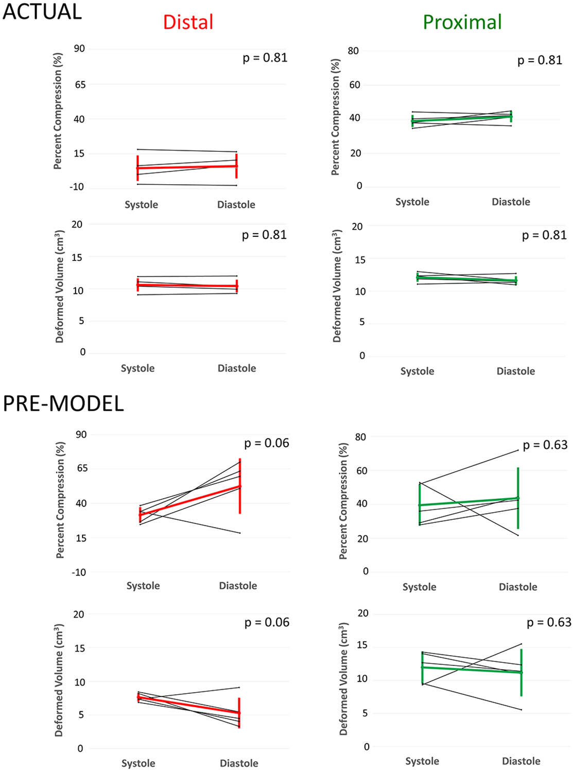
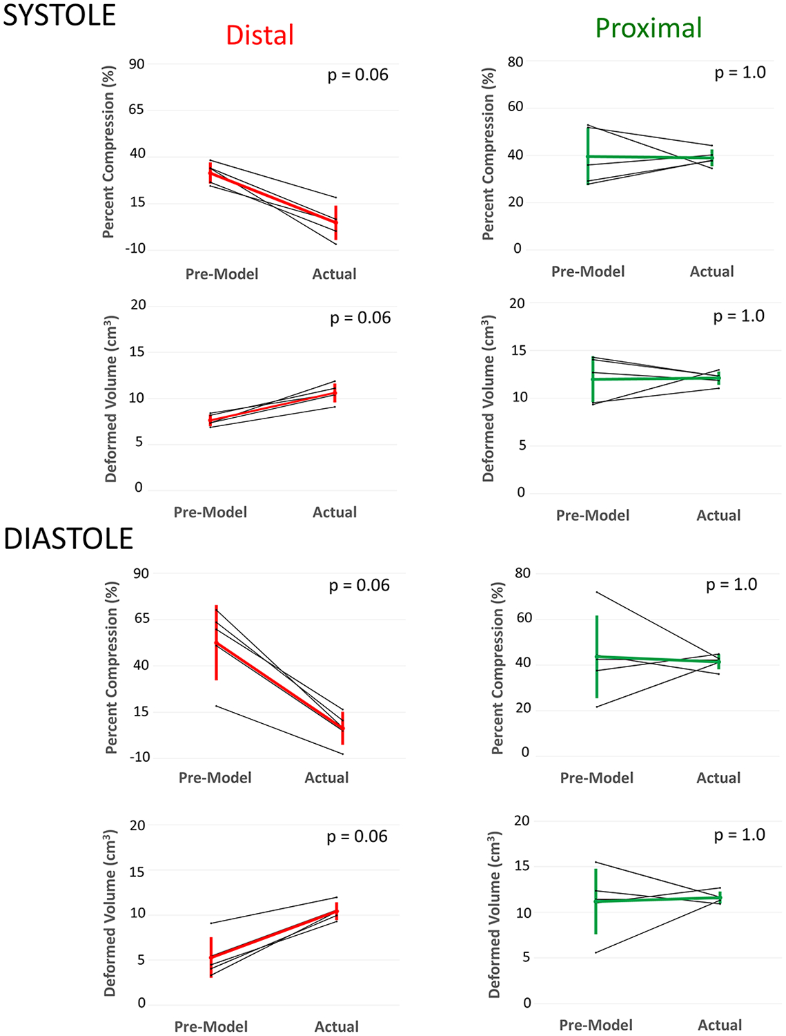
Background: Pulmonary insufficiency is a consequence of transannular patch repair in Tetralogy of Fallot (ToF) leading to late morbidity and mortality. Transcatheter native outflow tract pulmonary valve replacement has become a reality. However, predicting a secure, atraumatic implantation of a catheter-based device remains a significant challenge due to the complex and dynamic nature of the right ventricular outflow tract (RVOT). We sought to quantify the differences in compression and volume for actual implants, and those predicted by pre-implant modeling.

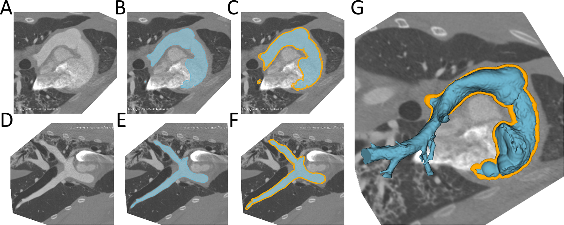
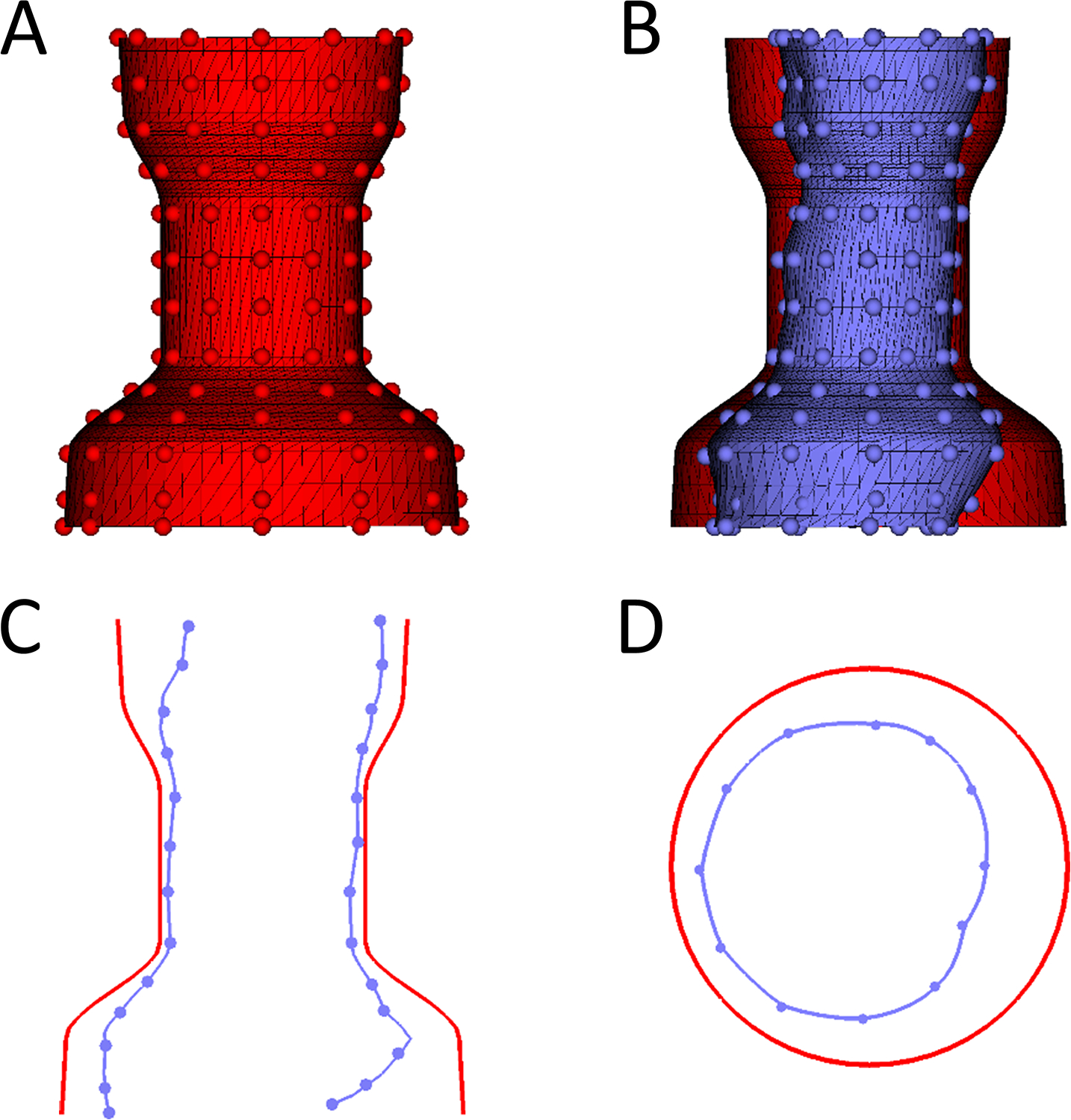
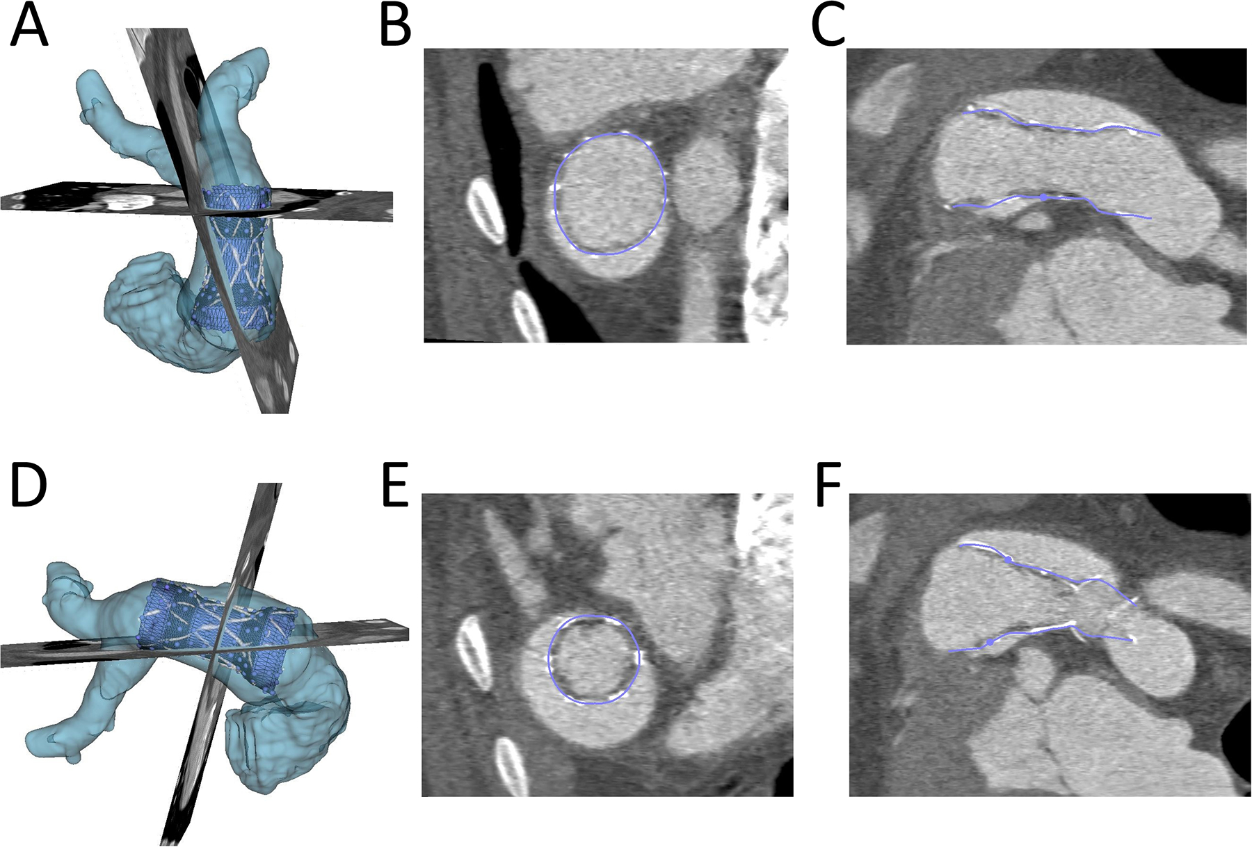
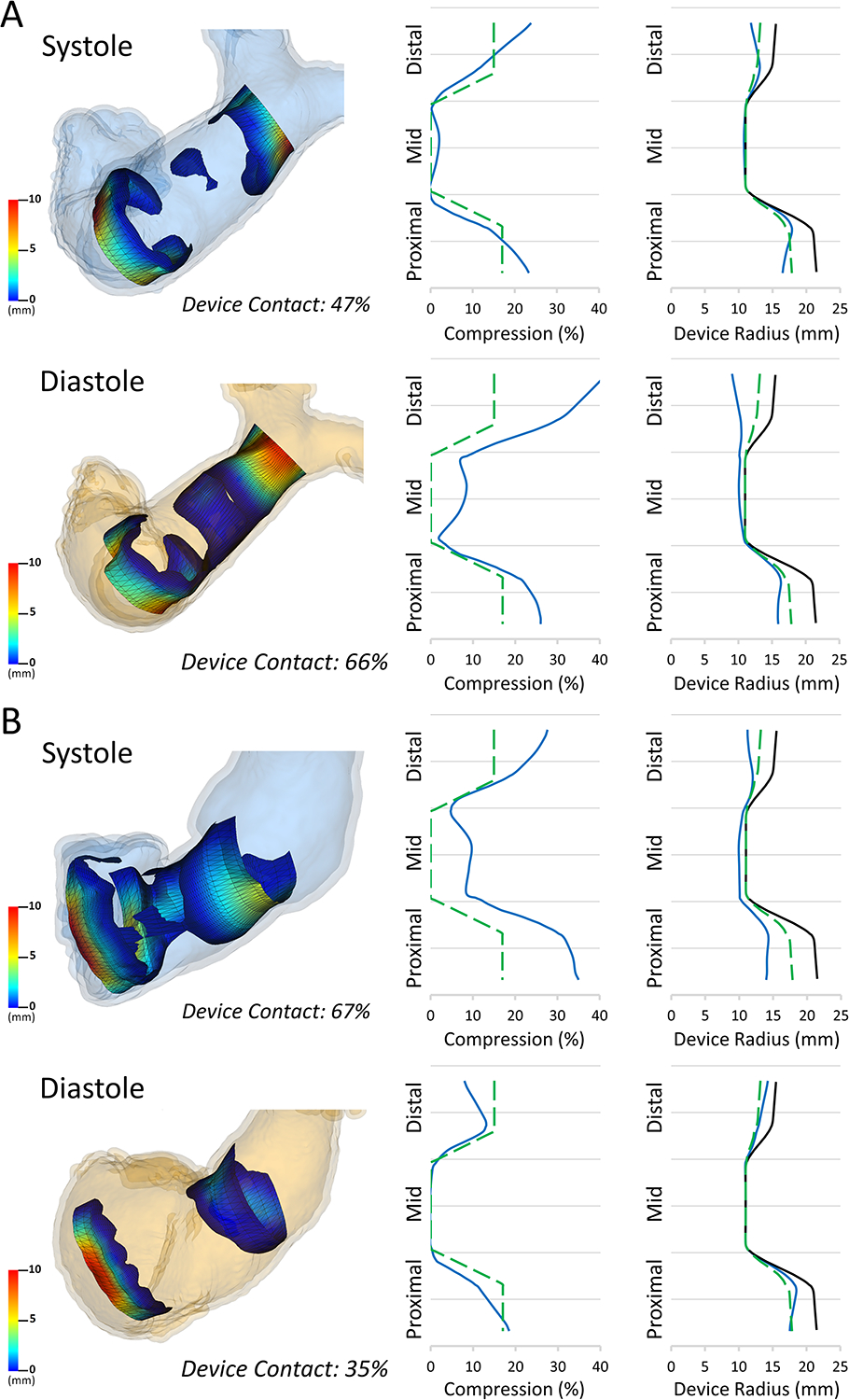
Methods: We used custom software to interactively place virtual transcatheter pulmonary valves (TPVs) into RVOT models created from pre-implant and post Harmony valve implant CT scans of 5 ovine surgical models of TOF to quantify and visualize device volume and compression.

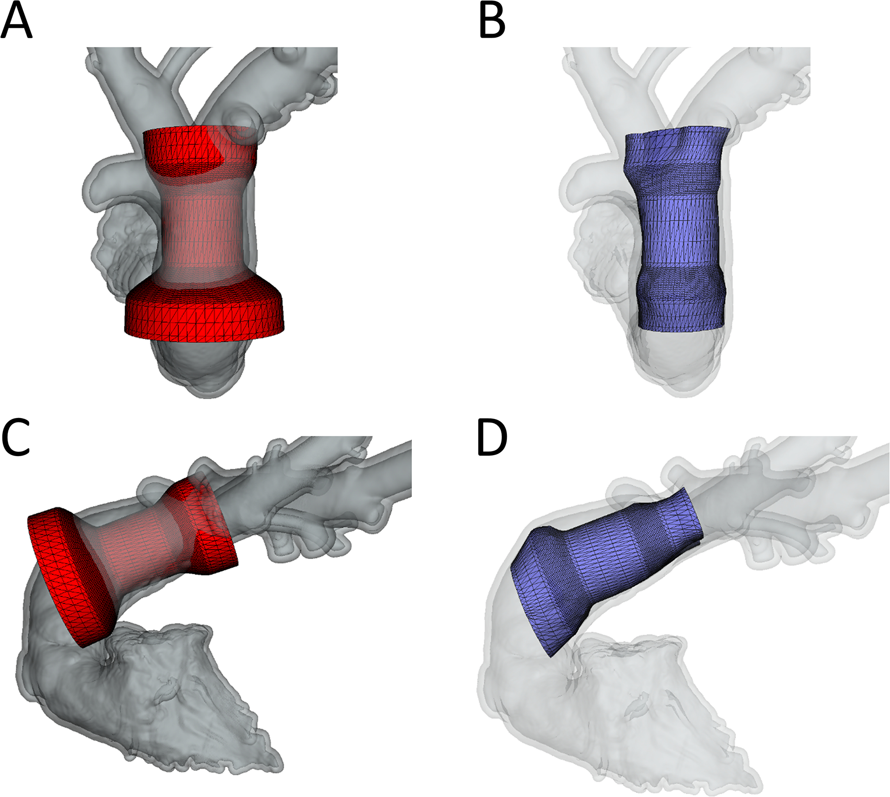
Results: Virtual device placement visually mimicked actual device placement and allowed for quantification of device volume and radius. On average, simulated proximal and distal device volumes and compression did not vary statistically throughout the cardiac cycle (P = 0.11) but assessment was limited by small sample size. In comparison to actual implants, there was no significant pairwise difference in the proximal third of the device (P > 0.80), but the simulated distal device volume was significantly underestimated relative to actual device implant volume (P = 0.06).

Conclusions: This study demonstrates that pre-implant modeling which assumes a rigid vessel wall may not accurately predict the degree of distal RVOT expansion following actual device placement. We suggest the potential for virtual modeling of TPVR to be a useful adjunct to procedural planning, but further development is needed.

Keywords: magnetic resonance imaging; percutaneous pulmonary valve implantation; prosthetic heart valve; tetralogy of Fallot.


Conflict of interest statement

DISCLOSURES

Drs. Gillespie and Morray serve as consultants for Medtronic, the manufacturer of the Harmony Device. The other authors report no conflicts.

Figures

Figure 1.
Figure 1.. Creation of RVOT Model (Segmentation) from CT data.
A) Native CT slice plane, B)RVOT model created in slice plane, C) RVOT (blue) surrounded by RVOT shell (orange) in slice plane; D-F) Same as above from side view; G: 3D rendered RVOT segmentation surrounded by RVOT Shell. RVOT=Right ventricular outflow tract.
Figure 2.
Figure 2.. Device Conformation Workflow.
A. Example of unconformed device in 3D (Red) with handles depicted as small spheres which can moved to conform the device; B. Conformed device (blue) compared to unconfirmed device (red); C. Saggital plane comparing uncomformed device (Red) to conformed device (Blue). D. Transverse cutting plane comparing uncomformed device (Red) to conformed device (Blue).
Figure 3.
Figure 3.. Demonstration of Ability of Virtual Device to Model Actual Device Placement in Post-Implant CT Scan:
The actual volume rendered device (white), RVOT(light blue), the conformed virtual device (dark blue) are shown. A) and D): Device conformation in 3D from anterior and lateral view; B) and E) Device conformation in two axial planes; C) and F) Device conformation in two sagittal planes.
Figure 4.
Figure 4.. Demonstration of Conforming Virtual Device to the Right ventricular Outflow Tract from Pre-Implant CT Scan.
A)Frontal view of unconformed device; B) Frontal view of conformed device; C) Lateral view of unconformed device; D) Lateral view of conformed device.
Figure 5.
Figure 5.. Comparison of Pre-Modeling vs. Actual Implant in Systole and Diastole.
Computer models shown with olor scale on representing amount of compression: Blue, no compression through red significant compression. White is the volume rendered actual device implant overlayed on device segmentation with underlying model used for quantification of volumes and compression.
Figure 6.
Figure 6.. Changes in Device Volume and Compression from Systole and Diastole for Actual Implants (Top) and Pre-Model Virtual Implants (Bottom).
Distal: Distal third of device; Proximal: Proximal third of device. * Minimum p-value achievable with Wilcoxon signed-rank test with n=5.
Figure 7.
Figure 7.. Comparison of Actual Model Volumes and Compression to Pre-Model Volumes and Compression in Systole (Top) and Diastole (Bottom).
Total: Whole device; Distal: Distal third of device; Proximal: Proximal third of device. * Minimum p-value achievable with Wilcoxon signed-rank test with n=5.
Figure 8.
Figure 8.. Estimated Contact Visualization and Graphical Summary Data in Pre-Implant Model.
Left: Estimated contact maps for A)Device Primarily in the Pulmonary Artery(Sheep 5) and B)Device Primarily in the Right Ventricle(Sheep 4) in both Systole and Diastole. Middle: Example plot of the mean % compression (blue) relative to ideal minimal compression (dashed green) across device length for each model; Right: Example plot of mean device radius (blue) compared to ideal minimal compression (dashed green) and fully expanded device (black) for each model.

References

    1. Al Habib HF, Jacobs JP, Mavroudis C, Tchervenkov CI, O’Brien SM, Mohammadi S, Jacobs ML. Contemporary patterns of management of tetralogy of Fallot: data from the Society of Thoracic Surgeons Database. Ann Thorac Surg 2010;90(3):813–9; discussion 819–20. - PubMed
    1. Apitz C, Webb GD, Redington AN. Tetralogy of fallot. The Lancet 2009;374(9699):1462–1471. - PubMed
    1. Schoonbeek RC, Takebayashi S, Aoki C, Shimaoka T, Harris MA, Fu GL, Kim TS, Dori Y, McGarvey J, Litt H and others. Implantation of the Medtronic Harmony Transcatheter Pulmonary Valve Improves Right Ventricular Size and Function in an Ovine Model of Postoperative Chronic Pulmonary Insufficiency. Circ Cardiovasc Interv 2016;9(10). - PMC - PubMed
    1. Geva T. Repaired tetralogy of Fallot: the roles of cardiovascular magnetic resonance in evaluating pathophysiology and for pulmonary valve replacement decision support. J Cardiovasc Magn Reson 2011;13:9. - PMC - PubMed
    1. Geva T. Indications for pulmonary valve replacement in repaired tetralogy of fallot: the quest continues. Circulation 2013;128(17):1855–7. - PMC - PubMed

MeSH terms