. 2019 Mar;52(2):e12544.
doi: 10.1111/cpr.12544. Epub 2018 Nov 15.

TGF-β1 promotes gap junctions formation in chondrocytes via Smad3/Smad4 signalling

Affiliations

TGF-β1 promotes gap junctions formation in chondrocytes via Smad3/Smad4 signalling

Qingxuan Wang et al. Cell Prolif. 2019 Mar.

Abstract

Objectives: Connexin-mediated functional gap junction intercellular communication (GJIC) has a vital role in development, homeostasis and pathology. Transforming growth factor-β1 (TGF-β1), as one of the most vital factors in chondrocytes, promotes cartilage precursor cell differentiation and chondrocyte proliferation, migration and metabolism. However, how TGF-β1 mediates GJIC in chondrocytes remains unclear. This study aims to determine the influence of TGF-β1 on GJIC in mouse chondrocytes and its underlying mechanism.

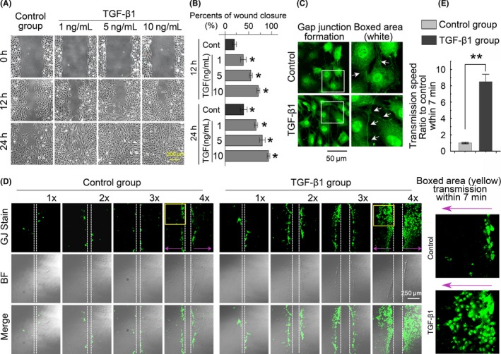
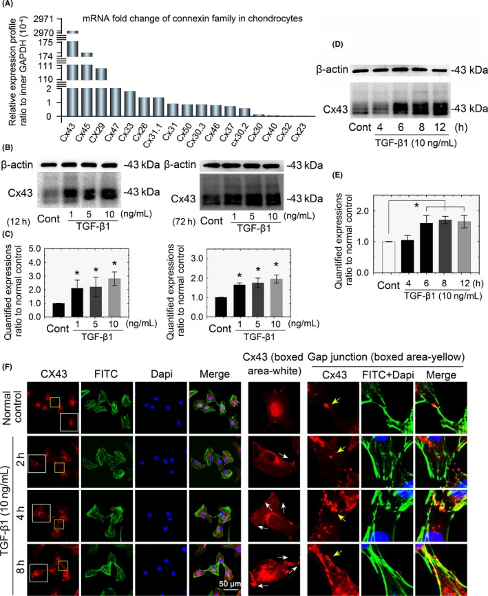
Methods: qPCR and mRNA microarray were used to verify the expression of genes in the TGF-β and connexin families in cartilage and chondrocytes. A scrape loading/dye transfer assay was performed to explore GJIC. Western blot analysis was used to detect connexin43 (Cx43) and Smad signalling components. Immunofluorescence staining was performed to characterize protein distribution.

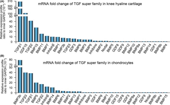
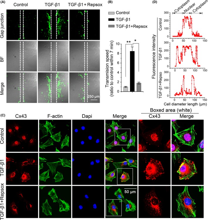
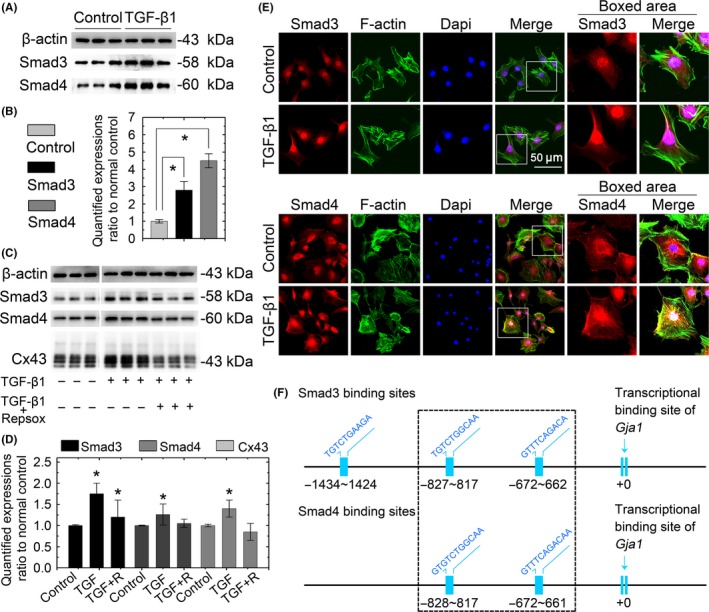
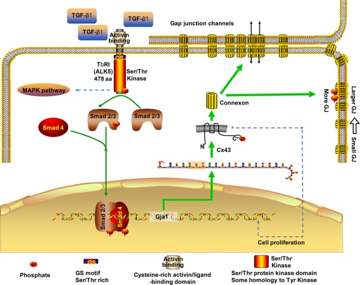
Results: The TGF-β1 mRNA was the highest expressed member of the TGFβ super family in cartilage. TGF-β1 promoted functional GJIC through increased expression of Cx43. TGF-β1-mediated GJIC required the participation of TGF-β type I receptor. TGF-β1 activated Smad3 and Smad4 signalling to facilitate their nuclear translocation. The Smad3 and Smad4 signalling proteins bound to the promoter of Gja1 and thus initiated Cx43 gene expression.

Conclusions: For the first time, these results revealed a vital role of TGF-β1 in cell-cell communication in chondrocytes via gap junction formation. We describe the regulatory mechanism, the involvement of TGF-β type I receptor and the nuclear translocation of Smad3/4.

Keywords: GJIC; Smad3; Smad4; TGF-β1; chondrocyte; connexin43.


Conflict of interest statement

The authors declare that no competing interests exist.

Figures

Figure 1
Figure 1
Expression of the TGF‐β super family in knee hyaline cartilage tissue and chondrocytes. A, qPCR confirmed gene profiles of the TGF‐β super family members in mouse knee hyaline cartilage tissue. The gene profiles of the TGF‐β super family were presented as the fold change ratio to the internal GAPDH control. B, qPCR confirmed gene profiles of the TGF‐β super family in mouse chondrocytes. The gene profiles of TGF‐β super family members were presented as the fold change ratio to internal GAPDH control. The fold change values were displayed in descending order. Data shown are representative of three independent experiments (n = 3). qPCR, quantitative real‐time PCR; TGF‐β, Transforming growth factor‐β1
Figure 2
Figure 2
TGF‐β1 promotes gap junction intercellular communication in chondrocytes. A, Scratch assay showing that TGF‐β1 increased chondrocyte migration in a dose‐dependent manner. The images were based on three independent experiments (n = 3). B, Monolayer cells were scraped in the presence of 1 mg/mL of Lucifer yellow. LYdye assay (transferred for 30 minutes to trace functional cell‐cell connections among chondrocytes) showing functional gap junction formation was significantly enhanced in the TGF‐β1 (10 ng/mL)‐treated group relative to the control group. The white arrows denote long gap junction formations among chondrocytes induced by TGF‐β1. The images were based on three independent experiments (n = 3). C, The scrape loading/dye transfer (SL/DT) assay further demonstrated a cell density‐dependent increase in gap junctions in chondrocytes induced by TGF‐β1 (10 ng/mL). Images of chondrocyte cells positive for intercellular gap junction transfer as detected by Lucifer yellow dye in different phases of cell cultures experiments. The control groups refer to different densities of cells (1×: 2 × 105 cells/mL; 2×: 4 × 105 cells/mL; 3×: 6 × 105 cells/mL; 4×: 8 × 105 cells/mL). The TGF‐β1 group refers to the corresponding cell densities with 10 ng/mL TGF‐β1 treatment for 24 hours. The Lucifer yellow dye enters cells at the scratch (white dotted line) and is transferred to cells distant from the scratch (purple arrow). Intercellular gap junction transfer was calculated by measuring the distance from the scratching edge to the most distant cells with Lucifer yellow uptake. The images were based on three independent experiments observed with CLSM (n = 3). D, Quantification was performed to show different transmission speeds within 7 minutes of TGF‐β1 treatment compared to the control groups. Data are presented as the mean ± SEM (n = 3). ** P < 0.01. CLSM, confocal laser‐scanning microscopy; TGF‐β, Transforming growth factor‐β1
Figure 3
Figure 3
TGF‐β1 induced an increase in Cx43 in chondrocytes. mRNA microarray showing the gene profiles of the connexin family in chondrocytes. Seventeen connexin family members were detected in mouse chondrocytes. Primary chondrocytes isolated from articular cartilage were analysed. Data are presented as ratios to the internal GAPDH control. Fold change values were displayed in descending order. B, Western blots showing that TGF‐β1 promoted Cx43 expression in a dose‐dependent manner in early (12 hours) and late (72 hours) stages after treatment. The gels shown are representative of three different experiments (n = 3). C, Western blots showing that TGF‐β1 promoted Cx43 expression in a time‐dependent manner after the 10 ng/mL treatment. The gels shown are representative of three different experiments (n = 3). D, Immunofluorescence showing the TGF‐β1‐mediated change in localization of Cx43 in chondrocytes and gap junction sites between cells. Cytoskeleton stained with FITC‐ phalloidine (green) and nuclei stained with DAPI (blue). The TGF‐β1‐induced translocation of Cx43 from the nucleus towards the membrane border is indicated with white arrows (middle boxed area). The TGF‐β1‐induced accumulation of Cx43 at gap junction sites is indicated in yellow arrows (right boxed area). The images shown are representative of three different experiments (n = 3). Cx43, connexin43; TGF‐β, Transforming growth factor‐β1
Figure 4
Figure 4
Repsox pre‐incubation attenuated TGF‐β1‐induced gap junction formation and Cx43 expression. A, Repsox pre‐incubation attenuated TGF‐β1‐induced gap junction formation. The concentration of TGF‐β1 was 10 ng/mL. The repsox used was at 100 μmol/L for a 12 hours pre‐incubation with primary chondrocytes. The images were based on three independent experiments observed with CLSM (n = 3). B, Quantitative analysis of the cell transmission speed of Lucifer yellow dye with respect to the control and TGF‐β1‐induced group (n = 3). *P < 0.05; **P < 0.01. C, Immunofluorescence staining of Cx43 (red) in chondrocytes after a 12‐hour treatment with TGF‐β1 (10 ng/mL) in the presence or absence of repsox (100 μmol/L). The chondrocytes were counterstained with markers of the nucleus (DAPI, blue) and actin cytoskeleton (phalloidin, green). The images were based on three independent experiments observed by CLSM (n = 3). D, Fluorescence optical density (OD) was analysed in Image Pro Plus 6.0 to measure the specific distribution of Cx43 cross the cell body. Data analysis was performed on at least 40 cells per group. CLSM, confocal laser‐scanning microscopy; Cx43, connexin43; TGF‐β, Transforming growth factor‐β1
Figure 5
Figure 5
TGF‐β1 modulates expression of Cx43 in chondrocytes via Smad3 and Smad4 signalling. A, Western blots showing that TGF‐β1 increased the expressions of Smad3 and Smad4 in chondrocytes. Cell lysates were collected after a 2‐hour treatment with TGF‐β1 (10 ng/ml). The gels shown are representative of three independent experiments (n = 3). B, Quantification was performed to analyse the changes of Smad3 and Smad4 in (A). *Significant difference with respect to the untreated control chondrocytes (P < 0.05). C, Western blots showing that the repsox pre‐incubation attenuated the Smad3, Smad4 and Cx43 induction caused by TGF‐β1. Lysates were collected after 12‐hour TGF‐β1 (10 ng/mL) treatment with or without a repsox (100 μmol/L) pre‐incubation. The gels shown are representative of three independent experiments (n = 3). D, Immunofluorescent images showing the translocation of Smad3 and Smad4 in chondrocytes after induction with 10 ng/mL TGF‐β1. The chondrocytes were counterstained with markers of the nucleus (DAPI, blue) and actin cytoskeleton (phalloidin, green). The images are based on three independent experiments observed by CLSM (n = 3). E, Bioinformatics analysis by PROMO resource showing the putative Smad3‐ and Smad4‐binding sites in the promoter of Cx43 (Gja1, GeneBank name). Smad3 had three potential binding sites in the promoter of Gja1, and those sites were located at 1434‐1424 bp, 827‐817 bp and 672‐622 bp upstream of the Gja1 transcriptional start site. Smad4 had two binding sites in the Gja1 promoter located 828‐817 bp and 672‐621 bp upstream of the Gja1 TSS. CLSM, confocal laser‐scanning microscopy; Cx43, connexin43; TGF‐β, Transforming growth factor‐β1
Figure 6
Figure 6
Schematic diagram illustrates the mechanism of TGF‐β1 regulation of Cx43 in chondrocytes. The green lines point to Smad‐dependent pathways elucidated by the current study, and the blue dotted lines are potential mechanisms not included in the present study. TGF‐β1 induced the canonical Smad signalling pathway, the activated TβRI phosphorylates the R‐Smads (Smad2, Smad3), transform them into transcriptional co‐regulator together with Smad4, and dock to potential binding site of promoter of Gja1 which encodes Cx43. As a result, Cx43 accumulated more on cell border and phosphorylated into functional gap junctions with larger size. Cx43, connexin43; TGF‐β, Transforming growth factor‐β1

Similar articles

Cited by

References

    1. Giepmans B. Gap junctions and connexin‐interacting proteins. Cardiovasc Res. 2004;62:233‐245. - PubMed
    1. Hervé JC, Derangeon M. Gap‐junction‐mediated cell‐to‐cell communication. Cell Tissue Res. 2013;352:21‐31. - PubMed
    1. Willecke K, Eiberger J, Degen J, et al. Structural and functional diversity of connexin genes in the mouse and human genome. Biol Chem. 2002;383:725‐737. - PubMed
    1. Cherian PP, Sillerjackson AJ, Gu S et al. Mechanical strain opens connexin 43 hemichannels in osteocytes: a novel mechanism for the release of prostaglandin. Mol Biol Cell. 2005;16:3100‐3106. - PMC - PubMed
    1. Ishikawa M, Williams GL, Ikeuchi T, Sakai K, Fukumoto S, Yamada Y. Pannexin 3 and connexin 43 modulate skeletal development via distinct functions and expression patterns. J Cell Sci. 2016;129:1018‐1030. - PMC - PubMed