. 2018 Nov;23(12):1-9.
doi: 10.1117/1.JBO.23.12.121615.

Depth dependence of coherent hemodynamics in the human head

Affiliations

Depth dependence of coherent hemodynamics in the human head

Kosar Khaksari et al. J Biomed Opt. 2018 Nov.

Abstract

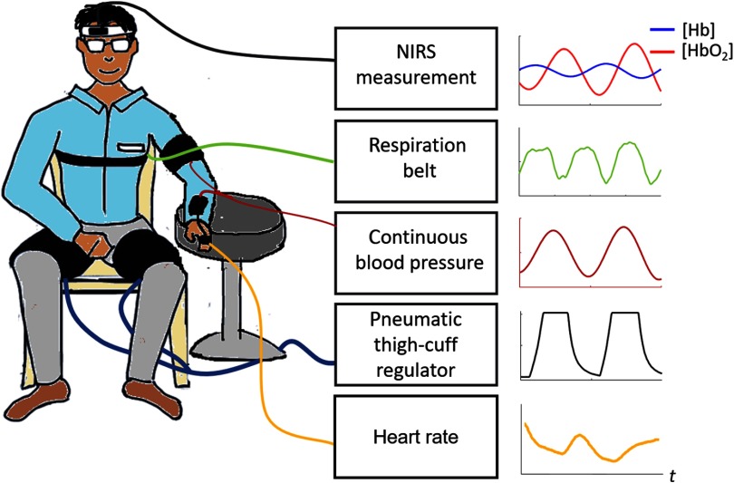
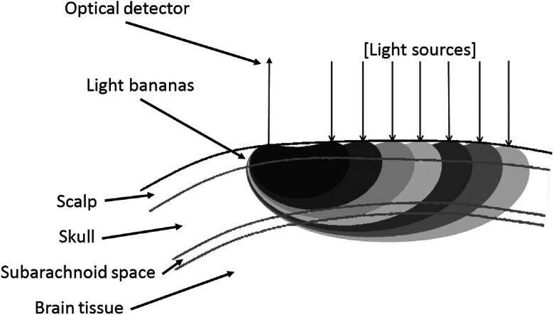
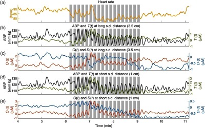
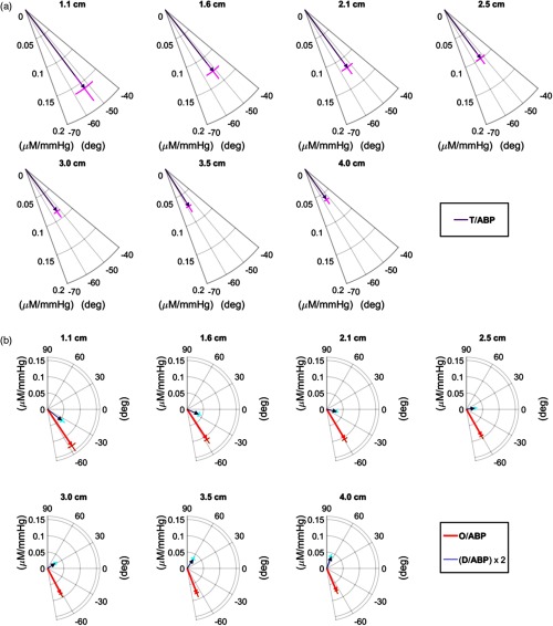
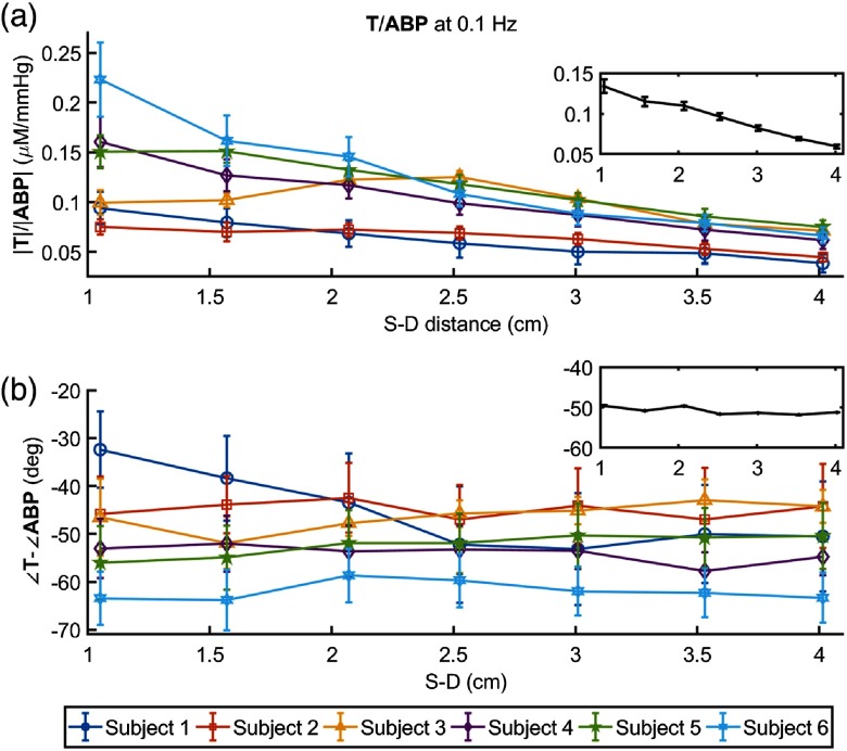
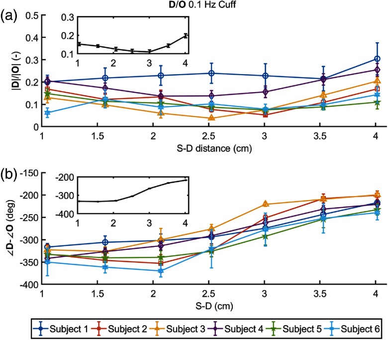
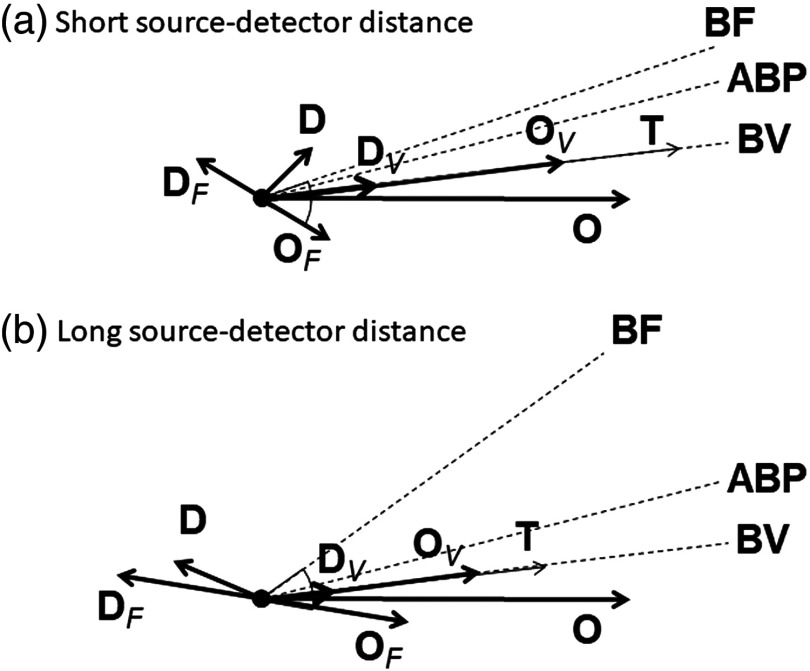
We report a near-infrared spectroscopy (NIRS) study of coherent hemodynamic oscillations measured on the human forehead at multiple source-detector distances (1 to 4 cm). The physiological source of the coherent hemodynamics is arterial blood pressure oscillations at a frequency of 0.1 Hz, induced by cyclic inflation (to a pressure of 200 mmHg) and deflation of two thigh cuffs wrapped around the subject's thighs. To interpret our results, we use a recently developed hemodynamic model and a phasor representation of the oscillations of oxyhemoglobin, deoxyhemoglobin, and total hemoglobin concentrations in the tissue (phasors O, D, and T, respectively). The increase in the phase angle between D and O at larger source-detector separations is assigned to greater flow versus volume contributions and to a stronger blood flow autoregulation in deeper tissue (brain cortex) with respect to superficial tissue (scalp and skull). The relatively constant phase lag of T versus arterial blood pressure oscillations at all source-detector distances was assigned to competing effects from stronger autoregulation and smaller arterial-to-venous contributions in deeper tissue with respect to superficial tissue. We demonstrate the application of a hemodynamic model to interpret coherent hemodynamics measured with NIRS and to assess the different nature of shallow (extracerebral) versus deep (cerebral) tissue hemodynamics.

Keywords: brain perfusion; cerebral autoregulation; coherent hemodynamics; hemoglobin concentration; near-infrared spectroscopy; phasors.


Figures

Fig. 1
Fig. 1
Experimental setup and typical temporal signals collected.
Fig. 2
Fig. 2
Cross-section of optical probe with representative light bananas in relation to multi-layer tissue structure.
Fig. 3
Fig. 3
Representative time series of the data collected on subject no. 1. (a) Heart rate, (b) ABP and T at a source–detector (s.d.) distance of 3.5 cm, (c) O and D at 3.5 cm, (d) ABP and T at 1 cm, and (e) O and D at 1 cm. The shaded areas indicate the times of thigh cuffs inflation.
Fig. 4
Fig. 4
(a) Phasor ratio T/ABP and (b) phasor ratios D/ABP and O/ABP for different source-detector separations (specified in the figure) for subject no. 4. Note that the phasor ratio D/ABP is multiplied by 2 for better visualization.
Fig. 5
Fig. 5
(a) Amplitude ratio |T|/|ABP| and (b) phase difference TABP versus source-detector distance at 0.1 Hz for all subjects. The inset in each panel shows the grand average, with standard error, over the six subjects.
Fig. 6
Fig. 6
Grand average of amplitude ratio |T|/|ABP| versus source–detector distance at the heart rate for all subjects.
Fig. 7
Fig. 7
(a) Amplitude ratio and (b) phase difference of D versus O as a function of source–detector distance at 0.1 Hz for all subjects. The inset in each panel shows the grand average, with standard error, over the six subjects.
Fig. 8
Fig. 8
Phasor diagrams representative of the oscillatory hemodynamics induced by ABP oscillations as measured with NIRS on the forehead of human subjects at (a) short and (b) long source–detector distance. Dashed lines indicate the phase of BF, ABP, and BV oscillations. Subscripts V and F indicate volume and flow contributions, respectively, to oxyhemoglobin (O) and deoxyhemoglobin (D) concentration oscillations. Key phasor relationships are: T=O+D, O=OV+OF, D=DV+DF, and OF=DF.

References

    1. Fantini S., “Dynamic model for the tissue concentration and oxygen saturation of hemoglobin in relation to blood volume, flow velocity, and oxygen consumption: implications for functional neuroimaging and coherent hemodynamics spectroscopy (CHS),” NeuroImage 85(1), 202–221 (2014).NEIMEF10.1016/j.neuroimage.2013.03.065 - DOI - PMC - PubMed
    1. Katura T., et al. , “Quantitative evaluation of interrelations between spontaneous low-frequency oscillations in cerebral hemodynamics and systemic cardiovascular dynamics,” NeuroImage 31(4), 1592–1600 (2006).NEIMEF10.1016/j.neuroimage.2006.02.010 - DOI - PubMed
    1. Taga G., et al. , “Spontaneous oscillation of oxy- and deoxy-hemoglobin changes with a phase difference throughout the occipital cortex of newborn infants observed using non-invasive optical topography,” Neurosci. Lett. 282(1–2), 101–104 (2000).10.1016/S0304-3940(00)00874-0 - DOI - PubMed
    1. Watanabe H., et al. , “Hemoglobin phase of oxygenation and deoxygenation in early brain development measured using fNIRS,” Proc. Natl. Acad. Sci. U. S. A. 114(9), E1737–E1744 (2017)10.1073/pnas.1616866114 - DOI - PMC - PubMed
    1. Obrig H., et al. , “Spontaneous low frequency oscillations of cerebral hemodynamics and metabolism in human adults,” NeuroImage 12(6), 623–639 (2000).NEIMEF10.1006/nimg.2000.0657 - DOI - PubMed

Publication types