Review
. 2019 Jan;248(1):98-117.
doi: 10.1002/dvdy.1. Epub 2018 Dec 10.

Developmental roles of microglia: A window into mechanisms of disease

Affiliations
Review

Developmental roles of microglia: A window into mechanisms of disease

Sarah R Anderson et al. Dev Dyn. 2019 Jan.

Abstract

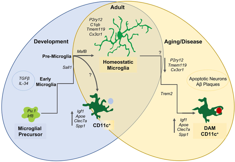
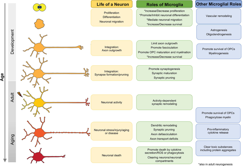
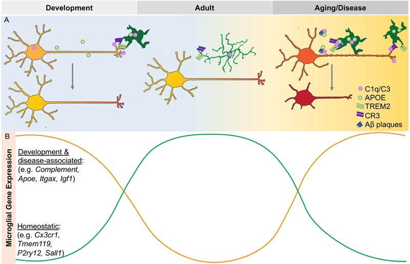
Microglia are engineers of the central nervous system (CNS) both in health and disease. In addition to the canonical immunological roles of clearing damaging entities and limiting the spread of toxicity and death, microglia remodel the CNS throughout life. While they have been extensively studied in disease and injury, due to their highly variable functions, their precise role in these contexts still remains uncertain. Over the past decade, we have greatly expanded our understanding of microglial function, including their essential homeostatic roles during development. Here, we review these developmental roles, identify parallels in disease, and speculate whether developmental mechanisms re-emerge in disease and injury. Developmental Dynamics 248:98-117, 2019. © 2018 Wiley Periodicals, Inc.

Keywords: CNS development; aging; disease; microglia; neurodegeneration; neurogenesis.


Figures

Figure 1.
Figure 1.. Microglia have distinct functional states in development, health, and disease.
During early development, microglial precursors migrate to the brain and mature in a stepwise process into early, pre-, and adult microglia. During the early postnatal period, a subpopulation of CD11c-expressing microglia with a distinct profile localize to developing white matter tracts. During aging and disease, microglia downregulate homeostatic genes and upregulate disease-associated genes, a subset of which are common to developmental CD11c+ microglia. (DAM = disease-associated microglia).
Figure 2.
Figure 2.. Neurons are remodeled by microglia over their lifetime.
Microglia enter the neuroepithelium and influence progenitors, neuronal differentiation, migration, and survival. They then remodel neuronal structures throughout development, adulthood and in aging, injury, and disease. Microglia also regulate gliogenesis and vascular development, and clear damaging entities and release cytokines in aging and disease.
Figure 3.
Figure 3.. Genes implicated in disease are highly expressed in development and drive parallel functions.
Microglia utilize the same signaling pathways to regulate synaptic pruning and phagocytosis in development and disease (A). Genes implicated in disease are developmentally regulated and have inverse expression with homeostatic genes (B).

References

    1. Aarum J, Sandberg K, Haeberlein SL, Persson MA. 2003. Migration and differentiation of neural precursor cells can be directed by microglia. Proc Natl Acad Sci U S A 100:15983–15988 - PMC - PubMed
    1. Ahmed F, Brown KM, Stephan DA, Morrison JC, Johnson EC, Tomarev SI. 2004. Microarray analysis of changes in mRNA levels in the rat retina after experimental elevation of intraocular pressure. Invest Ophthalmol Vis Sci 45:1247–1258. - PubMed
    1. Ajami B, Bennett JL, Krieger C, Tetzlaff W, Rossi FM. 2007. Local self-renewal can sustain CNS microglia maintenance and function throughout adult life. Nat Neurosci 10:1538–1543. - PubMed
    1. Al-Zamil WM, Yassin SA. 2017. Recent developments in age-related macular degeneration: a review. Clinical Interventions in Aging 12:1313–1330. - PMC - PubMed
    1. Alizadeh A, Karimi‐Abdolrezaee S. 2016. Microenvironmental regulation of oligodendrocyte replacement and remyelination in spinal cord injury. The Journal of Physiology 594:3539–3552. - PMC - PubMed

Publication types

MeSH terms

LinkOut - more resources