. 2019 May;29(3):437-450.
doi: 10.1111/bpa.12677. Epub 2018 Dec 27.

CLARITY reveals a more protracted temporal course of axon swelling and disconnection than previously described following traumatic brain injury

Affiliations

CLARITY reveals a more protracted temporal course of axon swelling and disconnection than previously described following traumatic brain injury

Maura T Weber et al. Brain Pathol. 2019 May.

Abstract

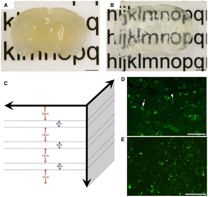
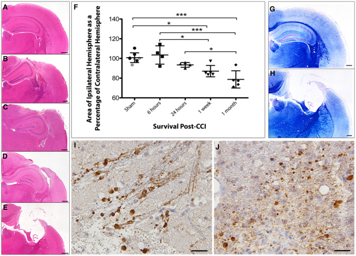
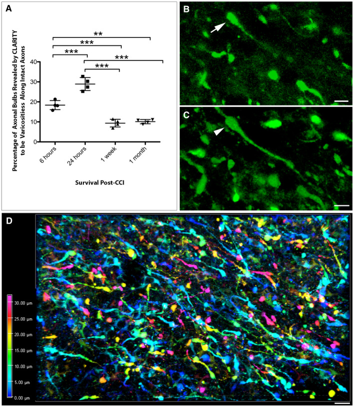
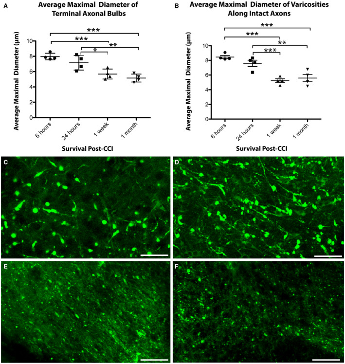
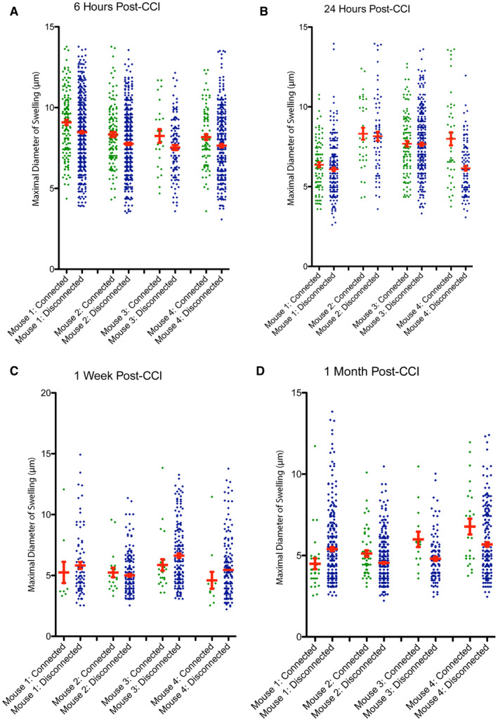
Diffuse axonal injury (DAI) is an important consequence of traumatic brain injury (TBI). At the moment of trauma, axons rarely disconnect, but undergo cytoskeletal disruption and transport interruption leading to protein accumulation within swellings. The amyloid precursor protein (APP) accumulates rapidly and the standard histological evaluation of axonal pathology relies upon its detection. APP+ swellings first appear as varicosities along intact axons, which can ultimately undergo secondary disconnection to leave a terminal "axon bulb" at the disconnected, proximal end. However, sites of disconnection are difficult to determine with certainty using standard, thin tissue sections, thus limiting the comprehensive evaluation of axon degeneration. The tissue-clearing technique, CLARITY, permits three-dimensional visualization of axons that would otherwise be out of plane in standard tissue sections. Here, we examined the morphology and connection status of APP+ swellings using CLARITY at 6 h, 24 h, 1 week and 1 month following the controlled cortical impact (CCI) model of TBI in mice. Remarkably, many APP+ swellings that appeared as terminal bulbs when viewed in standard 8-µm-thick regions of tissue were instead revealed to be varicose swellings along intact axons when three dimensions were fully visible. Moreover, the percentage of these potentially viable axon swellings differed with survival from injury and may represent the delayed onset of distinct mechanisms of degeneration. Even at 1-month post-CCI, ~10% of apparently terminal bulbs were revealed as connected by CLARITY and are thus potentially salvageable. Intriguingly, the diameter of swellings decreased with survival, including varicosities along intact axons, and may reflect reversal of, or reduced, axonal transport interruption in the chronic setting. These data indicate that APP immunohistochemistry on standard thickness tissue sections overestimates axon disconnection, particularly acutely post-injury. Evaluating cleared tissue demonstrates a surprisingly delayed process of axon disconnection and thus longer window of therapeutic opportunity than previously appreciated. Intriguingly, a subset of axon swellings may also be capable of recovery.

Keywords: CLARITY; TBI; amyloid precursor protein; axon degeneration; axonal pathology; diffuse axonal injury; traumatic brain injury.


Conflict of interest statement

No authors have any conflicts of interest.

Figures

Figure 1
Figure 1
Evaluation of axonal pathology using the CLARITY technique. Representative example of 2‐mm‐thick block of uninjured mouse brain sectioned in the coronal plane (A) before and (B) after tissue clearing. (C) Diagram demonstrating the approach to analysis of cleared tissue. Specifically, 8‐µm regions of tissue were flattened (maximum‐projection function) to simulate traditional formalin‐fixed paraffin‐embedded (FFPE) tissue sections. Bulbs identified within these regions were re‐evaluated with the full 3D of tissue up to 100 µm to determine their true morphology and connectivity status. Fourteen micrometer gaps were included to avoid measurement of the same bulbs twice (based on maximal bulb size). (D) Representative example of standard 8µm‐thick FFPE mouse tissue section immunofluorescently labeled with APP. Axonal swellings can be observed in the peri‐contusional region at 24 h following CCI. Note, while the vast majority of APP swellings appear as disconnected, terminal axonal bulbs (arrow), occasional varicosities can be observed with two clear projections from a single swelling along an intact axon (arrowhead). (E) APP immunoreactive axonal pathology observed peri‐contusionally at 24 h post‐CCI on 8‐µm‐thick maximum projection images using cleared tissue appears indistinguishable from standard FFPE immunofluorescent sections in (D). Scale bars: (A,B) 2 mm, (D,E) 50 µm.
Figure 2
Figure 2
Progressive atrophy and associated axonal pathology post‐CCI in standard FFPE tissue. (A) H&E staining on sham animals reveals an absence of any focal pathology and a morphologically normal cortical surface. (B–E) H&E staining performed (B) 6 h, (C) 24 h, (D) 1 week and (E) 1 month after CCI injury reveals acute cortical contusion and intraparenchymal hemorrhage at the site of impact as well as progressive atrophy of the ipsilateral hemisphere. (F) Tissue atrophy quantified by measuring the area of the injured ipsilateral hemisphere as a percentage of the contralateral hemisphere. All individual data are presented in addition to the mean percentage area ± SD. In the sham group, gray data points indicate mice sacrificed 24 h after sham injury and black data points indicate mice sacrificed 1 month following sham injury, indicating an absence of change caused by craniectomy. (G–H) Luxol fast blue staining indicates loss of white matter and major cortical tissue loss at (H) 1 month following injury compared to (G) sham. (I–J) Swollen axonal profiles and varicosities immunroeactive for APP at (I) 24 h and (J) 1‐month post‐CCI. Scale bars: (A–E, G–H) 400 µm, (I–J) 25 µm. *P < 0.05, ***< 0.001.
Figure 3
Figure 3
CLARITY reveals less axonal disconnection than previously thought following CCI. (A) Graph showing the percentage of APP+ swellings that appeared terminally disconnected when viewed in standard 8‐µm‐thick regions of tissue that were revealed to be connected varicose axonal swellings when all three dimensions were fully visible using CLARITY (**P ≤ 0.01, ***P ≤ 0.001). (B) Representative example of an APP+ apparent terminal axonal bulb (arrow) observed on an 8‐µm‐thick region of cleared tissue (maximum projection) at 24 h post‐CCI. (C) The same region of tissue as (B) re‐evaluated with all three dimensions now visible reveals that the apparent bulb is in fact a swelling that has two clear projections and thus represents a varicose swelling along an intact region of axon (arrowhead). (Alpha‐blended image) (D) Depth encoded three‐dimensional region of extensive APP+ axonal pathology in the peri‐contusional region 24 h post‐CCI. Extensive and large axonal swellings can be observed with complex morphologies comprising both disconnected axonal bulbs and varicosities along the intact length of axons. Scale bars: (B,C) 10 µm, (D) 50 µm.
Figure 4
Figure 4
The size of axonal swellings decreases with increased survival post‐CCI. The average maximal diameter of axonal swellings of both (A) terminally disconnected bulbs and (B) axonal varicosities along intact axons decreases over time following CCI. (*P ≤ 0.05, **P ≤ 0.01, ***P ≤ 0.001). (C–F) Representative examples of the relative size of axonal swellings in the peri‐contusional region in cleared tissue at (C) 6 h, (D) 24 h, (E) 1‐week and (F) 1‐month post‐CCI. Scale bars: (C–F) 20 µm.
Figure 5
Figure 5
The size of axonal swellings and connection status with survival. The maximal diameter for all individual disconnected, terminal axonal bulbs and connected axonal varicosities shown for each mouse at (A) 6 h, (B) 24 h, (C) 1‐week and (D) 1‐month post‐CCI. [Color figure can be viewed at wileyonlinelibrary.com]

Similar articles

Cited by

References

    1. Adams JH, Doyle D, Ford I, Gennarelli TA, Graham DI, McLellan DR (1989) Diffuse axonal injury in head injury: definition, diagnosis and grading. Histopathology 15:49–59. - PubMed
    1. Adams JH, Doyle D, Graham DI, Lawrence AE, Mclellan DR (1984) Diffuse axonal injury in head injuries caused by a fall. Lancet 2:1420–1422. - PubMed
    1. Adams JH, Graham DI, Gennarelli TA, Maxwell WL (1991) Diffuse axonal injury in non‐missile head injury. J Neurol Neurosurg Psychiatry 54:481–483. - PMC - PubMed
    1. Adams JH, Graham DI, Murray LS, Scott G (1982) Diffuse axonal injury due to nonmissile head injury in humans: an analysis of 45 cases. Ann Neurol 12:557–563. - PubMed
    1. Anderson CV, Bigler ED (1995) Ventricular dilation, cortical atrophy, and neuropsychological outcome following traumatic brain injury. J Neuropsychiatry Clin Neurosci 7:42–48. - PubMed

Publication types

Substances